Keywords
birth defects, micromelia, skeletal dysplasia, thanatophoric dysplasia
This article is included in the Rare diseases collection.
birth defects, micromelia, skeletal dysplasia, thanatophoric dysplasia
Thanatophoric dysplasia (TD) is rare, sporadic and lethal skeletal dysplasia attributed to a de novo mutation in the fibroblast growth factor receptor 3 gene (FGFR3) located on chromosome 4p 16.3.1 We encountered a 19-year-old primigravida at 26 weeks and three days of gestation presenting with ultrasonographic findings suggestive of thanatophoric dysplasia. She delivered a dead fetus with macrocephaly, a narrow bell-shaped thorax, a protruding abdomen and shortened limbs. Only a few cases of this devastating condition have been reported from Nepal. Here, we report a case of a dead female fetus born with TD at a tertiary hospital in rural Nepal.
This case report describes a case of a 19-year-old primigravida housewife in a non-consanguineous marriage. She had attended three antenatal visits before presenting for a routine antenatal check-up at our tertiary hospital in rural Nepal at 26 weeks and three days of gestation. She has no known chronic medical conditions or family history of congenital anomalies. She is a non-smoker and non-alcoholic, with no history of fever, skin rashes, spotting per vaginum, any drug intake, or radiation exposure.
Upon arrival, the patient’s vital signs were within the normal range. General physical examination revealed absence of pallor, edema, lymphadenopathy, or thyroid swelling. Systemic examination findings were not significant. Per abdomen examination revealed uterus at 28 weeks of gestation, non-tender with no contractions. However, fetal lie, presentation and heart rate could not be appreciated. Routine blood investigations including complete blood count, blood sugar, blood group and Rh typing and serum thyroid stimulating hormone (TSH) were within normal limits. HIV, HBsAg, HCV, and rapid plasma reagin (RPR) were non-reactive.
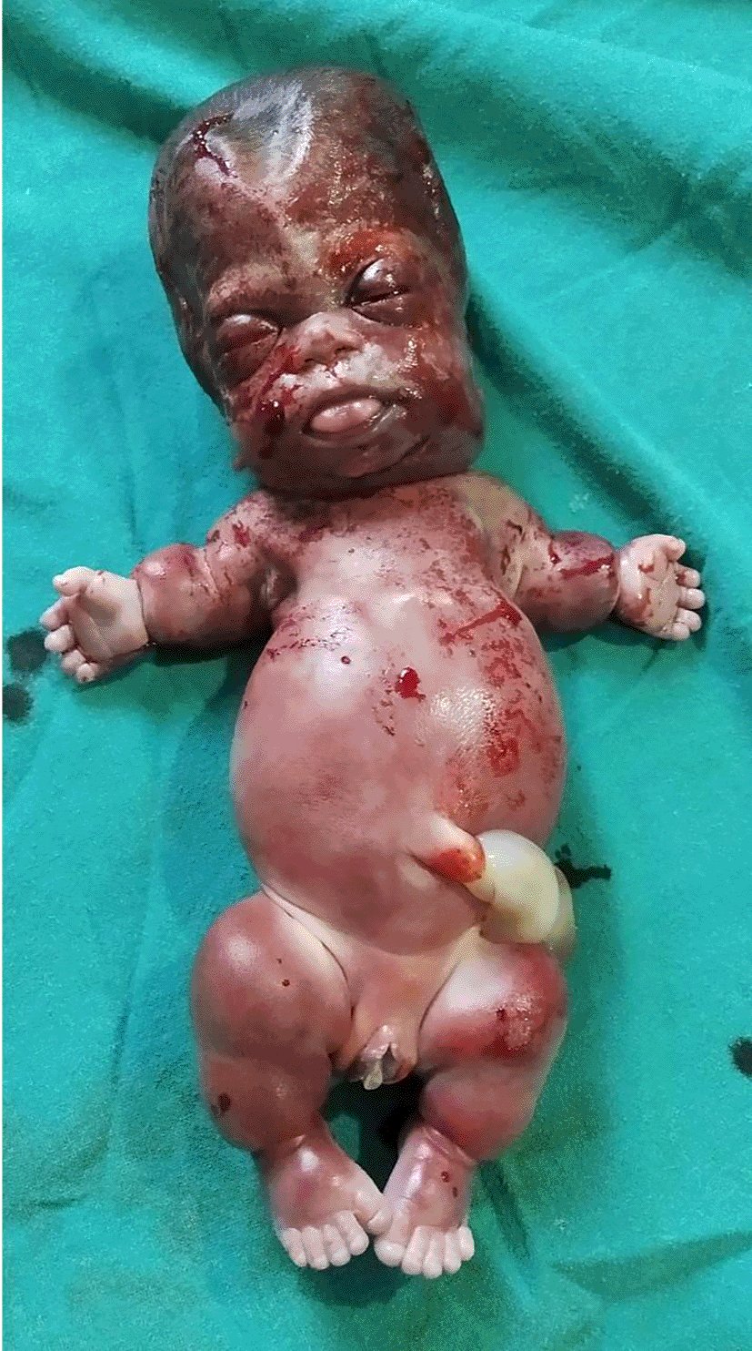
Ultrasonography revealed a single live fetus with an unstable lie at 26 weeks three days of gestation with all four limbs significantly shorter than expected for the gestational age, suggestive of micromelia (Figure 1). A diagnosis of thanatophoric dysplasia was made and the patient was counseled for termination of pregnancy, which she consented to. Labor was induced with Tab Mifepristone 200mg orally followed by Tab Misoprostol 200mcg vaginally three doses on the next day. Subsequently, she delivered a dead female fetus weighing 800 grams with coarse edematous facies, frontal bossing, long forehead with a saddle nose, low-set ears, short neck, a bell-shaped thorax with protruded abdomen, and shortened upper and lower limbs with stubby fingers and deep skin creases (Figure 2). Additionally, there was flat back with a small dimpling over the sacral region (Figure 3).

Based on the clinical features, the newborn was diagnosed as a case of TD type 1. The couple was counseled regarding the severity and lethal nature of this condition and advised for routine ultrasonography screening in subsequent pregnancies for the timely identification and management of this lethal disorder.
Thanatophoric dysplasia (TD) is the most common lethal neonatal skeletal dysplasia, with an approximate incidence of one in 20,000 to 50,000. The term “thanatophoric” is derived from the Greek words “thanatos” meaning ‘death’ and “phoros” meaning ‘bearing’. Marotux initially coined this term to describe dwarf newborns who tragically passed away within the first hour of their lives.1 Thanatophoric dysplasia is attributed to a de novo mutation in the FGFR3 gene located on chromosome 4p16.3, which encodes for one of the members of the FGFR3 protein. This protein influences the development and maintenance of bone and brain tissues.2
Based on the shape of skull and morphology of femur, thanatophoric dysplasia is classified into two types: type I and type II. Type I is the more common form and is characterized by the typical short and bowed “telephone receiver” shape of the femur with no clover-leaf skull. Type II is a less common variety characterized by a straight femur and a trilobal clover-leaf skull.1 Both types share a set of features, including short ribs, a narrow bell-shaped thorax, relative macrocephaly, specific facial traits, short fingers and toes, hypotonia, and redundant skin folds on the limbs.3 Most of the fetuses with TD die in utero. Respiratory insufficiency, primarily attributed to the constrained chest cavity and underdeveloped lungs, as well as the brainstem compression resulting from a narrow foramen magnum or a combination thereof, is the primary underlying cause of mortality in both types of this condition.4
Thanatophoric dysplasia requires accurate prenatal diagnosis to facilitate counseling and enable parents to make informed decision-making regarding whether to process with or terminate the pregnancy. Antenatal ultrasonography in the second trimester aids in diagnosis and also helps to differentiate it from other non-lethal dysplasias.3 Scans done in the third-trimester help to distinguish between the types based on fetal skeletal morphology. Diagnosis can be further confirmed with autopsy and histopathological examination.5 Unfortunately, these confirmatory examinations were not conducted in our case due to the lack of consent from the parent.
Long-term survival with thanatophoric dysplasia is rare, but it is more common in type 1 than in type 2.6 A case report illustrates a nine-year-old male with TD who survived with the assistance of advanced medical and surgical interventions. The report emphasizes the need for pediatric palliative care providers to approach the labeling of TD as “lethal” cautiously as the prognosis of the condition remains uncertain. This highlights the importance of adopting an individualized approach in providing care and support to individuals and families affected by TD.7
We report a rare case of thanatophoric dysplasia leading to termination of pregnancy in a 19-year-old primigravida residing in rural Nepal. Accurate prenatal diagnosis with comprehensive counselling are paramount for families affected by this devastating condition. Furthermore, incorporating routine ultrasonography screening during subsequent pregnancies facilitates prompt identification and effective management of this lethal disorder.
All data underlying the results are available as part of the article and no additional source data are required.
Figshare: CARE checklist for ‘Case Report: Thanatophoric dysplasia’. https://doi.org/10.6084/m9.figshare.24107604.
Data are available under the terms of the Creative Commons Attribution 4.0 International license (CC-BY 4.0).
We would like to thank Department of Obstetrics and Gynaecology and Department of Radiology of Lumbini Medical College and Teaching Hospital.
| Views | Downloads | |
|---|---|---|
| F1000Research | - | - |
|
PubMed Central
Data from PMC are received and updated monthly.
|
- | - |
Is the background of the case’s history and progression described in sufficient detail?
No
Are enough details provided of any physical examination and diagnostic tests, treatment given and outcomes?
Partly
Is sufficient discussion included of the importance of the findings and their relevance to future understanding of disease processes, diagnosis or treatment?
Partly
Is the case presented with sufficient detail to be useful for other practitioners?
Partly
Competing Interests: No competing interests were disclosed.
Reviewer Expertise: Fetal Medicine consultant, Experience in Genetics (Done the ICMR-approved course on medical Genetics)
Is the background of the case’s history and progression described in sufficient detail?
Partly
Are enough details provided of any physical examination and diagnostic tests, treatment given and outcomes?
Partly
Is sufficient discussion included of the importance of the findings and their relevance to future understanding of disease processes, diagnosis or treatment?
Partly
Is the case presented with sufficient detail to be useful for other practitioners?
Partly
Competing Interests: No competing interests were disclosed.
Reviewer Expertise: Perinatology
Is the background of the case’s history and progression described in sufficient detail?
Partly
Are enough details provided of any physical examination and diagnostic tests, treatment given and outcomes?
No
Is sufficient discussion included of the importance of the findings and their relevance to future understanding of disease processes, diagnosis or treatment?
Partly
Is the case presented with sufficient detail to be useful for other practitioners?
Partly
References
1. Ozdemir O, Aksoy F, Sen C: Fetal autopsy for the diagnosis of skeletal dysplasia and comparison with prenatal ultrasound findings over a 16-year period.J Perinat Med. 2022; 50 (9): 1239-1247 PubMed Abstract | Publisher Full TextCompeting Interests: No competing interests were disclosed.
Reviewer Expertise: maternal fetal medicine, fetal abnormalities, fetal autopsy; fetal skeletal dysplasia; pregnancy termination; prenatal ultrasonography.
Alongside their report, reviewers assign a status to the article:
| Invited Reviewers | |||
|---|---|---|---|
| 1 | 2 | 3 | |
|
Version 1 14 Dec 23 |
read | read | read |
Provide sufficient details of any financial or non-financial competing interests to enable users to assess whether your comments might lead a reasonable person to question your impartiality. Consider the following examples, but note that this is not an exhaustive list:
Sign up for content alerts and receive a weekly or monthly email with all newly published articles
Already registered? Sign in
The email address should be the one you originally registered with F1000.
You registered with F1000 via Google, so we cannot reset your password.
To sign in, please click here.
If you still need help with your Google account password, please click here.
You registered with F1000 via Facebook, so we cannot reset your password.
To sign in, please click here.
If you still need help with your Facebook account password, please click here.
If your email address is registered with us, we will email you instructions to reset your password.
If you think you should have received this email but it has not arrived, please check your spam filters and/or contact for further assistance.
Comments on this article Comments (0)