Keywords
Ridge Augmentation, Autogenous Bone Block, Anterior-Implant, Titanium-prepared platelet rich fibrin
This article is included in the Datta Meghe Institute of Higher Education and Research collection.
Ridge Augmentation, Autogenous Bone Block, Anterior-Implant, Titanium-prepared platelet rich fibrin
Contrary to missing a posterior tooth, most patients have an emotional response regarding a maxillary anterior missing tooth. Because the premaxillary teeth are directly within the smile line, no question exists regarding the need to replace the tooth, and financial considerations are usually less important. However, it should be noted, the maxillary anterior single-tooth replacement is often the most difficult procedure in all of implant dentistry. The highly esthetic zone of the premaxilla often requires both hard (bone and teeth) and soft tissue restoration. The soft tissue drape is usually the most difficult aspect of treatment to develop and maintain. As a consequence, maxillary anterior single-tooth replacement is often a significant challenge, regardless of the experience and skill of the clinician.1
Dental implants provide a novel method of successful treatment and have been a highly predictable surgical procedure for replacing single or multiple teeth (Nevins et al., 1995). Implants must be placed with at least 1 mm of bone on the buccal and lingual aspects in order to maintain crestal bone levels and to achieve primary stability. Insufficient height or width of alveolar bone at implantation site hinders the feasibility of implant therapy. Dental implants placed in deficient ridges have higher failure rates & unesthetic results. Therefore, augmentation of alveolar ridge defect becomes more important in terms of providing sufficient bone volume for the placement of implants and for aesthetic considerations.2
Ridge augmentation types may be Horizontal or Vertical. There are various ridge augmentation techniques such as Particulate bone grafting, Distraction osteogenesis, Ridge split technique, Tissue engineering and Bone block grafting. There are various types bone grafts available and they are; autografts, allografts, isografts, xenografts, alloplasts and composite grafts.3,4,5
Autogenous bone graft is the most predictable and is considered as gold standard for ridge augmentation procedure (Rissolo and Bennett et al., 1998). Autogenous bone grafts are the only bone grafts with an osteogenic potential. Autogenous bone grafts are either cortical blocks, corticocancellous blocks, bone chips or compressed cancellous bone cakes. Studies have shown that autogenous cancellous bone produces successful and predictable results (Marx 1994).6
A 27-year-old Indian male patient presented to the department of periodontics and implantology with a chief complain of poor aesthetics owing to missing tooth in the upper front region (11) due to trauma that occurred 3 years back. The pre-op OPG (Figure 1) revealed no abnormality with the adjacent teeth (21). The treatment options that were given to the patient were Removable prosthesis, Fixed prosthesis (bridge) and Implant supported fixed prosthesis.

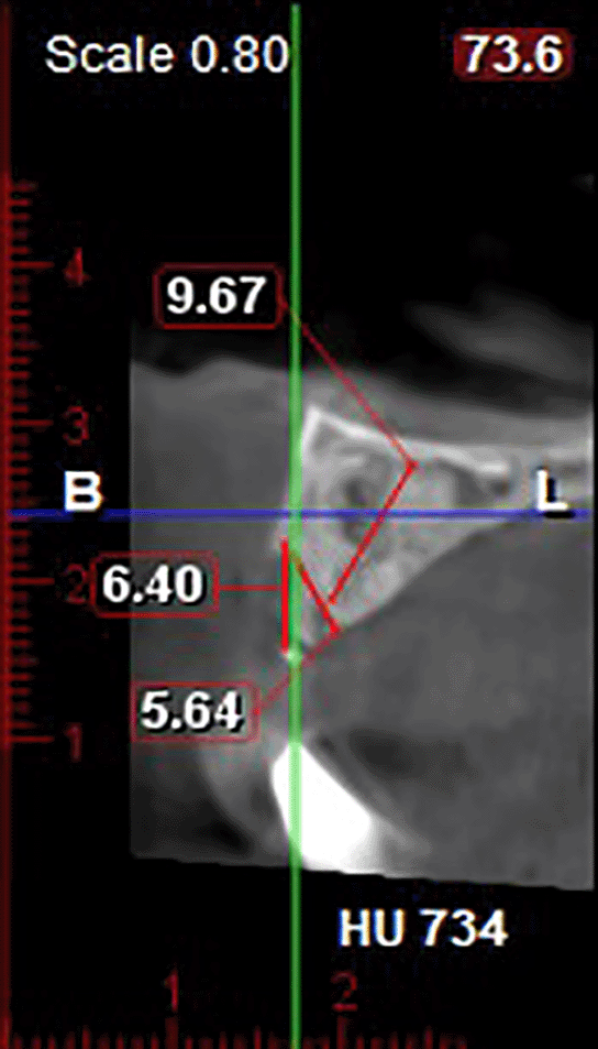
The patient opted for implant supported fixed prosthesis. Owing to this the patient was then asked to get a pre-op CBCT done. Computed tomography imaging showed extensive bone loss and when measured, a labial wall defect of 6.40 mm and apico-coronal width of 9.67 mm was seen (Figure 2). Hence it was imperative to reconstruct the labial wall.
Ridge augmentation
The procedure was performed under local anesthesia. Figure 3.1 shows the pre-op clinical view. A crevicular incision was made extending from 21-12 region (adjacent teeth), followed by a midcrestal incision in 11 region (Figure 3.2). As the augmentation was required in a localized area using a bone block graft, adequate mucoperiosteal flap reflection was required to accommodate the graft. Therefore, vertical incisions were made at the line angles of adjacent teeth (12 and 21) with a broad base, extending into the vestibule (Figure 3.2). Adequate mucoperiosteal flap reflection was done and the defect site was confirmed clinically, consistent with the CBCT (Figure 3.3).
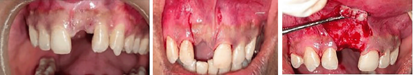
Figure 3.2: Crevicular, Midcrestal and Vertical incisions.
Figure 3.3: Adequate mucoperiosteal flap reflection.
A surgical tin foil template was made over the defect in order to guide the harvest of bone block graft from the symphysis region (Figure 4).

At the donor site the initial incision was planned in the mucosal tissue i.e. beyond the mucosal junction to get the harvest below 5mm from the apices of the teeth. A horizontal incision was made at the vestibule extending from 32-42 region (Figure 5). Adequate reflection at the donor site was done to ensure a proper placement of the template in the safe zone following ‘The 5 rule’ (Figure 6).


The template was placed (Figure 7) and outline was made using surgical pencils to guide the harvest (Figure 8). The outline was connected using a piezoelectric apparatus and the graft of size 6.5x4.0x5.5 mm was harvested (Figure 9), which was immediately placed in NS (normal saline) solution for hydration (Figure 10).


No additional graft was needed at the donor site since the size of the harvest was small (Figure 11). Simple interrupted sutures (3.0 silk non-absorbable) were placed at the donor site for primary approximation (Figure 12).


The harvested bone block was fixed at the recipient site with a titanium screw (Figure 13). Particulate Demineralized Freeze Dried Bone Allograft (DFDBA) of size 1000-1500 μ was placed (Figure 14), as it will help in achieving confluence of both soft and hard tissue dimension during closure which will produce better results. Simple Interrupted Sutures Placed for approximation (Figure 15).


A 5 day antibiotic therapy was prescribed along with analgesics. Cold packs were used in order to reduce post-operative oedema. There were no post operative complications and the sutures were removed 14 days after the surgery.
Implant placement
A follow-up CBCT was done 6 months post-operatively. The CBCT indicated a labio-palatal width of 4.66 mm at the crestal level and apico-coronal width of 11.05 mm (Figure 16). Intraorally the mucosa appeared normal with adequate alveolar ridge height. Table 1 shows the difference of labiopalatal and apicocoronal width at baseline and post augmentation.

| Labiopalatal width | Apicocoronal width | |
|---|---|---|
| Baseline | 0 mm | 9.67 mm |
| Post Augmentation | 4.66 mm | 11.05 mm |
| Difference | 4.66 mm | 1.38 mm |
The second phase of the treatment plan was commenced (under local anaesthesia). A combination of crevicular, midcrestal and vertical incision was given, just like in the first phase, to completely expose the bone block (Figure 17). On reflection of the mucoperiosteal flap, substantial resorption of bone block was seen (Figure 18). The titanium screws were removed and osteotomy site was prepared (Figure 19).


An implant of size 3.5×10 mm was placed and pre-suturing was done using 3.0 silk suture (Figure 20). For soft tissue augmentation, Titanium prepared platelet rich fibrin (T-PRF) was placed and sutures were placed (Figure 21). An immediate post-operative IOPA suggested that the implant placement is correct (Figure 22).


Just as in the first phase, the patient was prescribed antibiotics and analgesics for 5 days. There were no post operative complications and the sutures were removed 14 days after the surgery. The second stage implant surgery was done following 3 months of osteointegration and the patient was referred to the department of Prosthodontics.
Prosthetic procedure
The healing abutment was removed and the Impression coping was screwed into the implant for making implant level impression. The impression was made with a polyvinyl siloxane material using closed tray impression technique and poured after securing the implant analogue in order to produce the working cast. The shade was selected, an interocclusal record and impression of the opposing teeth were made. At the last appointment the anterior definitive crown was cemented over definitive implant abutment (Figure 23).

The authors of this case report, along with many scientists and dental professionals, concur that autograft is the ideal grafting material. (Acocella et al. 2010; Hyeon-Jung et al. 2007; Draenert et al. 2013).7,8 There is no rejection reaction, the technique has the utmost efficacy and predictability possible, and there is no chance of infectious diseases like HIV infection or prions spreading between the donor and the receiver. It is readily available from local or distant sites, sterile, and has Osteoinductive and conductive properties. Basel Elnayef et al. in his Meta-Analysis assessed the stability of bone grafting material between augmentation procedures and final healing, in terms of resorption rate. He concluded that block grafts seemed to maintain the volume of the initial augmentation site more than GBR techniques. This procedure is what was used in the discussed case.9
The authors decided to use the piezoelectric (PZ) method over the conventional method to harvest the graft as the perception by the patient of PZ chin harvest is less traumatic as compared to rotary burs and the increased temperature during the bone cutting was avoided which reduces the bone damage. The PZ osteotomy also makes a narrow cut with little bone wastage (Moutamed et al 2011).
The size of the grafted block should only be slightly larger than necessary to embed implants, according to the authors' own experience, as bone atrophy was only observed along the allograft's edge and in the vicinity of the fastening screws. The authors want to emphasize how important it is to achieve normal stability of the graft, which includes durable fixation with screws, sealing the donor/recipient site with particulate bone grafts, and covering the operational site with PRF membranes, which are membranes concentrated in growth factors. In order to prevent abrasion and the exposure of the autograft to the oral environment, it is also crucial to cover the wound with an unstretched mucous membrane.10
The replacement of missing teeth in the premaxilla is very challenging because of the highly specific soft and hard tissue criteria, in addition to all other esthetic, phonetic, functional, and occlusal requirements. Implant diameter, compared with that of natural teeth, results in challenging cervical esthetics. Unique surgical and prosthetic concepts were implemented in this case for proper and successful results.
In spite of all the technical difficulties that the surgical and restoring clinician may face, the anterior single-tooth implant is the ideal modality of choice to replace a missing anterior maxillary tooth. However, the clinician must have a strong foundation for the inherent complications that are involved with replacing maxillary anterior teeth.
| Views | Downloads | |
|---|---|---|
| F1000Research | - | - |
|
PubMed Central
Data from PMC are received and updated monthly.
|
- | - |
Provide sufficient details of any financial or non-financial competing interests to enable users to assess whether your comments might lead a reasonable person to question your impartiality. Consider the following examples, but note that this is not an exhaustive list:
Sign up for content alerts and receive a weekly or monthly email with all newly published articles
Already registered? Sign in
The email address should be the one you originally registered with F1000.
You registered with F1000 via Google, so we cannot reset your password.
To sign in, please click here.
If you still need help with your Google account password, please click here.
You registered with F1000 via Facebook, so we cannot reset your password.
To sign in, please click here.
If you still need help with your Facebook account password, please click here.
If your email address is registered with us, we will email you instructions to reset your password.
If you think you should have received this email but it has not arrived, please check your spam filters and/or contact for further assistance.
Comments on this article Comments (0)