Keywords
Actinomyces meyeri, Actinomycosis, Actinomyces empyema, COVID-19
This article is included in the Coronavirus (COVID-19) collection.
Actinomyces meyeri, Actinomycosis, Actinomyces empyema, COVID-19
Actinomyces meyeri (A. meyeri) is one of the novel species of actinomyces that have been identified in the last three decades due to new advances in clinical microbiological methods.1 It was found to be a member of the normal flora of the oral, gastrointestinal, and genital tract.2 Pulmonary actinomycosis is thought to occur due to aspiration of the microorganism with oropharyngeal secretions. Other possible causes include hematogenous or direct spread from local infection.
A. meyeri is an uncommon cause of actinomycosis in humans. Other species of actinomycosis, such as A. israelii, predominantly affect the facial area. In contrast, pulmonary is the most common organ system involved in A. meyeri infection.3 Approximately 40 cases of A. meyeri infection have been published; however, empyema is extremely rare and only 15 cases have been documented.3–15 In most of the cases of actinomycosis, periodontal disease and alcoholism have been described as key risk factors.
Prognosis for a patient with empyema is favorable if treated early and aggressively from the time of diagnosis. Penicillin is the antibiotic of choice in the treatment of actinomycosis, but other antibiotics have been used extensively, such as ceftriaxone, clindamycin, erythromycin, and tetracycline.9,12,14 We present a patient in his early 30s who presented with chest pain. In spite of being diagnosed with actinomycosis empyema and undergoing chest tube placement, the patient left the hospital two days later against the advice of his physicians.
A Hispanic man in his early 30s with a past medical history of a mixed germ cell testicular tumor treated with left inguinal radical orchiectomy, presented to our institution with one week of left posterior chest wall pain with radiation to the left flank. The pain was described as constant, sharp, pleuritic in nature, and improved when laying on his left side. He reported shortness of breath as the pain increased in severity. The patient has no family history of cancer, heart, nor lung disease. He was unemployed at the time of admission but had previously worked in construction. He was married with 5 children. He used tobacco and alcohol occasionally. He denied any recent travel history or pets at home. Upon physical examination, the patient was tachycardic and tachypneic. Examination of the mouth revealed adequate dental hygiene with no evidence of a dental infection. Chest examination revealed decreased breath sounds over the left middle to lower lung fields.
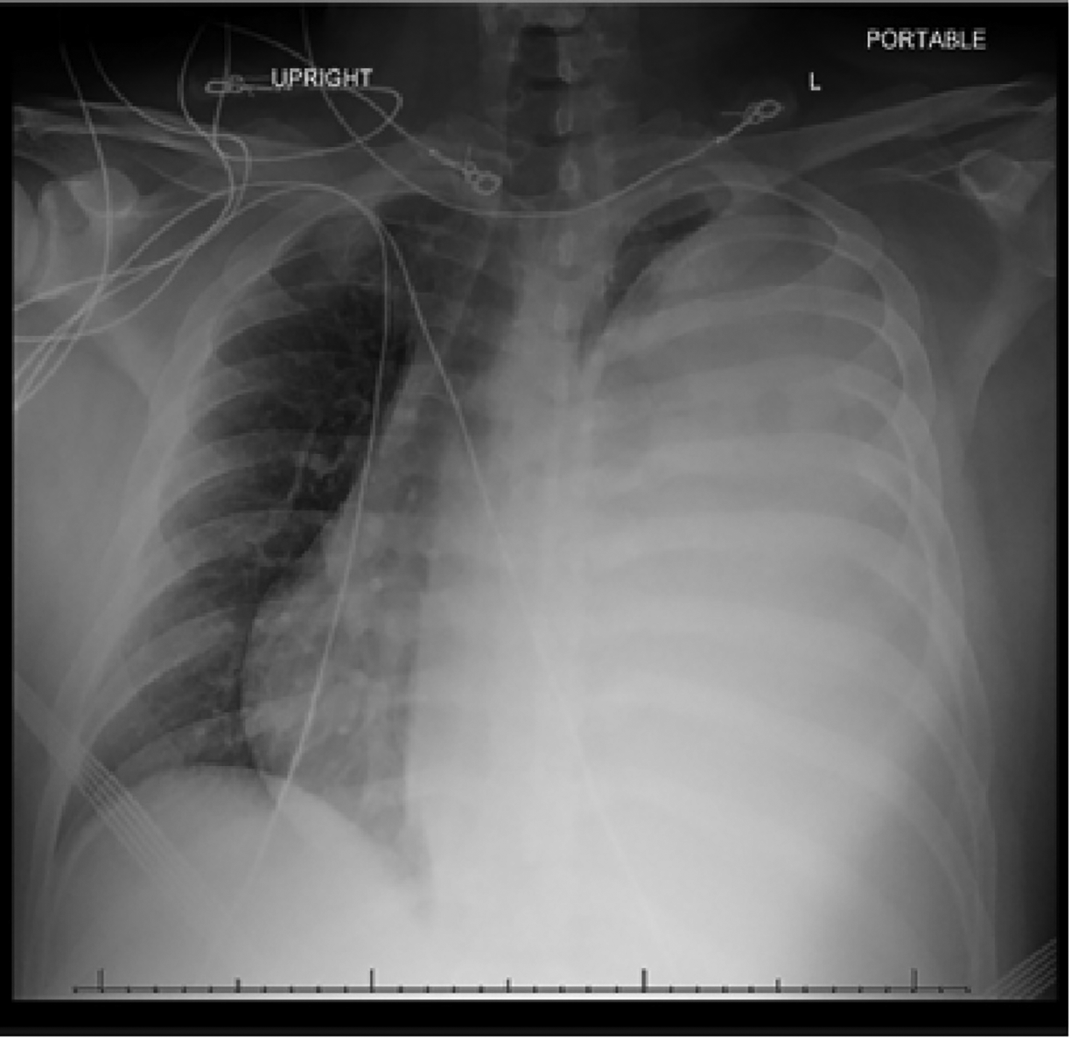
During the visit, reverse transcriptase-polymerase chain reaction analysis of coronavirus disease 2019 (COVID-19) was positive. On chest radiograph, there was complete opacification of the left hemithorax with mediastinal shift to the right (Figure 1). The computed tomography (CT) without contrast of the chest revealed a loculated large low-density left pleural effusion with complete collapse of most of the left lung with shift of the mediastinum to the right (Figure 2).

The patient’s lactate dehydrogenase was 499 IU/L (normal range (NR) 105-333), beta human chorionic gonadotropin <2.4 mIU/mL (NV<5), and alpha-fetoprotein 2.2 ng/mL (NV <40). He underwent an ultrasound guided thoracentesis, and 1 litre of exudative fluid was removed. The pleural fluid studies revealed a pH of 7.00, glucose <2 mg/dL, protein <2 mg/dL, red blood cell count of 28,143, and total nucleated cell count of 28,143 with 98% neutrophils, suggestive of an empyema. Microbiology confirmed the presence of actinomycosis, whereas cytology was negative for the presence of malignant cells.
Once the actinomyces empyema was confirmed, the patient began treatment with 2 grams of ampicillin intravenously every 6 hours. A mandible panorex radiography was obtained with no evidence of dental infection. A 14 French pigtail catheter was inserted into the left pleural collection under ultrasound guidance and another 1200 mL of pus was drained. A follow up chest radiograph revealed nearly complete drainage of the pleural effusion. Alteplase cathflo 2 mg intrapleural injection for pleural effusion thrombolysis was used for two days and a follow up chest radiograph showed complete resolution of the pleural effusion.
Two days after the pigtail placement, the patient complained of pain at the site of the chest tube. He requested the tube to be removed and left our institution against medical advice. One month later, he presented to the oncology clinic for follow up. He had no respiratory symptoms, and he had not taken any antibiotics. A CT scan of the chest was obtained and demonstrated minimal residual pleural effusion. Upon this visit, he was prescribed amoxicillin-clavulanate 500 mg three times a day for 12 months. He visited the clinic again one month later reporting that he was asymptomatic and compliant with the antibiotic. The patient was subsequently lost to follow-up.
Actinomycosis is a chronic infection caused by an organism of the genus Actinomyces, predominantly caused by A. israelii. Recent microbiological detection methods isolated new Actinomyces species such as A. meyeri. Formerly known as Streptothrix and Actinobacterium meyeri, A meyeri is a rare pathogen involved in human actinomycosis.16,17 It has a predilection for disseminated disease likely secondary to pulmonary infection.
Initially, our patient was presumed to have a malignant pleural effusion due to his medical history of a testicular tumor. The testicular biopsy results were consistent with mixed germ cell tumor. The percentages of the tumor were concluded to be 70% embryonal carcinoma, 10% teratoma, 10% choriocarcinoma, 5% yolk sac tumor, and 5% seminoma with invasion of the vasculature. In our patient, the tumor markers were negative, and the effusion fluid cytology analysis was negative for malignancy.
While, to this date, there have been no other reports of patients with concurrent COVID-19 and Actinomyces meyeri infection within the literature, empyema in relation with COVID-19 positive patients have primarily been seen in hosts who are immunocompromised, have autoimmune diseases, or have had prior antibiotic exposure.18 SARS-CoV studies have demonstrated that the virus infects epithelial cells in the lung which induces interleukin-8 (IL-8) production.18 IL-8, a prominent interleukin in attracting T-cells and neutrophils, recruits a large number of inflammatory cells which are able to further induce damage to weaken the lung parenchyma.19 We considered that in our patient with adequate oral hygiene and no history of alcoholism, the recent hospitalization and the concurrent COVID-19 infection were the most important risk factors for the A. meyeri empyema infection. Thoracentesis followed by chest tube placement and the treatment with alteplase were very instrumental in the prompt resolution of the patients’ symptoms.
Review of the literature showed around 40 cases with A. meyeri infection, and the most frequent site of infection was pulmonary. A meyeri empyema has been reported in English literature in 15 previous cases, some of them more than 20 years ago (Table 1). It was more common in males with ages ranging from 16 to 83 years. The most common treatment method was thoracotomy with or without debridement.3–15 The most common antibiotic used was penicillin for a period of up to 12 months.
| Case | Gender | Age (years) | Treatment | Antibiotic | Antibiotic duration (months) | Reference |
|---|---|---|---|---|---|---|
| 1 | Male | 45 | Thoracotomy and decortication | Penicillin (PNC) | 12 | 3 |
| 2 | Male | 61 | Thoracotomy and decortication | PNC/Doxycycline | 8.5 | 4 |
| 3 | Male | 62 | Chest tube | NA | 13 | 5 |
| 4 | Male | 16 | Thoracotomy | NA | 6 | 6 |
| 5 | Female | 64 | Thoracotomy and decortication | PNC | 6.5 | 7 |
| 6 | Male | 49 | Chest tube | PNC | 4 | 8 |
| 7 | Male | 44 | Chest tube | Clindamycin | 4 | 9 |
| 8 | Male | 38 | Soft tissue and intrapleural debridement | PNC | 12 | 10 |
| 9 | Female | 61 | Soft tissue and intrapleural debridement | PNC | 6 | 10 |
| 10 | Male | 52 | Thoracotomy and decortication | PNC | 2 | 11 |
| 11 | Male | 54 | Thoracentesis | Ceftriaxone | 6 | 12 |
| 13 | Female | 66 | Thoracotomy and decortication | PNC | NA | 13 |
| 14 | Male | 83 | Chest tube | Clindamycin | 3 | 14 |
| 15 | Male | 57 | Thoracotomy and decortication | PNC | NA | 15 |
| 16 | Male | 30s | Chest tube | PNC | 12 | Present |
A. meyeri should always be considered as the cause of a pleural effusion in patients who are immunocompromised, alcoholics, or have dental infections. Our patient was COVID-19 positive and had a respiratory infection with a rare bacterium. Having two respiratory insults coupled with the possibility of lung metastasis, presented a complex case with many avenues to explore. Our patient required the specialized support of internists, pulmonologists, interventional radiologists, infectious disease, cardiothoracic surgeons, and oncologists. To date, this is the only case reported in the literature with concurrent COVID-19 and A. meyeri empyema infection.
Although the patient left the hospital against medical advice, he followed up in the clinic and resumed antibiotics. There were several limitations to our case report due to the patients’ absence, which made it difficult to monitor the disease course, treatment effectivity, side effects, and possible complications. Despite these factors, ultimately, the patient had a favorable outcome.
Written informed consent for publication of the clinical details and clinical images was obtained from the patient.
All data underlying the results are available as part of the article and no additional source data are required.
| Views | Downloads | |
|---|---|---|
| F1000Research | - | - |
|
PubMed Central
Data from PMC are received and updated monthly.
|
- | - |
Provide sufficient details of any financial or non-financial competing interests to enable users to assess whether your comments might lead a reasonable person to question your impartiality. Consider the following examples, but note that this is not an exhaustive list:
Sign up for content alerts and receive a weekly or monthly email with all newly published articles
Already registered? Sign in
The email address should be the one you originally registered with F1000.
You registered with F1000 via Google, so we cannot reset your password.
To sign in, please click here.
If you still need help with your Google account password, please click here.
You registered with F1000 via Facebook, so we cannot reset your password.
To sign in, please click here.
If you still need help with your Facebook account password, please click here.
If your email address is registered with us, we will email you instructions to reset your password.
If you think you should have received this email but it has not arrived, please check your spam filters and/or contact for further assistance.
Comments on this article Comments (0)