Keywords
Cyclophosphane, Disseminated strongyloidiasis, hyperinfection, Immunosuppression, Strongyloidiasis, SLE, Strongyloides stercoralis, colitis.
This article is included in the Health Services gateway.
Cyclophosphane, Disseminated strongyloidiasis, hyperinfection, Immunosuppression, Strongyloidiasis, SLE, Strongyloides stercoralis, colitis.
GPIA: Gelatin particle indirect agglutination
IFA: Indirect fluorescent antibody
IHA: Indirect haemagglutination
LIPS: Luciferase immuno-precipitation system
PAP: Papanicolaou
PCR: Polymerase chain reaction
SLE: Systemic lupus erythematosus
SsIR: Strongyloides stercoralis immuno-reactive antigen
UGIE: Upper gastrointestinal tract endoscopy
USG: Ultrasonogram
Strongyloidiasis is a common parasitic opportunistic infection affecting millions of people worldwide, especially in tropical and sub-tropical zones. It has a unique ability to complete autoinfection and remains in dormant larval form for many years. Some of the factors leading to the immunocompromise state like corticosteroid therapy, human T-cell lymphotropic virus type-1 infection (HTLV-1), hematological malignancy, malnutrition, human immunodeficiency virus (HIV), among others, can activate and transform the dormant larval form into to widespread disseminated form with the end result of mortality in an unexpected manner. The practice of early screening for such parasitic infection in an immunocompromised patient has been neglected for many years unless and until some suspicious clinical presentation has drawn attention to the clinician. Therefore, in view of the above, the study of our case report draws the attention of many clinicians to the fact that early diagnosis of strongyloidiasis is one of the essential clinical practices in order to prevent the high risk of mortality due to negligence of parasitic screening in an immunocompromise patient.
Duodenal strongyloidiasis is caused by a parasite, Strongyloides stercoralis, a soil-transmissible nematode. Strongyloidiasis is an infection caused by Strongyloides stercoralis (and rarely Strongyloides fuelleborni), which is a helminth present mainly in tropical and subtropical regions, as well as in temperate climates.1 The three different definitive hosts for Strongyloides stercoralis are humans, dogs, and cats. Around 3-100 million people are estimated to be infected worldwide.2 Strongyloidiasis has varied manifestations ranging from asymptomatic to potentially fatal hyper infection or disseminated infection. The female worm produces eggs by parthenogenesis that hatch to larvae, which may also cause auto-infection as well.3
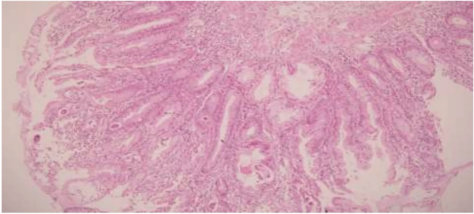
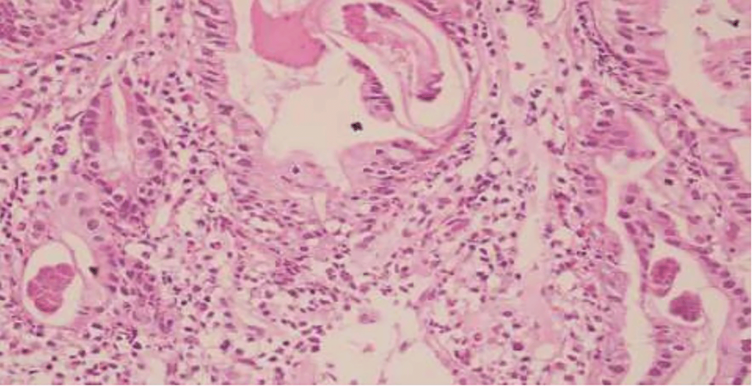
A 50-year-old female, a known case of systemic lupus erythematous (SLE) under cyclophosphane, presented to emergency services with repeated bouts of watery vomiting for one week. She had repeated loose stools for one month. She was admitted for severe dehydration and dyselectrolytemia, for which she had been managed with an intravenous fluid like ringer lactate and antibiotics (metronidazole and ceftriaxone) for seven days. She was a housewife involved in agriculture occupation in the past. There was no family history of similar illnesses and other genetic diseases so far. Stool examination revealed cysts of Entamoeba histolytica. Ultrasonography (USG) of abdomen showed features suggestive of colitis and mild hydroureteronephrosis. Upper gastrointestinal endoscopy (UGIE) showed antral erosions, pangastritis, loss of mucosal folds in D2, esophageal candidiasis, and urease-positive Helicobacter pylori-induced chronic fundal gastritis. Computed tomography (CT) of the abdomen showed ill-defined, diffuse irregular luminal narrowing of ascending colon with a diffuse thickened wall. Endoscopic duodenal biopsy from the second part of the duodenum was done with suspicion of celiac disease. Histopathological examination showed duodenal mucosa revealing numerous larval forms and eggs of Strongyloides stercoralis within crypts and glandular epithelium in a background of dense eosinophilic infiltrates in lamina propria with no evidence of celiac disease or malignancy, as shown in Figures 1 and 2. After the diagnosis and constant lack of improvement in the condition, the patient opted for changing treatment and went to another hospital. Resection anastomosis of the intestine was done at another institute. Unfortunately, the patient died without any signs of cure.
Strongyloidiasis is a much-neglected health problem.4 Until the diagnosis of this ailment, most of the time, it has already been disseminated in the body. The causative organism for strongyloidiasis, Strongyloides stercoralis, is a helminth that gets transmitted by transdermal migration of filariform larvae, which is the infective form with hematogenous dissemination to the lungs. Through the tracheobronchial tree, it then enters the alveolar sacs, and the larvae gain access to the gastrointestinal tract.5 In the duodenum and jejunum, it hatches into rhabditiform larvae, which is the pathogenic form. Rhabditiform larvae, when passed in feces, can develop directly into free-living adult males and females that can copulate and release eggs or can instead molt two times to become infective filariform larvae and penetrate the skin to start a new parasitic cycle.3,6
Patients are usually asymptomatic; however, gastrointestinal manifestations may mimic coeliac disease. The common symptoms are dyspepsia, abdominal discomfort, diarrhea, nausea, and small bowel obstruction. Intestines, particularly the duodenum and jejunum, are commonly affected sites that can present as small bowel obstruction.7 In patients with disseminated strongyloidiasis, pulmonary manifestations are also common, with the lung being the most common extraintestinal site to be involved. Pulmonary symptoms like dry cough, progressive dyspnoea, and wheezing are common in patients with pulmonary strongyloidiasis.5 Skin rash and pruritus are the dermal manifestations, and systemic symptoms of weight loss may also be noted in most cases.4 Immunocompromised conditions, HTLV-1, HIV infection, as well as patients under corticosteroids, present with more severe forms of infection.6 Some studies even indicate that corticosteroids accelerate the transformation of rhabdtitiform larvae to filariform larvae.1 The helminthic manifestation, though most common in the small intestine, can also occur at any level from the esophagus and stomach to the rectum.8 Duodenal aspirate or biopsy, though more sensitive for the diagnosis, is an invasive procedure, is less desirable.8,9
S. stercoralis infection is difficult to differentiate from the infection caused by Ancylostoma duodenale, further requiring serological tests, stool examination, and culture.3,10 Adult worms or larvae are seen on stool examination. Detection of larvae in a smear of stool in saline is a definitive diagnostic feature; however, Papanicolaou (PAP) smears of sputum may also reveal the organism.5 Both S. stercoralis and A. duodenale are transmitted via soil.6 Eggs of S. stercoralis are oval, thin-shelled, resembling that of A. duodenale but are rarely seen in the stool. The non-infective form known as rhabditiform larva is the pathogenic form, which is the feeding stage with a length of 0.2-0.25 mm. They have a prominent genital primordium, a short buccal cavity, a clear esophagus, and a notched buccal opening.
In contrast, A. duodenale has an inconspicuous genital primordium and a long buccal canal. Filariform larva is the infective stage in both parasites, with a similar length of approximately 600 μ. However, S. stercoralis has a notched tail, and A. duodenale has a pointed tail. The esophagus to intestine ratio is 1:1 in S. stercoralis and 1:3 in A. duodenale.9,11 Enzyme-linked immunosorbent assays (ELISA), along with stool studies, are done together to increase the sensitivity of the investigation.6 Various immunodiagnostic methods like indirect haemagglutination (IHA), indirect fluorescent antibody (IFA), Western blot, ELISA, luciferase immuno-precipitation system (LIPS), gelatin particle indirect agglutination (GPIA), S. stercoralis immuno-reactive antigen (SsIR), and molecular methods like PCR are available.9,12 Culture methods for the proper identification of S. stercoralis, like Baermann and Koga agar plate techniques, are also available. The Kato Katz technique is employed for demonstration of A. duodenale.10
Strongyloidiasis is one of the common parasitic infestations and is neglected in many developing countries. The infection is more prevalent among people with an immunocompromised state and clinically presents as nonspecific symptoms ranging from nonspecific gastrointestinal symptoms like abdominal pain, diarrhea, to fatal disseminated infection, thereby increasing the risk of unexpected mortality. Hence, while reviewing the cases, scrutiny is required not to miss out on the parasites. Also, before commencing the treatment with corticosteroids and other immunosuppressive drugs, the possibility of already existing parasites should be kept in mind so as to prevent the disease's activation, evolution, and progression to life-threatening or fatal complications. Hence, there is much need to transform the early screening of neglected common parasites into good clinical practice in immunocompromised patients in tropical and sub-tropical zones across the globe.
| Views | Downloads | |
|---|---|---|
| F1000Research | - | - |
|
PubMed Central
Data from PMC are received and updated monthly.
|
- | - |
Provide sufficient details of any financial or non-financial competing interests to enable users to assess whether your comments might lead a reasonable person to question your impartiality. Consider the following examples, but note that this is not an exhaustive list:
Sign up for content alerts and receive a weekly or monthly email with all newly published articles
Already registered? Sign in
The email address should be the one you originally registered with F1000.
You registered with F1000 via Google, so we cannot reset your password.
To sign in, please click here.
If you still need help with your Google account password, please click here.
You registered with F1000 via Facebook, so we cannot reset your password.
To sign in, please click here.
If you still need help with your Facebook account password, please click here.
If your email address is registered with us, we will email you instructions to reset your password.
If you think you should have received this email but it has not arrived, please check your spam filters and/or contact for further assistance.
Comments on this article Comments (0)