Keywords
Uniprot ID Q9NUM4, TMEM106B, Transmembrane protein 106B, antibody characterization, antibody validation, Western Blot, immunoprecipitation, immunofluorescence
This article is included in the Cell & Molecular Biology gateway.
This article is included in the YCharOS (Antibody Characterization through Open Science) gateway.
Uniprot ID Q9NUM4, TMEM106B, Transmembrane protein 106B, antibody characterization, antibody validation, Western Blot, immunoprecipitation, immunofluorescence
Transmembrane protein 106B (TMEM106B) is a genetic risk variant for many neurodegenerative diseases. The presence of the TMEM106B major risk allele, rs1990622, is suspected to be a risk factor and disease modifier for Frontotemporal Dementia (FTD), with few studies investigating its potential role in Amyotrophic Lateral Sclerosis (ALS) pathogenesis.1–5
TMEM106B is a transmembrane endosomal and lysosomal glycoprotein. The protein has garnered interest lately, with the discovery that a 135 amino acid portion of the protein from its luminal C-terminal domain forms fibrils in the brains of patients with frontotemporal lobar degeneration, progressive supranuclear palsy, and dementia with Lewy bodies.6,7
The roles of TMEM106B fibrils in normal lysosomal function or disease pathogenesis are not known, nor is the mechanism by which the protein is proteolyzed, or forms fibrils.7 Mechanistic studies would be greatly facilitated with the availability of high-quality validated antibodies. Here, we compared the performance of a range of commercially available antibodies for TMEM106B and characterized several high-quality antibodies for Western blot, immunoprecipitation and immunofluorescence, enabling biochemical and cellular assessment of TMEM106B properties and function.
Our standard protocol involved comparing readouts from wild-type (WT) and knockout (KO) cells.8,9 The first step was to identify a cell line(s) that expresses sufficient levels of TMEM106B to generate a measurable signal. To this end, we examined the DepMap transcriptomics databases to identify all cell lines that express the target at levels greater than 2.5 log2 (transcripts per million “TPM” +1), which we have found to be a suitable cut-off (Cancer Dependency Map Portal, RRID:SCR_017655). Commercially available HAP1 cells expressed the TMEM106B at RNA levels above the average range of cancer cells analyzed. The parental and KO HAP1 cell lines were obtained from Horizon Discovery (Table 1).
| Institution | Catalog number | RRID (Cellosaurus) | Cell line | Genotype |
|---|---|---|---|---|
| Horizon Discovery | C631 | CVCL_Y019 | HAP1 | WT |
| Horizon Discovery | HZGHC005877c010 | CVCL_XU38 | HAP1 | TMEM106B KO |
For Western blot experiments, we resolved proteins from WT and TMEM106B KO cell extracts and probed them side-by-side with all antibodies in parallel (Figure 1).
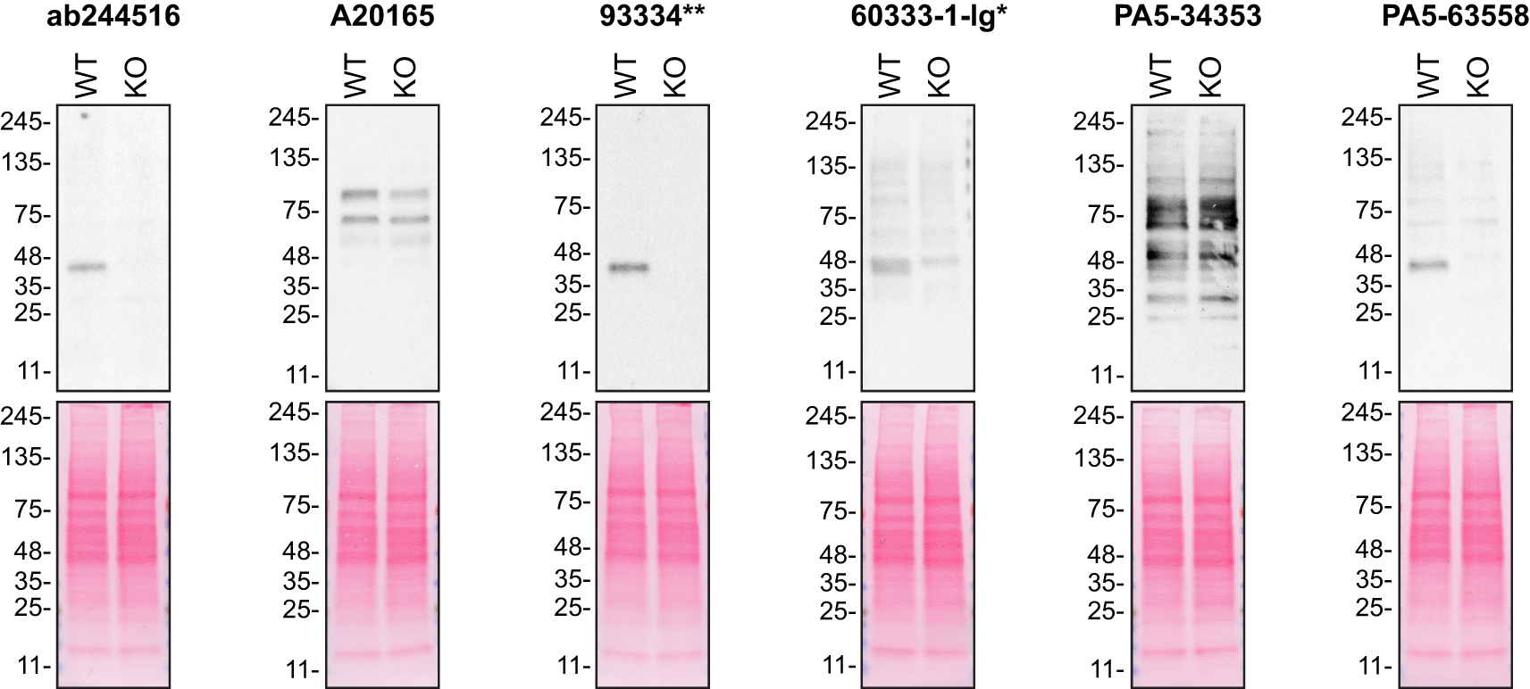
Lysates of HAP1 (WT and TMEM106B KO) were prepared and 15 μg of protein were processed for Western Blot with the indicated TMEM106B antibodies. The Ponceau stained transfers of each blot are presented are shown equal loading of WT and KO lysates and protein transfer efficiency from the acrylamide gels to the nitrocellulose membrane. Antibody dilutions were chosen according to the recommendations of the antibody supplier. Exceptions were given for antibodies 93334** and 60333-1-Ig*, which were titrated to 1/500 and 1/2000, respectively, as the signal was too weak when following the supplier’s recommendations. Antibody dilution used: ab244516 at 1/500, A20165 at 1/2000, 93334** at 1/500, 60333-1-lg* at 1/2000, PA5-34353 at 1/500, and PA5-63558 at 1/200.Predicted band size: 31 kDa. *= monoclonal antibody, **= recombinant antibody.
For immunoprecipitation experiments, we used the antibodies to immunopurify TMEM106B from HAP1 cell extracts. The performance of each antibody was evaluated by detecting the TMEM106B protein in extracts, in the immunodepleted extracts and in the immunoprecipitates (Figure 2).
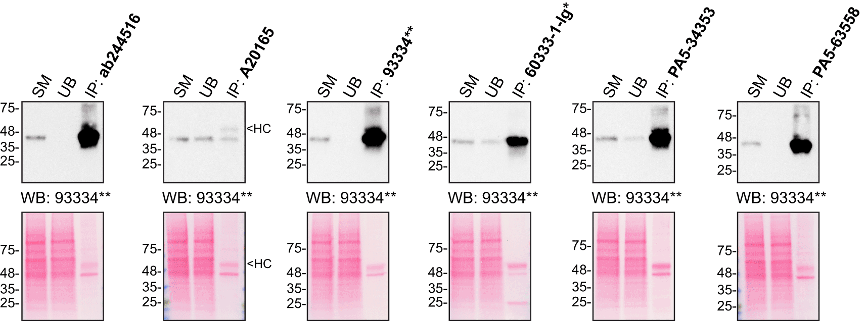
HAP1 lysates were prepared, and IP was performed using 2.0 μg of the indicated TMEM106B antibodies pre-coupled to Dynabeads protein G or protein A. Samples were washed and processed for Western Blot with the indicated TMEM106B antibody. For Western Blot, 93334** was used at 1/500. The Ponceau stained transfers of each blot are shown for similar reasons as in Figure 1. SM=2% starting material; UB=2% unbound fraction; IP=immunoprecipitate; HC=antibody heavy chain. *= monoclonal antibody, **= recombinant antibody.
For immunofluorescence, as described previously, antibodies were screened using a mosaic strategy.10 In brief, we plated WT and KO cells together in the same well and imaged both cell types in the same field of view to reduce staining, imaging and image analysis bias (Figure 3).
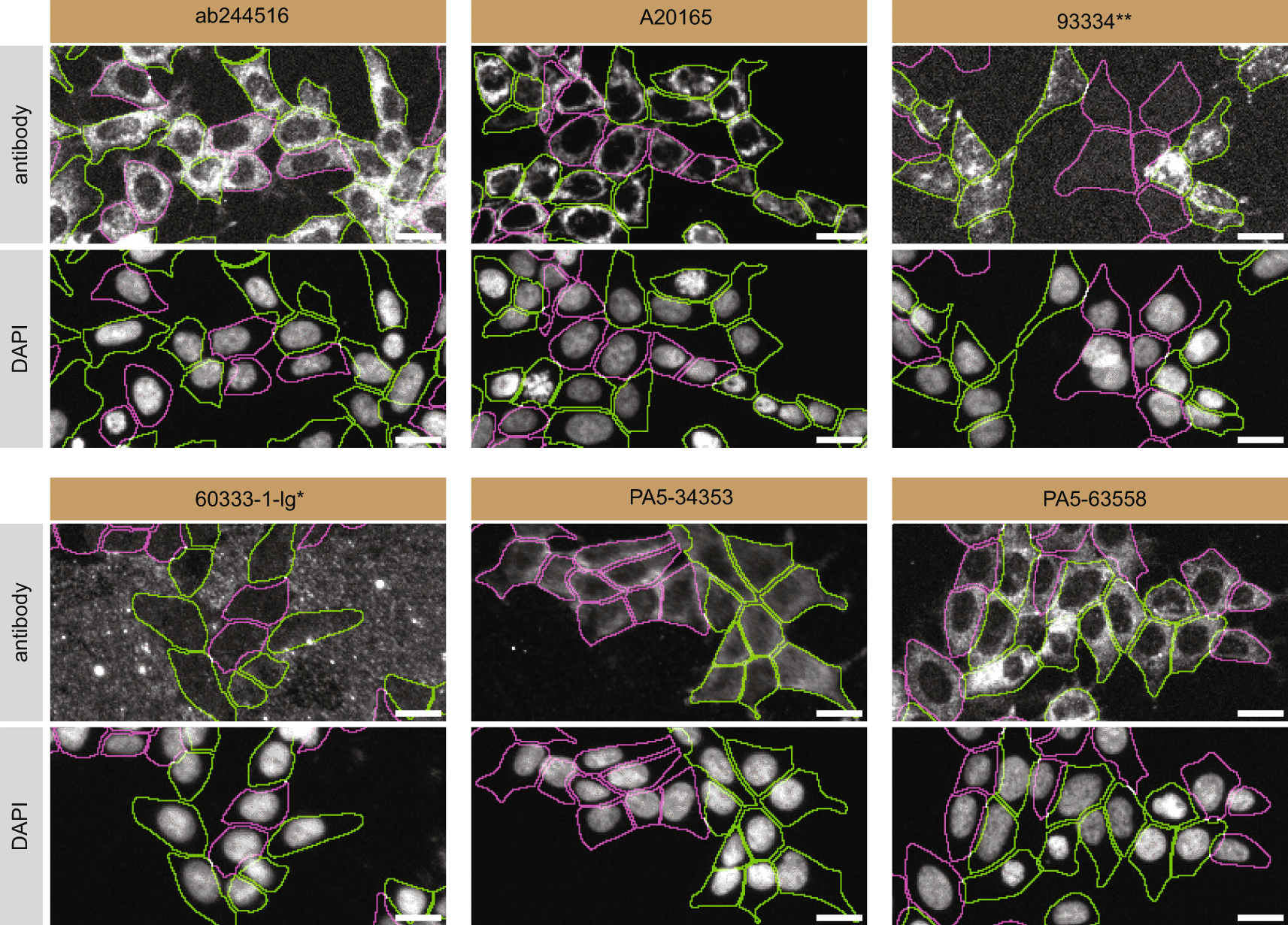
HAP1 WT and TMEM106B KO cells were labelled with a green or a far-red fluorescent dye, respectively. WT and KO cells were mixed and plated to a 1:1 ratio in a 96-well plate with a glass bottom. Cells were stained with the indicated TMEM106B antibodies and with the corresponding Alexa-fluor 555 coupled secondary antibody including DAPI. Acquisition of the blue (nucleus-DAPI), green (identification of WT cells), red (antibody staining) and far-red (identification of KO cells) channels were performed. Representative images of the merged blue and red (grayscale) channels are shown. WT and KO cells are outlined with green and magenta dashed line, respectively. When the concentration was not indicated by the supplier, we tested antibodies at 1/1000 or 1/2000. At this concentration, the signal from each antibody was in the range of detection of the microscope used. Antibody dilution used: ab244516 at 1/100, A20165 at 1/2000, 93334** at 1/100, 60333-1-lg* at 1/2000, PA5-34353 at 1/1000, and PA5-63558 at 1/100. Bars = 10 μm. *= monoclonal antibody, **= recombinant antibody.
In conclusion, we have screened many TMEM106B commercial antibodies by Western blot, immunoprecipitation and immunofluorescence and identified several high-quality antibodies under our standardized experimental conditions.
All TMEM106B antibodies are listed in Table 2 together with their corresponding Research Resource Identifiers, or RRID, to ensure the antibodies are cited properly.11 Peroxidase-conjugated goat anti-mouse and anti-rabbit antibodies are from Thermo Fisher Scientific (cat. number 62-6520 and 65-6120). Alexa-555 conjugated secondary goat anti-rabbit and anti-mouse antibodies are from Thermo Fisher Scientific (cat. number A21429 and A21424)
| Company | Catalog number | Lot number | RRID (Antibody Registry) | Clonality | Clone ID | Host | Concentration (μg/μl) | Vendors recommended applications |
|---|---|---|---|---|---|---|---|---|
| Abcam | ab244516 | GR3421643 | AB_29242681 | polyclonal | - | rabbit | 0.10 | IF |
| ABclonal | A20165 | 131370201 | AB_2862952 | polyclonal | - | rabbit | 1.88 | Wb |
| Cell Signaling Technology | 93334** | 1 | AB_29242671 | recombinant-mono | E7H7Z | rabbit | 0.10 | Wb, IP, IF |
| Proteintech | 60333-1-lg* | 10002132 | AB_2881442 | monoclonal | 5D1F8 | mouse | 2.00 | Wb |
| Thermo Fisher Scientific | PA5-34353 | XD3572518 | AB_2551705 | polyclonal | - | rabbit | 1.00 | Wb |
| Thermo Fisher Scientific | PA5-63558 | XD3571635 | AB_2648556 | polyclonal | - | rabbit | 0.10 | IF |
Both HAP1 WT and TMEM106B KO cell lines used are listed in Table 1, together with their corresponding RRID, to ensure the cell lines are cited properly.12 Cells were cultured in DMEM high glucose (GE Healthcare, cat. number SH30081.01) containing 10% fetal bovine serum (Wisent, cat. number 080450), 2 mM L-glutamate (Wisent, cat. number 609065), 100 IU penicillin and 100 μg/mL streptomycin (Wisent, cat. number 450201).
Western blot experiments were performed as described in our standard operating procedure.13 HAP1 WT and TMEM106B KO were collected in RIPA buffer (25mM Tris-HCl pH 7.6, 150mM NaCl, 1% NP-40, 1% sodium deoxycholate, 0.1% SDS) from Thermo Fisher Scientific (cat number 0089901), supplemented with 1x protease inhibitor cocktail mix from MilliporeSigma (cat. number 78429). Lysates were sonicated briefly and incubated for 30 min on ice. Lysates were spun at ~110,000 x g for 15 min at 4°C and equal protein aliquots of the supernatants were analyzed by SDS-PAGE and Western Blot. BLUelf prestained protein ladder from GeneDireX (cat. number PM008-0500) was used.
Western blots were performed with pre-cast mini 4-15% gradient polyacrylamide gels from Bio-Rad (cat. number 4561084) and transferred on nitrocellulose membranes. Proteins on the blots were visualized with Ponceau staining which is scanned to show together with individual Western blots. Blots were blocked with 5% milk for 1 h, and antibodies were incubated overnight at 4°C with 5% bovine serum albumin (BSA) (Wisent, cat. number 800-095) in TBS with 0,1% Tween 20 (TBST) (Cell Signaling Technology, cat. number 9997). Following three washes with TBST, the peroxidase conjugated secondary antibody was incubated at a dilution of ~0.2 μg/mL in TBST with 5% milk for 1 hr at room temperature followed by three washes with TBST. Membranes are incubated with Pierce ECL from Thermo Fisher Scientific (cat. number 32106) prior to detection with HyBlot CL autoradiography films from Denville (cat. number 1159T41).
Immunoprecipitation was performed as described in our standard operating procedure.14 Antibody-bead conjugates were prepared by adding 2 μg of antibody to 500 μL of Pierce IP Lysis Buffer from Thermo Fisher Scientific (cat. number 87788) in a 1.5 mL microcentrifuge tube together with 30 μL of Dynabeads protein A- (for rabbit antibodies) or protein G- (for mouse antibodies) from Thermo Fisher Scientific (cat. number 10002D and 10004D, respectively). Tubes were rocked ~2 hours at 4°C followed by several washes to remove unbound antibodies.
HAP1 WT were collected in Pierce IP buffer (25 mM Tris-HCl pH 7.4, 150 mM NaCl, 1 mM EDTA, 1% NP-40 and 5% glycerol) from Thermo Fisher Scientific (cat. number 87788), supplemented with protease inhibitor from MilliporeSigma (cat. number P8340). Lysates were rocked for 30 min at 4°C and spun at 110,000 x g for 15 min at 4°C. 0.5 mL aliquots at 2.0 mg/mL of lysate were incubated with an antibody-bead conjugate for ~2 hrs at 4°C. The unbound fractions were collected, and beads were subsequently washed three times with 1.0 mL of IP lysis buffer and processed for SDS-PAGE and Western blot on a pre-cast mini 4-15% polyacrylamide gel. Prot-A:HRP (MilliporeSigma, cat. number P8651) was used as a secondary detection system at a dilution of 0.4 μg/mL.
Immunofluorescence was performed as described in our standard operating procedure.10 HAP1 WT and TMEM106B KO were labelled with CellTracker green (Thermo Fisher Scientific, cat. number C2925) or CellTracker deep red (Thermo Fisher Scientific, cat. number C34565) fluorescence dye, respectively. The nuclei were labelled with DAPI (Thermo Fisher Scientific, cat. number D3571) fluorescent stain. WT and KO cells were plated in 96 well glass plates (Perkin Elmer, cat. number 6055300) as a mosaic and incubated for 24 hrs in a cell culture incubator at 37oC, 5% CO2. Cells were fixed in 4% paraformaldehyde (PFA) (Beantown chemical, cat. number 140770-10mL) in phosphate buffered saline (PBS) (Wisent, cat. number 311-010-CL) for 15 min at room temperature and then washed three times with PBS. Cells were permeabilized in PBS with 0.1% Triton X-100 (Thermo Fisher Scientific, cat. number BP151-500) for 10 min at room temperature and blocked with PBS containing 5% BSA, 5% goat serum (Gibco, cat. number 16210-064) and 0.01% Triton X-100 for 30 min at room temperature. Cells were incubated with IF buffer (PBS, 5% BSA, 0.01% Triton X-100) containing the primary TMEM106B antibodies overnight at 4°C. Cells were then washed 3 × 10 min with IF buffer and incubated with the corresponding Alexa Fluor 555-conjugated secondary antibodies in IF buffer at a dilution of 1.0 μg/mL for 1 hr at room temperature with DAPI. Cells were washed 3 × 10 min with IF buffer and once with PBS.
Images were acquired on an ImageXpress micro widefield high-content microscopy system (Molecular Devices), using a 20x/0.45 NA air objective lens and scientific CMOS camera (16-bit, 1.97mm field of view), equipped with 395, 475, 555 and 635 nm solid state LED lights (Lumencor Aura III light engine) and bandpass emission filters (432/36 nm, 520/35 nm, 600/37 nm and 692/40 nm) to excite and capture fluorescence emission for DAPI, CellTracker Green, Alexa fluor 555 and CellTracker Red, respectively. Images had pixel sizes of 0.68 × 0.68 microns. Exposure time was set with maximal (relevant) pixel intensity ~80% of dynamic range and verified on multiple wells before acquisition. Since the IF staining varied depending on the primary antibody used, the exposure time was set using the most intensely stained well as reference. Frequently, the focal plane varied slightly within a single field of view. To remedy this issue, a stack of three images per channel was acquired at a z-interval of 4 microns per field and best focus projections were generated during the acquisition (MetaXpress v6.7.1, Molecular Devices). Segmentation was carried out on the projections of CellTracker channels using CellPose v1.0 on green (WT) and far-red (KO) channels, using as parameters the ‘cyto’ model to detect whole cells, and using an estimated diameter tested for each cell type, between 15 and 20 microns.15 Masks were used to generate cell outlines for intensity quantification. Figures were assembled with Adobe Illustrator.
| Views | Downloads | |
|---|---|---|
| F1000Research | - | - |
|
PubMed Central
Data from PMC are received and updated monthly.
|
- | - |
Provide sufficient details of any financial or non-financial competing interests to enable users to assess whether your comments might lead a reasonable person to question your impartiality. Consider the following examples, but note that this is not an exhaustive list:
Sign up for content alerts and receive a weekly or monthly email with all newly published articles
Already registered? Sign in
The email address should be the one you originally registered with F1000.
You registered with F1000 via Google, so we cannot reset your password.
To sign in, please click here.
If you still need help with your Google account password, please click here.
You registered with F1000 via Facebook, so we cannot reset your password.
To sign in, please click here.
If you still need help with your Facebook account password, please click here.
If your email address is registered with us, we will email you instructions to reset your password.
If you think you should have received this email but it has not arrived, please check your spam filters and/or contact for further assistance.
Comments on this article Comments (0)