Keywords
venous leg ulcer, endovenous laser ablation, below the knee, laser power, outcome, complication
venous leg ulcer, endovenous laser ablation, below the knee, laser power, outcome, complication
Active venous leg ulcer (VLU) is the most severe manifestation of chronic venous disease (CVD), classified as clinical C6 on clinical, etiological, anatomical, and pathophysiological (CEAP) classification with several contributing factors including venous reflux, obstruction outflow, pump failure of calf muscles, and obesity that causes chronic venous hypertension.1 The prevalence ranges between 0.8 to 2.2 patients for every 1000 people/year and with 19.0±12.1 weeks healing time.2 Active VLU alone represents 3.5% of patient populations with CVD.3 Despite the small number, the quality of life of patients with active VLU is affected in various dimensions, especially on the emotional level and regarding body image, with pain and ulcer severity contributing to a decrease in patients’ quality of life.4 VLU not only impacts patients’ health but also delivers an economic burden with approximately £7706 per patient per year direct cost to manage VLU patients, and more than £2 billion annually (representing 1.2% of the annual budget) in a study on the British population.5 Patients with an advanced form of chronic venous insufficiency (CVI), including lipodermatosclerosis, healed VLU, and active VLU have a complex venous disease with a superficial, perforator, and deep vein involvement.6 Treatment of superficial venous reflux with early endovenous laser ablation (EVLA) has been proven to reduce ulcer recurrence levels and promote faster VLU healing.7 In a study by Montminy et al.,8 the VLU healing rate after 12 months in the patient group treated with EVLA was 82% versus 36% in the compression therapy only group. Here we report three cases of patients with active VLU undergoing EVLA using 1470-nano meter (nm) wavelength laser device and ELVeS radial fiber (Biolitec, Bonn, Germany) in National Cardiovascular Center Harapan Kita, Jakarta, Indonesia with different approaches.
A 60-year-old woman, who worked as a baker, presented with a complaint of a wound in her right ankle accompanied by pain (visual analog scale (VAS) 3/10) in the past three months. The complaint began with swelling in both lower limbs that got worse one year earlier. There was a change of skin color on both the patient’s leg and foot which became darker over time. The patient had a history of hypertension. On physical examination of the malleolus medial dextra region, there was a solitary ulcer, sized 3×4 cm with irregular edges, erythema ulcer base accompanied by purulent discharge, dry distant tissue, edema (+), and hyperpigmentation on 1/3 medial cruris until dorsum pedis (Figure 1A). There was severe CVI in above the knee (ATK) and below the knee (BTK) great saphenous vein (GSV) and moderate CVI in the deep vein of both lower limbs from duplex ultrasound (DUS) examination of the lower extremity. The patient was diagnosed with CVI on both lower limbs with active VLU on malleolus medial dextra (C6sEpAsdPr). The venous clinical severity score (VCSS) was 15. The patient agreed to EVLA treatment on the right lower limb and received ceftriaxone antibiotics for five days. The procedure was done after the patient finished the antibiotics course.
EVLA was performed under spinal anesthesia. Mapping was done on the right GSV. The diameter of the saphena-femoral junction (SFJ) was au11.0 mm, proximal ATK GSV 9.2 mm, medial ATK GSV 6.0 mm, distal ATK GSV ATK 6.1 mm, proximal GSV BTK GSV 5.2 mm, media BTK GSV 4.5 mm, and distal BTK GSV 3.0 mm. Double puncture was used on distal BTK and distal ATK GSV because of tortuosity and branching at distal ATK. Puncture and application of tumescent anesthesia were done by guided DUS. EVLA started 3 cm from SFJ with laser power of 6 W linear energy density (LEED) 50 J/cm at proximal until media ATK, 5 W LEED 40 J/cm at media ATK until proximal BTK, and 2W LEED 20 J/cm at proximal until distal BTK. The total ablated GSV length was 60 cm with a total of 850 mL tumescent. Lower extremity DUS showed perfectly obliterated ablated GSV, no thrombus found in the deep vein, and a patent epigastric vein. The patient was discharged one day after the procedure.
The patient’s condition was assessed one week after EVLA, and she reported no more pain, significant rapid improvement of the ulcer, granulation tissue (+), and no more discharge (Figure 1B). At follow up six months post-EVLA, the patient didn’t have any complaints and had a healed ulcer. There was a 14-point improvement in VCSS score down to a score of 1.
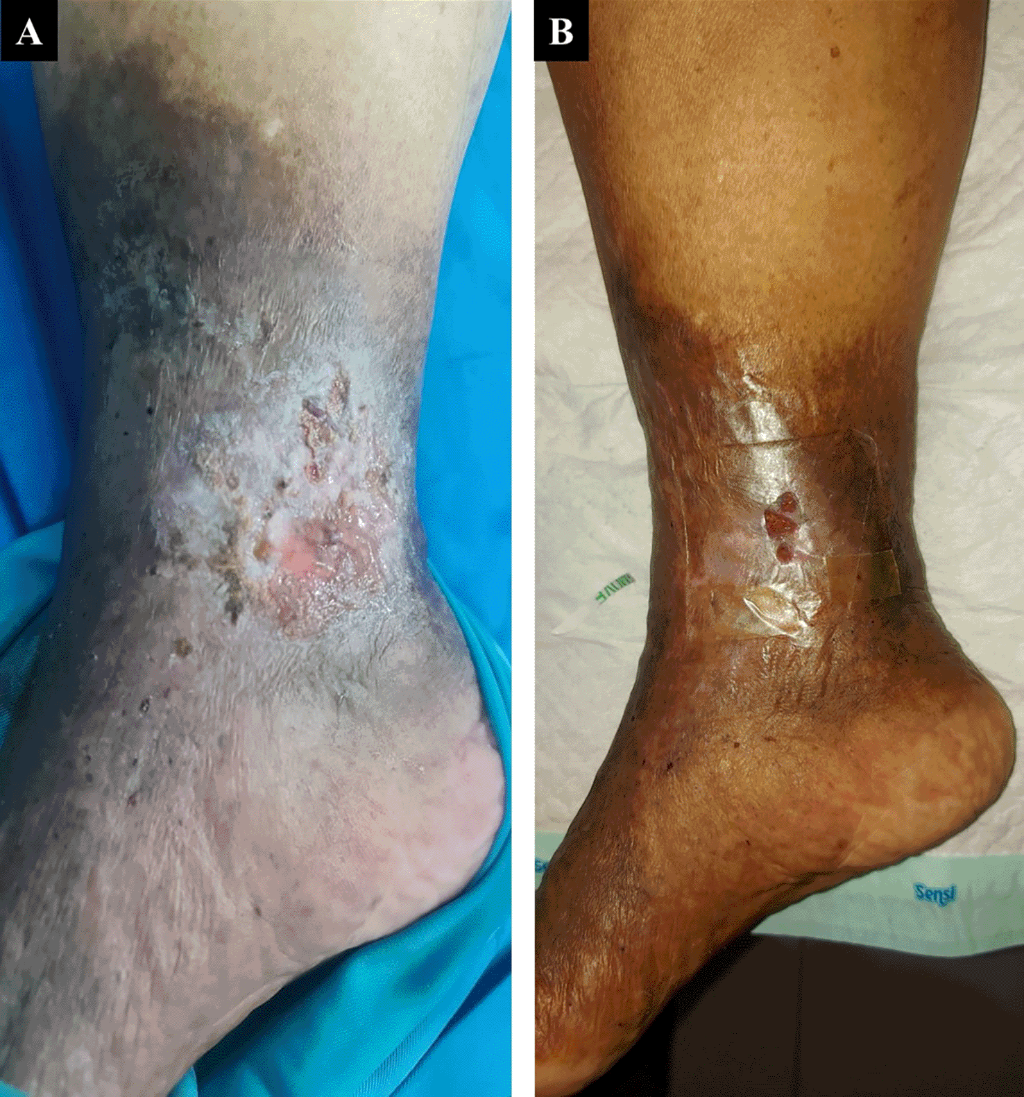
A. pre-EVLA; B. 1-week post-EVLA.
A 50-year-old male chef presented with complaints of wounds on his left ankle in the last two years accompanied by pain aggravated by walking (VAS 4/10). The patient had undergone EVLA on the left ATK GSV with phlebectomy eight months earlier, but there wasn’t any improvement of the ulcer. The patient had wound care with a surgeon who suggested doing a lower extremity vessel evaluation. The patient had a history of hypertension, type 2 diabetes mellitus, and obesity with body mass index (BMI) of 39.45 kg/m2. Physical examination of the malleolus sinistra region showed multiple ulcers sized 18×10 cm on the medial side, 8×5 cm, and 4×2 cm on the lateral side with irregular edges (Figure 2A). The ulcer had an erythema base, purulent discharge, dry distant tissue, and edema (+). Hyperpigmentation of 1/3 of the medial cruris sinistra until dorsum pedis sinistra. Lower extremity DUS revealed perfectly obliterated proximal until distal ATK left GSV, severe CVI on BTK left GSV, deep veins and accessory veins of both lower limbs, and moderate CVI in right GSV (Figure 3). The patient was diagnosed with CVI on both lower limbs and active VLU on malleolus medial and lateral sinistra (C6sEpAsdPr) with VCSS 23. The patient decided to undergo EVLA on the left lower limb. A culture of the ulcer base swab reported Pseudomonas aeruginosa (which is resistant to cefazoline). The patient received ceftriaxone and metronidazole for five days. The procedure was done after the patient finished the antibiotics course.
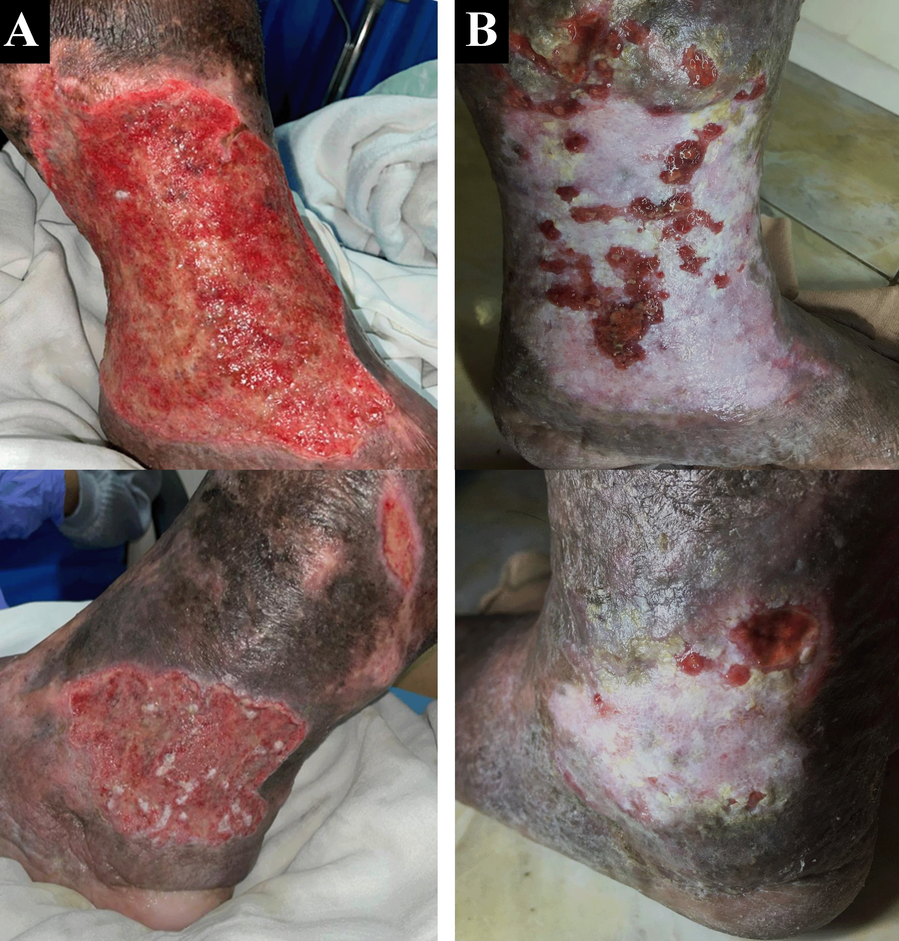
A. pre-EVLA; B. 6-months post EVLA.
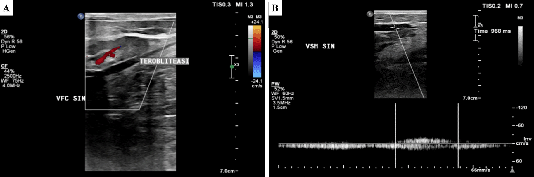
A. Obliterated segment of above-the-knee left GSV;
B. persistent reflux of below-the-knee GSV (968 ms).
EVLA was done under spinal anesthesia. Mapping of left GSV and accessory vein was done resulting in proximal ATK accessory vein diameter 6.0 mm, medial ATK 8.3 mm, and distal ATK 6.5 mm. The diameter of distal ATK GSV was 8.00 mm, proximal BTK GSV 7.4 mm, and media BTK GSV 5.4 mm. The puncture was initially started from distal ATK GSV retrogradely, but the laser catheter couldn’t pass because of branching in the proximal BTK area. Access was moved to the left dorsalis pedis, and a laser catheter was introduced until distal ATK. The total tumescent amount was 350 mL. EVLA was started in distal ATK GSV until proximal BTK with laser power of 5 W LEED 40 J/cm, followed by 2 W LEED 20 J/cm in proximal until media BTK GSV. The second puncture was done on distal ATK accessory veins with laser power 6 W LEED 50 J/cm from proximal until distal ATK of the left accessory vein. The total amount of the tumescent was 500 mL. The total ablated GSV length was 33 cm and the accessory vein was 29 cm. Lower extremity DUS showed ATK until BTK left GSV and accessory vein were perfectly obliterated, no DVT found, and patent epigastric vein.
The patient was discharged two days after the procedure with optimal medical treatment and wound care by the nursing team with modern wound dressing. At follow up after six months, the patient did not have any complaints. Skin condition on malleolus sinistra region showed healed ulcer with granulation tissues, no purulent discharge, minimal swelling until below the knee, and hyperpigmentation (Figure 2B). There was a 16-point reduction of VCSS down to 7.
A 65-year-old male farmer presented with complaints of ulcers with smelly discharge at the right ankle accompanied by pain (VAS 4/10) in the past two years which became worse and enlarged in the past six months. Complaints begin with swelling of both lower limbs for 11 years. The patient had undergone various traditional treatments before such as the use of a flour mixture. The patient had a history of hypertension and diabetes mellitus, was an ex-smoker, and was obese with a BMI of 33.3 kg/m2. Physical examination showed multiple ulcers sized 10×8 cm at malleolus lateral dextra and 8×3 cm at malleolus medial dextra with irregular edge, wet base with purulent discharge, erythema, edema (+), and hyperpigmentation from 1/3 proximal cruris dextra until dorsum pedis dextra (Figure 4A). Lower extremity DUS showed severe CVI of bilateral GSV, small saphenous vein (SSV), and deep vein. The patient was diagnosed with bilateral CVI with active VLU at malleolus medial and lateral dextra (C6sEpAsdPr), and VCSS was 22. The patient decided to undergo EVLA on right lower limb first and received ceftriaxone and metronidazole five days before the procedure. The procedure was done after the patient finished the antibiotics course.
EVLA was done under spinal anesthesia. Mapping was done along the right GSV and SSV. The diameter of SFJ was 12.0 mm, proximal ATK GSV was 7.5 mm, media ATK GSV was 7.0 mm, distal ATK GSV was 7.8 mm, proximal BTK GSV was 6.0 mm, media BTK GSV was 4.0 mm, and distal BTK GSV was 3.0 mm. The proximal SSV diameter was 7.0 mm, and the media SSV was 6.5 mm. Puncture by guided DUS was done at three locations for GSV: medial side of dorsalis pedis (distal from the ulcer), proximal BTK GSV with laser catheter introduced retrogradely, and proximal BTK GSV with laser catheter introduced until 3 cm passed SFJ. EVLA was done with laser power 6 W LEED 50 J/cm in proximal until media ATK GSV, 5 W LEED 40 J/cm in media ATK GSV until proximal BTK GSV, and 2 W LEED 20 J/cm in proximal until distal BTK. Puncture for SSV was done at media BTK, laser catheter was introduced until 5 cm passed Sapheno-poplitea Junction. EVLA at proximal until media SSV was done with laser power 4 W LEED 30 J/cm. The total ablated GSV length was 64 cm and the SSV length was 10 cm with a total of 850 mL of tumescent amount. Lower extremity DUS showed the ablated GSV and SSV were perfectly obliterated, no thrombus found, and a patent epigastric vein. Wound dressing of the ulcer and an elastic bandage was applied to the patient’s right lower limb.
The patient was discharged two days after the procedure with optimal medical therapy and the wound care was done by the nursing team with modern wound dressing. At follow up after six months, the patient did not have any complaints and the ulcer and skin condition had significantly improved. There was no ulcer left, hyperpigmentation was limited to 1/3 proximal cruris dextra, and there was a scar (+) on the previously ulcerous region (Figure 4B). There was a 17-point reduction of VCSS down to 5.
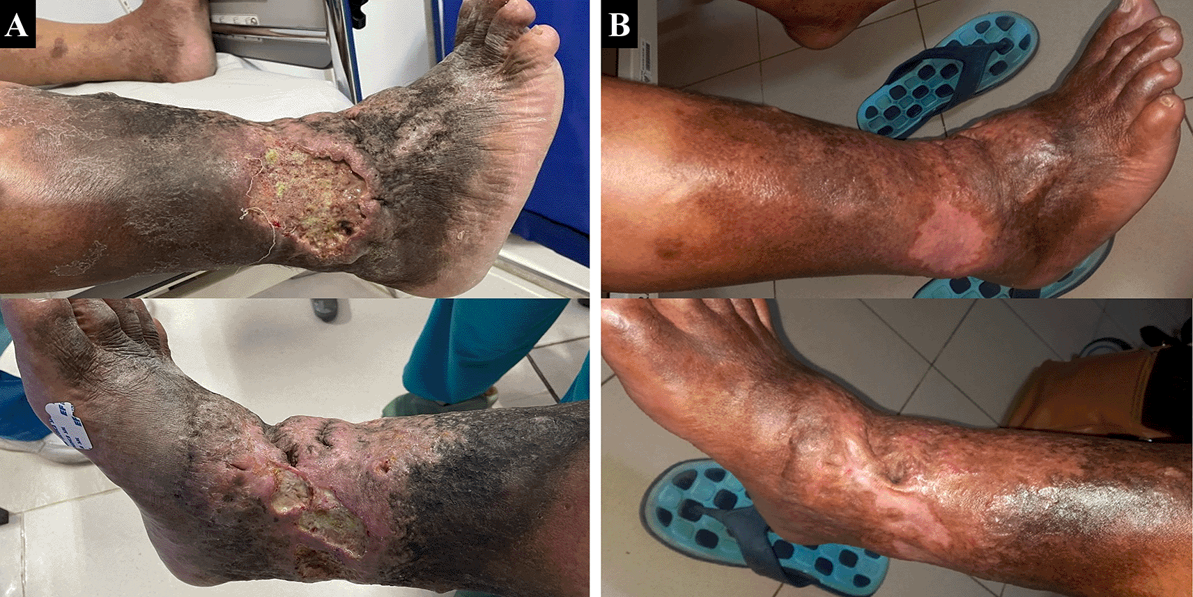
A. pre-EVLA; B. 6-months post EVLA.
According to the European Society for Vascular Surgery (ESVS) 2022 Clinical Practice Guidelines on the Management of Chronic Venous Disease of the Lower Limbs, it is recommended to do early endovenous ablation in patients presenting with active VLU and superficial vein incompetence to accelerate ulcer healing (Class 1 Recommendation with Level of Evidence).1 But the topic of how to do EVLA in active VLU patient remains to be discussed.
All of the patients we have reported here were classified as C6 CEAP clinical criteria with DUS examination revealing reflux of both ATK and BTK GSV, and one case each with accessory vein and SSV involvement. This clinical finding was correlated with superficial vein as mentioned in García-Gimeno et al.’s9 study that in CVD patients, involvement of ATK and BTK GSV without SSV (odds ratio (OR) 1.72, P<0.05) or with SSV (OR 2.62, P<0.05) significantly increases the possibility of the patient to be in C4 – C6 clinical class.
Numerous incidences of BTK GSV reflux after ATK GSV EVLA have been reported. A study by de Arujo et al.10 showed a 70% incidence of BTK reflux one year after ATK GSV ablation. The study was consistent with our second case which presented persistent symptoms and delayed ulcer healing even after the treatment of ATK GSV EVLA. Pihlaja et al.11 stated that persistent reflux of superficial vein after ablation was the main factor of delayed ulcer healing (multivariate hazard ratio (HR) 0.123, 0.0051-0.295 95% CI, P<0.001) with multifactorial etiology including missed reflux segment and incomplete therapy, reflux sources (GSV/SSV/perforator) which were normal at the earlier examination, recanalization of the treated segment, and history of deep vein thrombosis.
The decision to do GSV EVLA until BTK is preferred because it is supported by numerous pieces of evidence that provide significantly more advantages than only ATK GSV EVLA. Elimination of reflux throughout the length of the incompetent GSV segment from the SFJ to the most distal point will lead to significant clinical improvement and reduce the need for adjuvant therapy.10 A systematic review by Sussman et al.12 mentioned that in CVI with C4 to C6 disease, more aggressive treatment of the ATK and BTK GSV is justified if the DUS findings demonstrate groin to ankle reflux. These pieces of evidence support our decision to treat our patient with EVLA until BTK GSV.
However, there is another factor that should be taken into consideration which is the risk of complications. Reported complications of EVLA procedure are infection (0.5%), thromboembolic events such as deep vein thrombosis and endovenous heat-induced thrombosis (0.85%), hematoma (2.1%), skin burn (2.1%), short term paraesthesia (3.8%), superficial thrombophlebitis (5.6%), and bruising (39%).13 Nerve injury risk in EVLA is directly related to the distance of the ablated vein with the nerve through the needle stick mechanism or burn due to laser energy transfer. This means that BTK GSV EVLA had a higher risk of causing nerve injury since the saphenous nerve runs medially towards the GSV at the level of the upper calf to the ankle.14 Sinabulya et al.15 demonstrated a non-clinically significant permanent sensory loss at 8% of limbs that underwent BTK GSV EVLA after a mean of 41 months follow-up duration in VLU patients without any major nerve injury reported. Utoh and Tsukamoto observed the effect of ablation length on the incidence of post-EVLA neuropathy with an incidence of 0.8% if the ablation length was less than 40 cm from the SFJ and 4.6% if it was more or equal to 40 cm.16 All of the patients in our cases did not show any nerve injury symptoms after follow-up at six months.
Various efforts need to be made to minimize complications from EVLA. Tumescent anesthesia is used in EVLA to prevent thermal-induced tissue damage, compressing the size of the vein to be treated to allow better energy absorption, as well as allowing early mobilization and reducing the risk of DVT where EVLA without tumescence has an incidence of approximately 36.5% of nerve-related paresthesia.14,17,18 Reducing the number of punctures while administering tumescent anesthesia also logically reduces the incidence of nerve injury.10 We administered DUS-guided tumescent anesthesia in our patients and at follow-up, our patients did not show any signs or symptoms of nerve injury.
It’s hard to determine the cut-off of laser power in EVLA to perfectly obliterate the vein without complication. Reducing the laser energy along the distal segment of BTK GSV may reduce the incidence of paresthesia, but with consequences of decreased treatment success.10 The newer generation of EVLA with 1470-nm wavelength has more advantages with chromophore targeting only water in the endothelium compared to earlier laser generations with 940-nm and 980-nm wavelengths that targeted hemoglobin molecules.19,20 EVLA with higher wavelength is related to lower energy levels and minimal side effects.20 A study by Arslan et al.20 showed recanalization numbers were significantly lower (P=0.017) after 48 months of follow-up in a group of EVLA with 1470-nm radial-tip laser catheters using laser power LEED 45–120 J/cm (8.3%) compared to the group with 980-nm bare-tip laser catheter using laser power LEED 60–120 J/cm (15.9%). Post-procedural complications such as pain, induration, ecchymosis, paresthesia, and DVT were significantly lower in the 1470-nm group. In another study by Doganci and Demirkilic, 3.33% of the 1470-nm group reported paresthesia compared to 30% in the 980-nm group (P<0.0001). The symptoms experienced tended to be minor with an average duration of less than four weeks in both groups.21
Although a systematic review and meta-analysis by Malskat et al.22 showed that the commonly used EVLA parameters (wavelength, delivered energy, and outcome) did not affect the high success rate of the therapy (92%), the laser energy used in this study has a cut off of 50 J/cm. Mendes-Pinto et al.23 showed that closure rates at one year follow-up were lower in a group of 1920-nm EVLA with LEED 17.8±0.6 J/cm (87.5%) compared to the 1470-nm group with LEED 24.7±0.8 J/cm (94.7%) whereas Park et al.19 performed EVLA using 1470-nm laser with laser energy 6W LEED 30 J/cm on 267 limbs and obtained 84.5% completely obliterated GSV results in one month and 100% after six months. Utoh and Tsukamoto proposed EVLA with two-step ablation protocol in their trial that involved 90 limbs, using 1470-nm laser with energy 7W in ATK GSV (LEED 50 – 70 J/cm) and 5W in BTK GSV (LEED 20-25 J/cm) with mean ablated GSV length of 50.4 cm. The DUS follow-up resulted in perfect obliteration of the ablated vein without any saphenous nerve injuries.24 In our case, we were using a protocol with lower laser energy than some past literature because of anatomical consideration that the average GSV diameter of an Asian population was significantly lower than a Caucasian population (Median 2.9 mm vs 5.7 mm; P<0.01).25
We’ve reported three cases of patients with active VLU undergoing EVLA until BTK with significant results. The EVLA of GSV until BTK provides satisfactory results in patients with VLU. A combination of DUS-guided procedure, administration of tumescent anesthesia, use of newer generation device, minimal laser energy, and post-procedural management could minimize the risk of complications and produce a maximal outcome.
| Views | Downloads | |
|---|---|---|
| F1000Research | - | - |
|
PubMed Central
Data from PMC are received and updated monthly.
|
- | - |
Provide sufficient details of any financial or non-financial competing interests to enable users to assess whether your comments might lead a reasonable person to question your impartiality. Consider the following examples, but note that this is not an exhaustive list:
Sign up for content alerts and receive a weekly or monthly email with all newly published articles
Already registered? Sign in
The email address should be the one you originally registered with F1000.
You registered with F1000 via Google, so we cannot reset your password.
To sign in, please click here.
If you still need help with your Google account password, please click here.
You registered with F1000 via Facebook, so we cannot reset your password.
To sign in, please click here.
If you still need help with your Facebook account password, please click here.
If your email address is registered with us, we will email you instructions to reset your password.
If you think you should have received this email but it has not arrived, please check your spam filters and/or contact for further assistance.
Comments on this article Comments (0)