Keywords
T-PRF, Platelet Concentrates, Chronic Periodontitis, Infrabony Defects, Systematic Review, Meta-analysis
This article is included in the Datta Meghe Institute of Higher Education and Research collection.
T-PRF, Platelet Concentrates, Chronic Periodontitis, Infrabony Defects, Systematic Review, Meta-analysis
Plasma fractions with higher platelet concentrations, such as platelet-rich plasma (PRP) and platelet rich fibrin (PRF) are crucial to regeneration.1 Their use has been successfully linked to connective tissue and bone repair.2 PRP has been documented and used in a variety of applications with what appears to be clinical success. It is an autologous modification of fibrin glue that is created by techniques that concentrate autologous platelets.1 PRP supports bioactivity but exhibits no stimulant properties for activation of regenerative cells.3 Additionally, its preparation is technique sensitive.4 PRP preparation, on the other hand, takes time therefore various efforts have been made to modify and enhance the PRP’s characteristics and quality which include Leukocyte platelet-rich fibrin (L-PRF). It was first described by Choukroun as cited by Dohan DM et al. (2006).5 It is regarded as a second-generation platelet concentrate and has been used to speed up wound healing during several surgical operations. Choukroun et al. first created PRF in France in 2001 as an autologous biomaterial that includes leucocytes.6 The method does not require bovine thrombin or an anticoagulant, in contrast to other platelet-rich products (or any other gelling agent).5,7–9 Although PRF has been shown to produce positive therapeutic outcomes, some clinicians are concerned that the method’s use of glass-evacuated collection tubes for blood with silica activators may pose a health risk.10,11 O’Connell provided a description of the inevitable silica interaction.12 Although the silica in the tube is thick enough to settle with the red blood cells, some of the particles are still colloidally suspended in the buffy coat, fibrin, and platelet-poor layers of plasma. Therefore, when the product is used for therapy, the particles could come in contact with the patient. One of them is titanium platelet rich fibrin (T-PRF).13 In the glass vacuum containers used to prepare “Platelet Rich Fibrin,” silica is known to have a hazardous impact that this third generation platelet concentrate is known to eliminate (PRF).
Nonsurgical periodontal therapy, such as scaling and root planing (SRP) alone or SRP plus systemic or local anti-inflammatory or antibacterial drugs, to surgical flap debridement, are the approaches for treatment of periodontitis cases.14 However, in cases of soft and hard tissue defects, surgical correction of the defects is of prime importance. For correction of bony defects, the use of autografts and allografts have been advocated.15 Autologous platelet concentrates when used in conjunction with bone grafts results in better improvement. A study by Olgun et al. (2018) reported clinical, radiographic and histological outcomes of bone that accelerated to four months compared to six months for allografts when combined with T-PRF.16 The desired outcome was seen in four months as compared to six months. Tunalı et al. in the year 201217 discovered a mature fibrin network when T-PRF clots were examined. In the T-PRF membrane, they discovered islets of bone tissue and newly developing connective tissue. These findings demonstrate that T-PRF could, within 30 days of treatment, cause the development of new bone and connective tissue in an experimental rabbit model of wound healing.
As mentioned above, the novel product termed T-PRF was prepared by Tunali et al. (2014)13 utilising a modified L-PRF process. Authors took the advantage of Dohan Ehrenfest’s taxonomy, which divides platelet-rich products into four main groups to prevent any misunderstanding about how to obtain these products. These four categories are determined according to the number of leukocytes and fibrin they contain, platelet-rich products: just platelet-rich plasma products, platelet- and leukocyte rich plasma products, platelet-rich fibrin, and platelet and leukocyte rich fibrin. The creation of T-PRF, a new platelet concentration, was motivated by the idea that titanium tubes would be more potent at activating platelets than the glass tubes used in Chouckron’s approach. The distinguishing properties of T-PRF, particularly its improved biocompatibility, are produced by platelet activation with titanium as opposed to activation with silica particles.18 T-PRF fibrin network covers a larger area than L-PRF fibrin network, also fibrin seemed thicker in the T-PRF samples.13
Since the introduction of T-PRF by Tunali et al.,18 it has been assessed for various properties including its structural and biochemical characteristics. Tunalı et al. (2014)19 described the structural properties of T-PRF, and compared it to L-PRF. T-PRF samples appeared to have a highly ordered network with continuous integrity. The fibrin network in T-PRF samples appeared to be thicker and to cover a wider area than the network in L-PRF samples, according to histomorphometric analyses. T-PRF is a naturally occurring leukocyte- and PRF product that has been defined for the first time in a human investigation. Titanium platelet activation appears to provide T-PRF with certain high features. Since then T-PRF has been used for various periodontal procedures including treatment of various infrabony defects, gingival recession defects, in open flap debridement cases, periimplantitis.13,16,18,20–22
Systematic evaluation of the studies to assess the clinical efficacy have not been done to date. This systematic review focuses to provide the best possible evidence for the use of T-PRF in periodontal regeneration.
We searched PubMed, Cochrane, Embase database without language restrictions. We used Medical subject headings (MeSH) or equivalent and textword terms. We searched the metaRegister of controlled trials (mRCT), National clinical trials government website. Additionally, we checked the reference lists of reviews, retrieved articles for additional studies, and performed citation searches on key articles manually by going through each reference of the included articles. We intended to use randomised controlled trials (RCTs) that assessed outcomes in an open or blinded manner. Short abstracts (typically meeting reports), non-randomised research, experimental pain studies, animal model studies, case reports, and clinical observational studies were all omitted.
Search strategy
(“t prf”[Title/Abstract] OR “titanium prepared platelet rich fibrin”[Title/Abstract] OR ((“titanium”[MeSH Terms] OR “titanium”[All Fields] OR “titaniums”[All Fields]) AND “platelet rich fibrin”[MeSH Terms])) AND (“gingival recession”[Title/Abstract] OR “gingival recession”[MeSH Terms] OR “furcation defects”[MeSH Terms] OR “alveolar ridge augmentation”[MeSH Terms] OR “infrabony defect”[Title/Abstract] OR “furcation”[Title/Abstract] OR “periimplantitis”[Title/Abstract])
Systemically healthy patients (18+ years) clinically diagnosed with any type of periodontal disease (infrabony defects, gingival recession, osseous defects) were included irrespective of gender and race). Our outcome measures included change in the indices [Plaque index (PI) Gingival index (GI)], mean of periodontal pocket depth (PPD), mean of relative vertical clinical attachment loss (RVCAL), mean of relative horizontal clinical attachment loss (RHCAL) and mean of bone defect (BD). All the parameters were measured at baseline, 3 months and 6 months.
Selection of studies
An open source online screening tool Rayyan23 (RRID:SCR_017584) was used by review writers. They independently screened the search results (RO, PD, PB, KB). By reading through the abstracts of each study that the search produced, the eligibility of each research was established. The review writers excluded studies that did not distinctly meet the inclusion criteria. For all included studies’ complete texts were acquired. Primary reviewers independently screened the complete texts of these studies to pick the most pertinent studies (RO, PD, PB, KB). The respective authors were reached by phone or email to get the required clarification for any missing data or information in the studies that had an impact on the study selection criteria. In cases of disagreement or dispute, a fifth author was asked for a judgement (KD). Prior to evaluation, the trials were not anonymized. Any language restrictions in the study selection process were not taken into consideration as a constraint for carrying out this evaluation. The complete review includes a PRISMA flow chart32 that shows the precise status of all identified studies in accordance with the recommendation found in “Part 2, Section 11.2.1 of the Cochrane Guidelines for Systematic Reviews of Interventions”.24 Irrespective of the reporting of outcome data, studies were included in this review as shown in Figure 1.

The data extraction from “included studies” was performed by four reviewers (RO, CS, KB, and KD) and given in the “characteristics of studies table” using a pre-defined data extraction form (Table 1). Data were extracted based on the nature of research, participant information, intervention information, and reported results. The disagreement between the main reviewers was resolved by the third reviewer (PB). The fourth reviewer successfully addressed the “risk of bias evaluation” discrepancy (KB). We have included a total of nine studies for qualitative analysis and have been included in the characteristics of studies table (Table 1). All the studies were matched for type of intervention, type of defect and participant details. Three studies were included and matched for meta-analysis followed by quantitative analysis.20,22,25
Four reviewers (RO, PD, CS, and SH) from each included study independently evaluated the risk of bias (RoB) using the Cochrane domain-based, two-part tool as outlined in Chapter 8 of the Cochrane Handbook for Systematic Reviews of Interventions.24 The fourth reviewer was able to resolve the disagreement between the main reviewers (KB). Sequence generation, allocation concealment, participant and staff blinding, blinding of outcome evaluation, incomplete outcome data, biased outcome reporting, and other bias, such as baseline imbalance, were all areas in which we evaluated the RoB.
In parallel-group RCTs, the unit of analysis was the individual subject. The Elbourne-recommended method is used to integrate the cross-over designed trials into the meta-analysis.26 Measurements from experimental intervention periods and control intervention periods, respectively, were taken for these trials, and they were analysed under the assumption that it was a parallel group study of intervention versus control.
The Chi2 test (P value set at 0.10 for statistical significance) was used to examine clinical heterogeneity, and the I2 statistic was utilised to quantify heterogeneity in the outcomes of the included studies using RevMan manager version 5.0.27 Significant heterogeneity is defined as I2 over 75%; substantial heterogeneity is defined as I2 between 50% and 90%; moderate heterogeneity is defined as I2 between 30% and 60%; and mild heterogeneity is defined as I2 less than 40%. If statistical heterogeneity with I2 more than or equal to 50% is found, relevant causes were investigated using predefined subgroup analysis, and a random-effects model was used and reported.
Only when it was determined that the participants, interventions, comparisons, and results of the included studies were adequately comparable to yield a conclusion with clinical relevance and significance was a meta-analysis performed. We intended to carry out the meta-analysis using the Cochrane Collaboration’s open source RevMan 2014 statistical software (RRID: SCR_003581). If statistical heterogeneity with I2 higher than or equal to 50% is found, the sources of the heterogeneity were found, and a random-effects model meta-analysis was then carried out. In a meta-analysis, four papers were used. Table 2 presents data from all three investigations (Data Entry).
The analysis of the PI, PPD, CAL, DF was done separately for the interventional (OFD+T-PRF) and control groups (OFD alone). The details of the studies included for meta-analysis are given in Figures 2-5. All the included studies reported the use T-PRF combined with OFD. Three articles reported the use of T-PRF in infrabony defects,20,22,25 one study reported use of T-PRF combined with allograft for patients with chronic periodontitis.28 Another study reported its use in sinus lift procedures.16 Two studies reported its use in gingival recession21,29 and other in healing of extraction socket.29 Based on the overall similarities in participants, outcome and data three studies were found to be eligible for meta-analysis.20,22,25 The conclusion regarding the overall effect size estimates were made based on the meta-analysis.
All the included studies reported PI at baseline and after nine months.20,22,25 However, two studies reported data in the form of mean±standard deviation. All the studies showed statistically significant reduction in plaque index at nine months follow up period. The overall meta-analysis showed marginally significant reduction in plaque scores after nine months. (Mean Difference -0.52; 95% CI -1.30 to 0.27; p=0.03; I2 78%; two studies; 33 participants). However, in view of significant heterogeneity (I2=78%), the results need to be interpreted with caution because of increased heterogeneity as shown in Table 3 and Figure 2.

All the three included studies reported data on PPD at baseline and after nine months. All the studies reported significant reduction in PPD. The overall meta-analysis showed marginally significant reduction in PPD scores after nine months. (MD -0.85; 95% CI -1.26 to -0.45; p=0.01; four studies; 62 participants). However, in view of significant heterogeneity (I2=93%), the results need to be interpreted with caution as shown in Table 4, Figure 3.

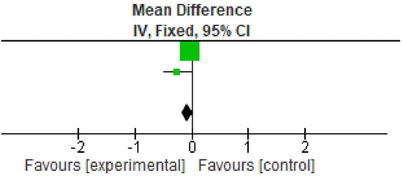
All the included studies reported clinical attachment loss (CAL) gain at baseline and after nine months. All the studies reported significant improvement in CAL gain. The overall meta-analysis showed marginally significant increase in CAL gain after nine months. (MD -0.12; 95% CI - 0.59 to -0.35; p<0.0001; three studies; 62 participants). However the percentage of variation (I2 – 55%) was found to be minimum in all the three included studies, therefore making the interpretation of result favourable as shown in Table 5, Figure 4.

All the included studies reported defect fill at nine months. However quantitative assessment was only possible for two studies because of difference in reporting of outcomes.20,22 Significant defect fill was observed in both the studies. The overall meta-analysis showed a marginally significant increase in defect fill after nine months (MD - 0.07; 95% CI -0.17 to -0.03; p=0.09; two studies; 47 participants). Overall defect fill was not statistically significant. However in spite of improvement in defect fill, in view of significant heterogeneity found the results needs to be interpreted with caution as shown in Table 6, Figure 5.
Allocation: One study reported use of the coin toss method for allocation hence was graded as low risk,22 the other two did not report the method and hence were graded as unclear.20,25 Blinding: all the studies were double blinded and were kept in low-risk bias.20,22,25 Missing result information: discretionary reporting In terms of reporting, we classified all three studies as low risk. The bias in the behaviour or observations: since all procedures and observations were carried out by a single trained professional in each research, we classified the study as low risk in terms of performance or observational bias. Figures 6 and 7 show, respectively, a summary of the risk of bias in the included studies and its graphical depiction.


There are seventeen pieces of literature published on T-PRF. Two in-vitro studies have been performed by Tunali et al.18,19 They discovered a mature fibrin network. In the T-PRF membrane, they discovered islets of bone tissue and newly developing connective tissue. These findings demonstrate that T-PRF could, within 30 days of treatment, cause the development of new bone and connective tissue in a rabbit model of wound healing. They have also described the structural properties of T-PRF, and it was compared to L-PRF. Another in-vitro study evaluated the regulation of gingival keratinocyte adherence, dissemination, and cytokine expressions on titanium and PRF surfaces.30 Ustaoğlu et al. (2016)29 in their clinical study assessed the T-PRF’s clinical effects on human palatal mucosal wound healing (PMWH), and its impact on “time-dependent variations in palatal soft-tissue thickness (PSTT), a novel idea, was discovered. Uzun et al. (2017)21 compared the results of connective tissue graft with autologous T-PRF (CTG). T-PRF (63 teeth) or CTG were utilized to treat 114 Miller Class I/II gingival recessions with abrasion defects utilizing a modified tunnel technique (51 teeth). Olgun et al. (2018)16 investigated the differences between using an allograft or totally autologous T-PRF in sinus-lifting surgeries on the clinical, radiological, and histological levels. Arabaci et al. (2018)20 T-PRF and open flap debridement (OFD) were examined for their effects on biological markers in gingival crevicular fluid (GCF) and periodontal results. TPRF+OFD or OFD alone were used to treat 29 subjects with chronic periodontitis. Ustaoğlu et al. (2019)31 analysis of extraction sockets preserved by L-PRF and T-Fractal PRF’s dimension (FD) and early soft tissue healing characteristics. Ustaoğlu et al.31 evaluated the impact of T-PRF and Connective tissue graft (CTG) on peri-implant soft tissue thickness, and crestal bone level. Thirty implants were inserted into 30 patients while the soft tissue was simultaneously augmented with either T-PRF or CTG. Gummaluri et al. (2020),22 based on clinical and radiographic criteria, assessed the efficacy of T-PRF and L-PRF in the management of intra-bony defects.
Out of the above-mentioned studies, three studies were included for meta-analysis.20,22,25 Results of meta-analysis supported that T-PRF is effective for correction of both hard and soft tissue defects. We included three studies that have reported data from 100 participants, aged 18+ years, that have used T-PRF in infrabony defects treated with open flap debridement. Studies have reported data on periodontal parameters like pocket depth, gingival bleeding, clinical attachment loss. Indices including plaque index and gingival index. Meta analysis was done for PPD, CAL, PI and defect fill parameters. The overall results of meta-analysis suggest that T-PRF is a better alternative to other platelet concentrates for both hard and soft tissue parameters.
This review focused on answering if T-PRF is a better alternative when compared with other platelet concentrates for periodontal regenerative procedures. The overall qualitative and quantitative analysis suggest that T-PRF have superior structural properties and thicker fibrin network. It provides superior results when used alone or combined with autograft or allograft. However, due to limited number of studies done so far, we recommend that more high quality RCTs to be conducted on this topic.
| Views | Downloads | |
|---|---|---|
| F1000Research | - | - |
|
PubMed Central
Data from PMC are received and updated monthly.
|
- | - |
Provide sufficient details of any financial or non-financial competing interests to enable users to assess whether your comments might lead a reasonable person to question your impartiality. Consider the following examples, but note that this is not an exhaustive list:
Sign up for content alerts and receive a weekly or monthly email with all newly published articles
Already registered? Sign in
The email address should be the one you originally registered with F1000.
You registered with F1000 via Google, so we cannot reset your password.
To sign in, please click here.
If you still need help with your Google account password, please click here.
You registered with F1000 via Facebook, so we cannot reset your password.
To sign in, please click here.
If you still need help with your Facebook account password, please click here.
If your email address is registered with us, we will email you instructions to reset your password.
If you think you should have received this email but it has not arrived, please check your spam filters and/or contact for further assistance.
Comments on this article Comments (0)