Keywords
acid base balance,SOFA score,sepis-3 criteria,critically ill patients,unmeasured anions,serum lactate,prognostic marker,cross sectional study.
This article is included in the Datta Meghe Institute of Higher Education and Research collection.
acid base balance,SOFA score,sepis-3 criteria,critically ill patients,unmeasured anions,serum lactate,prognostic marker,cross sectional study.
Severe sepsis and septic shock are chief health care complications, affecting millions of individuals around the globe per year, killing one in four (and frequently more), and increasing in occurrence.1 Sepsis, is a physiologic and pathologic condition, including biochemical abnormalities brought on by the infection, is a serious community health issue that cost the US healthcare system more than $20 billion (5.2%).2 According to the Global Burden of Diseases 2017, the percentage of sepsis associated deaths in each country was shown using mixed-effects for different ages, genders, locations, disease causes, using data from 8 million unique hospital records. A total of eleven million sepsis-associated deceases were documented, accounting for a projected 48.9 million cases of sepsis and 19.7 percent of all death cases globally. Between the years 1990 till 2017, age-standardized sepsis incidences declined by 37 percent, while death dropped by 52.8 percent. The occurrence and mortality of sepsis varied greatly by area, with Oceania, South Asia, East-Asia sub-Saharan Africa, Oceania, South Asia, and South-East Asia weighing the heaviest burdens.3 Sepsis remains to be a main source of health loss wide-reaching, with a particularly high health-associated burden in Sub-Saharan Africa, despite dropping age-standardized prevalence and mortality cases.3 A thorough evaluation of the literature by the World Health Organization estimated the hospital death rate from sepsis to be 27%,4 in patients receiving treatment for sepsis in critical care, mortality is expected to be 42%.
Sepsis incidence and mortality statistics are largely based on information from Western nations. In India, tropical illnesses like leptospirosis, malaria, dengue, tuberculosis, and enteric infections are also important reasons of severe sepsis or septic shock, in dissimilarity to the Western nations where septic infections by the Gram-negative organisms is the main origin of sepsis. Severe sepsis has a documented death rate >50% in the Indian setting.5
Novel definitions for sepsis and septic shock cases were released in the month of February (2016) by a Task Force accumulated by the European Society of Intensive Care Medicine and the Society of Critical Care Medicine.2 Earlier, the systemic inflammatory response syndrome (SIRS) criteria were not used in the new sepsis definitions, which associated Sepsis-3 sepsis to Sepsis-2 severe sepsis. Additionally, Sequential organ failure assessment (SOFA) score was used to operationalize organ dysfunction, and clear criteria were planned to diagnose septic shock, congruent with a number of clinical parameters in order to guide the clinical care, future studies, and healthcare planning. The task-force sepsis conditions, the necessity for a vasopressor to continue a Mean Arterial Pressure (MAP) of ≥65 mmHg and a Serum Lactate level of ≥2 mmol/L in the absence of hypovolemia.2
Sepsis patients may exhibit clinical presentations with the signs/symptoms hours before the situation gets worse. To screen these high-risk patients, early warning scores like the Modified Early Warning Score (MEWS), the National Early Warning Score (NEWS), or the Early Warning Scoring System (EWSS), were created. These results indicated a tendency toward better results, when emergency department (ED) teams enable prompt use of the most effective medicines when septic patients are identified.6 The formative evaluation and therapy of sepsis patients frequently take place in the ED. However, a few factors make it difficult to quickly determine which patients are utmost possible to develop severe illness, especially with septic infections due to the deficiency of the specificity of the systemic inflammatory response syndrome (SIRS) criteria for the infection; the heterogeneity of the clinical manifestations, including the clinical signs/symptoms, sites of infection, related complications, the etiologic pathogens; and the task of identifying patients most likely to develop to severe illness or death, especially among patients not severely ill during the initial evaluations.7 Early detection of high-risk individuals is necessary, either through a clinical decision rule that can be quickly evaluated or an easily accessible laboratory parameter.8 In the patients admitted to intensive care units (ICUs) who met severe sepsis criteria at twenty-four hours, the annual occurrences of severe sepsis was 51 per 10,000 patients, and the fatality rate was 47%. Sepsis patients made up 33% of hospital-bed days and 45 percent of ICU bed days.9
The initial and accurate identification of patients who benefit from the dynamic optimal medical interventions is critical if improved outcomes in terms of survival is needed to be attained. A substantial improvement in the survival can be achieved with an early goal-directed therapy (EGDT) in the emergency department prior to the ICU admissions. Detecting the time when pathophysiological processes can be stopped by the proper therapies is of immense importance for the final outcome.10 The diagnosis of sepsis is often confirmed by microbiological culture, which requires a 24 to 48-hour delay before antibiotics are administered. Early presentations may include an infectious source and aberrant biology and analytical data. Chest X-rays and computed tomography scans cannot reliably distinguish between different sepsis etiologies and permit the abuse of antibiotics. This is especially true for elderly individuals, for whom non-specific symptoms or organ malfunction may take the place of the typical sepsis presentation. This necessitates the use of sensitive sepsis biomarkers in these unusual presentations.11
Recent years have seen an increase in interest in research on lactate in severe sepsis. According to current theories, increased lactate in sepsis settings is linked to anaerobic cell metabolism because of disproportion between the body's mandate and supply for ATP and oxygen under circumstances like hypoperfusion or organ malfunction in sepsis patients. Several studies in literature have shown a greater mortality rate in people with hyperlactatemia who have severe sepsis and/or septic shock presentation. Because the lactate is generated from the tissues in the hypoperfusion condition before actual hypotension, it is a critical component in the diagnosis of the sepsis. Compared to revival without lactate monitoring or central venous oxygen saturation treatment, the lactate-guided recovery reduced mortality in the patients with septic shock. Lactate, a biomarker of shock, suggests the need for rapid fluid resuscitation, in addition to the diagnosis.12 Patients with blood lactate levels below 4 mmol/L are classified as in need of fluid resuscitation as per Surviving Sepsis Campaigns, 2012 and 2016.13 Normalizing lactate is often a resuscitation endpoint. However, due to the deficiency of access to readily available investigational equipment, particularly in poorer nations, point-of-care (POC) lactate measurement is not commonly employed.14,15
Another trending concept is of the serum anion gap (AG). It has been detected and it has also been used to spot paraproteins, assess patients with suspected acid-base diseases, such as sepsis, and find errors in electrolyte measurement.16 A larger AG is often recognized when it reaches the upper limit of normal (ULN). An increase in the anion concentration can exist without increase in AG, though, due to the wide range of typical values (commonly 8–10 mEq/L). Additionally, lactic acidosis is more severe than ketoacidosis in terms of the degree of increase in AG relative to the variation in the serum bicarbonate depending on the kind of retained anion.16
Current evidence is supportive to the fact that serum lactate is the predictor of mortality in sepsis.17,18 Serum lactate and AG can have a substantial link with one another in individuals with septic shock, but lactate and base excess demonstrated a modest correlation. Therefore, both indicators can be utilized interchangeably to assist in identifying septic shock in patients early.12 However, other reports identified that AG was not a sensitive indicator of an elevated lactate level within the initial hour of the onset of lactic acidosis. The AG increased more than the serum lactate, and only about 30% of the change in the AG could be attributed to rising serum lactate levels, indicating that other anions may be involved in the anion gap in lactic acidosis.19 Serum bicarbonate and AG are ineffective predictors of death and changes in lactate. Lactate is still the recommended biomarker, although AG levels of 20 mEq/L may be employed in situations with limited resources if lactate is not available to additional risk-stratify patients for the continued sepsis treatment.20 Between AG and the corrected AG (cAG) in terms of identifying hyperlactatemia, the cAG is not superior to the AG.21 Few studies reported high AG being a fairly sensitive and specific way to find individuals with sepsis who have elevated lactate levels.12,20–22 Despite these pieces of evidence, there’s still an excessive deal of interest in the biomarkers representing different pathogenic pathways. Given the general lack of studies in Indian settings with comparative evaluations of lactate and AG in sepsis, we performed this current study to determine the AG and serum lactate levels in the sepsis patients and their role in predicting the in-hospital mortality.
To study serum lactate vis-a-vis anion gap as prognostic markers in sepsis in rural tertiary care hospital.
This single-center, prospective, observational cross-sectional study with a cohort design was conducted in the Department of Medicine, Jawaharlal Nehru Medical College and its attached hospital Acharya Vinoba Bhave Rural Hospital (AVBRH), Sawangi (Meghe), Wardha. The suspicious sepsis patients were admitted to theICU, a 40-bed unit of the hospital under the Medicine Department of Acharya Vinoba Bhave Hospital, Sawangi (Meghe) associated to Datta Meghe Institute of Medical Sciences, Wardha.
The entire study was conducted as per the principles of the Declaration of Helsinki and local regulations. Relevant regulations and Good Clinical Practices and were followed throughout the study duration by each study team member. The present study was conducted after the clearance of the Institutional Ethical Committee (IEC number, DMIMS (DU)/IEC/2020-21/9300) of Acharya Vinoba Bhave Rural Hospital (AVBRH). Patients who showed their active interest to follow the projected research study design after a detailed written informed consent process were enrolled in the study by signing the informed consent.
This mid-term study was conducted during November 2020 to April 2022 as we found that peaks of infectious diseases leading to sepsis were at peak level during this time.
Patient enrollment was conducted at the Department of Medicine, Jawaharlal Nehru Medical College and its attached hospital Acharya Vinoba Bhave Rural Hospital (AVBRH), Sawangi (Meghe), Wardha. Subjects were screened in the ICU between Nov 2020 to Apr 2022 were eligible with a diagnosis of sepsis. Sepsis was diagnosed on SEPSIS-3 criteria.
1) Patients with either hepatic dysfunction, chronic kidney disease.
2) Patients with burns.
3) Patients with carbon monoxide intoxication, cyanide intoxication, and methanol intoxication.
4) Patients with collagen vascular diseases.
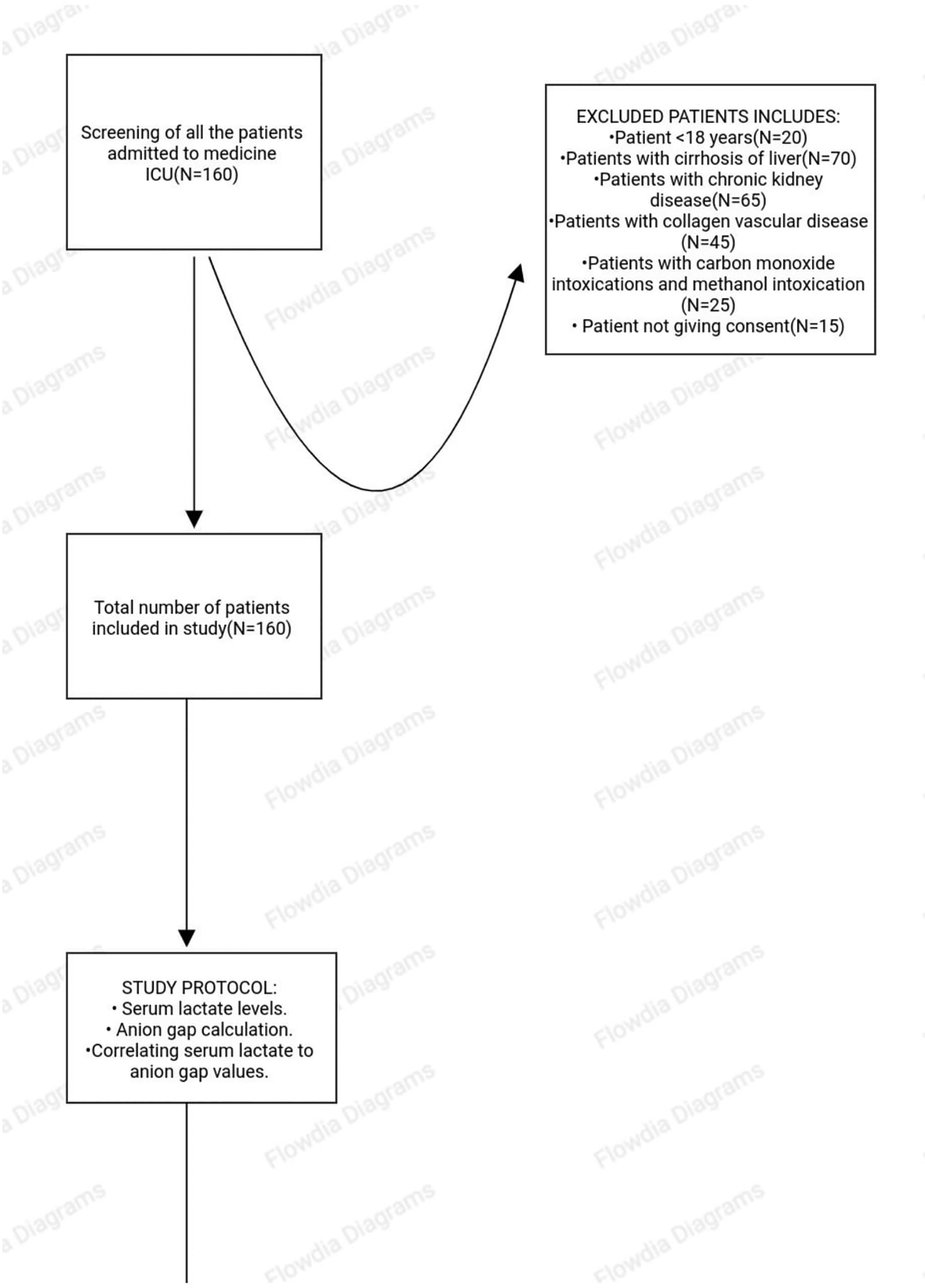
See Figure 1 for a plan of the study.
Medical residents under the direction of surgeons and clinicians from several medical disciplines made up the ICU's medical staff. Fellows with specialized training in detecting potential sepsis patients as per SOFA criteria went four times a day to the ICU to look for people who met the inclusionary criteria. Without being aware of the patients' subsequent hospital courses, we prospectively identified the patients and studied the ICU documents. In order to locate patients, we looked through the daily ICU log for admissions with an infection-related diagnosis (such as pneumonia, shortness of breath, etc). To confirm a suspect of infection based on ICU presentation as recorded in the medical decision-making section of the chart, all records of patients with infection or a probable infection underwent a confirmatory review.
According to institutional protocol, individuals with suspected of sepsis with SOFA score >2 had their venous lactate levels checked. The Glasgow Coma Score was assessed and recorded in the patient's record. Several other parameters like significant vitals, blood cultures, antibiotics, fluid therapy, resuscitation, length of hospital stay, complication, if any were also taken into account. The hospital ICU team's choices were unaffected by the researcher. We followed the study participants from initial admission to the hospital ICU until discharge or death.
The first venous serum lactate levels (mmol/L) obtained in the ICU were collected, together with the basic chemistry panel, estimated AG ([Na mEq/L] [Cl mEq/L] [HCO3 mEq/L]). The lactate levels were tested using a chemical analyzer from serum taken in a heparinized tube. According to normal reference range established by our institutional laboratory, we defined clinically relevant lactate to be >4 mmol/L and an anion gap of >12 was regarded as the highest limit of normal. Survival to hospital was referred to as a secondary outcome of in-hospital mortality.
The medical record number, baseline vital signs (BP, HR, RR), AG, and serum lactate, collected by venous sample, were obtained and documented by the ICU doctors in collaboration with the study team members upon ICU presentation and fulfilment of inclusion criteria. The following formula is used by to determine the anion gap:
Serum Lactate levels were measured by Lactate was measured by an enzymatic lactate oxidase assay using the Cobas 6000® analyzer, (Roche Diagnostics, IN, USA).
Sample size calculation:
Estimated specificity of anion gap=0.84
Prevalence of positive character (Prev)=0.283
Estimation Error 5%=7%
n=148
Minimum sample size required is 148, considering the drop out 10% sample size required was 163.3 samples drop out from the study, as per protocol analysis total 160 samples taken for observations.
The data from ICU admitted sepsis patient inclusionary of SOFA Score was entered into the Microsoft Excel sheet version 2016. Data were analyzed using SPSS software version 15 (SPSS IBM statistics, Chicago, USA), we can recommend R-studio software as a proprietary free alternative. We presented categorical variables as frequency and percentage. The continuous variables were presented as mean and standard deviation. In order to accurately characterize the results, demographic data were expressed as means with standard deviation, medians (interquartile range), or percentages. The student’s t-test was used to compare the means of two independent sets of quantitative variables. The Friedman and the Mann-Whitney test were used to gauge the non-symmetrical distributions of the variables. The Chi-square test was used to make proportional comparisons. Receiver operating curves were obtained to determine the area under curve with serum lactate and AG in prediction of mortality, P value was considered significant if p<0.05.
In this study, we included 160 patients with sepsis with SOFA score >2. Table 1 enlists the baseline characteristics in study population. Mean age of the population was 53.1±17.0 and 61.9% were males. Hypertension (41.3%) and diabetes (30%) were major comorbidities. Respiratory etiology (78.1%) was most common cause of sepsis. On admission, mean HR was 123.1±20.8 bpm, mean RR was 29.8±9.2 per min, mean SBP was 82.3±28.7 mmHg, and O2 saturation level was 84±12%. The mean SOFA score was 9.5±5.3. 53.8%, patients had alerted mental status. 58.8% required ventilator whereas 63.1% needed inotrope support.
Table 2 provides the details of various laboratory parameters assessed at baseline in study population. In hematological parameters, the mean Hb was 10.5±2.8 gm/dl, and leukocyte count was 23920±19050 cells/cmm. In liver function tests, the mean total bilirubin was 1.8±2.5 mg/dl, and the mean serum albumin was 3.0±0.8 mg/dl. The mean serum creatinine was 2.6±2.6 mg/dl. The mean serum lactate was 5.1±1.2 mmol/L and it was ≥4 mmol/L in 90% of patients. The mean AG was 14.0±3.9 and it was ≥12 in 75.6% of patients.
The mean hospital stay of 10.6±8.8 days and nearly 50% of patients had hospital stay duration of more than 10 days as shown in Table 3.
| Hospital stays (days) | Observation |
|---|---|
| Mean±SD | 10.6±8.8 |
| Median (IQR25-75) | 9 (4.3 to 15) |
| Hospital stay <10 days | 81 (50.6) |
| Hospital stay ≥10 days | 79 (49.4) |
Table 4 shows the outcome of patients during hospital stay. Mortality occurred in 45.6% of patients whereas 54.4% were discharged from the hospital.
| Outcome | Frequency (%) |
|---|---|
| Discharge (survivors) | 87 (54.4) |
| Death (non-survivors) | 73 (45.6) |
The mean lactate level was significantly higher in patients with high AG (5.6±1.1 vs. 4.1±0.8 mmol/L, p<0.0001). Similarly, the proportion of patients who had lactate levels ≥4 mmol/L was higher in those with AG ≥12 than AG <12 (95.9% vs. 71.8%, p<0.0001) as shown in Table 5. The sensitivity of AG ≥12 in detecting sepsis as defined by serum lactate ≥4 was 80.6% whereas specificity was 57.9%.
Among non-survivors, serum lactate level was significantly higher than survivors (5.8±1.1 vs. 4.5±0.9 mmol/L, odds ratio 4.55 (95% CI 2.68-7.70), p<0.0001). All non-survivors had lactate levels ≥4 mmol/L (Table 6). Among non-survivors, mean AG was significantly higher than survivors (15.8±3.7 vs. 11.9±3.4, odds ratio 1.39 (95% CI 1.23-1.56) p<0.0001). AG ≥12 was seen in significantly higher proportion non-survivors than survivors (89% vs. 64.4%, p<0.0001) (Table 7).
| Parameter | Survivors (n=87) | Non-survivors (n=73) | OR | 95% CI | P value |
|---|---|---|---|---|---|
| Serum lactate levels | 4.5±0.9 | 5.8±1.1 | 4.55 | 2.68 – 7.70 | <0.0001 |
| Lactate <4 | 16 (18.4) | 0 | - | <0.0001 | |
| Lactate ≥4 | 71 (81.6) | 73 (100.0) | |||
| Parameter | Survivors (n=87) | Non-survivors (n=73) | OR | 95% CI | P value |
|---|---|---|---|---|---|
| Anion gap | 11.9±3.4 | 15.8±3.7 | 1.39 | 1.23 – 1.56 | <0.0001 |
| AG <12 | 31 (35.6) | 8 (11.0) | - | <0.0001 | |
| AG ≥12 | 56 (64.4) | 65 (89.0) | |||
Figure 2 shows the ROC curves for AG and serum lactate.
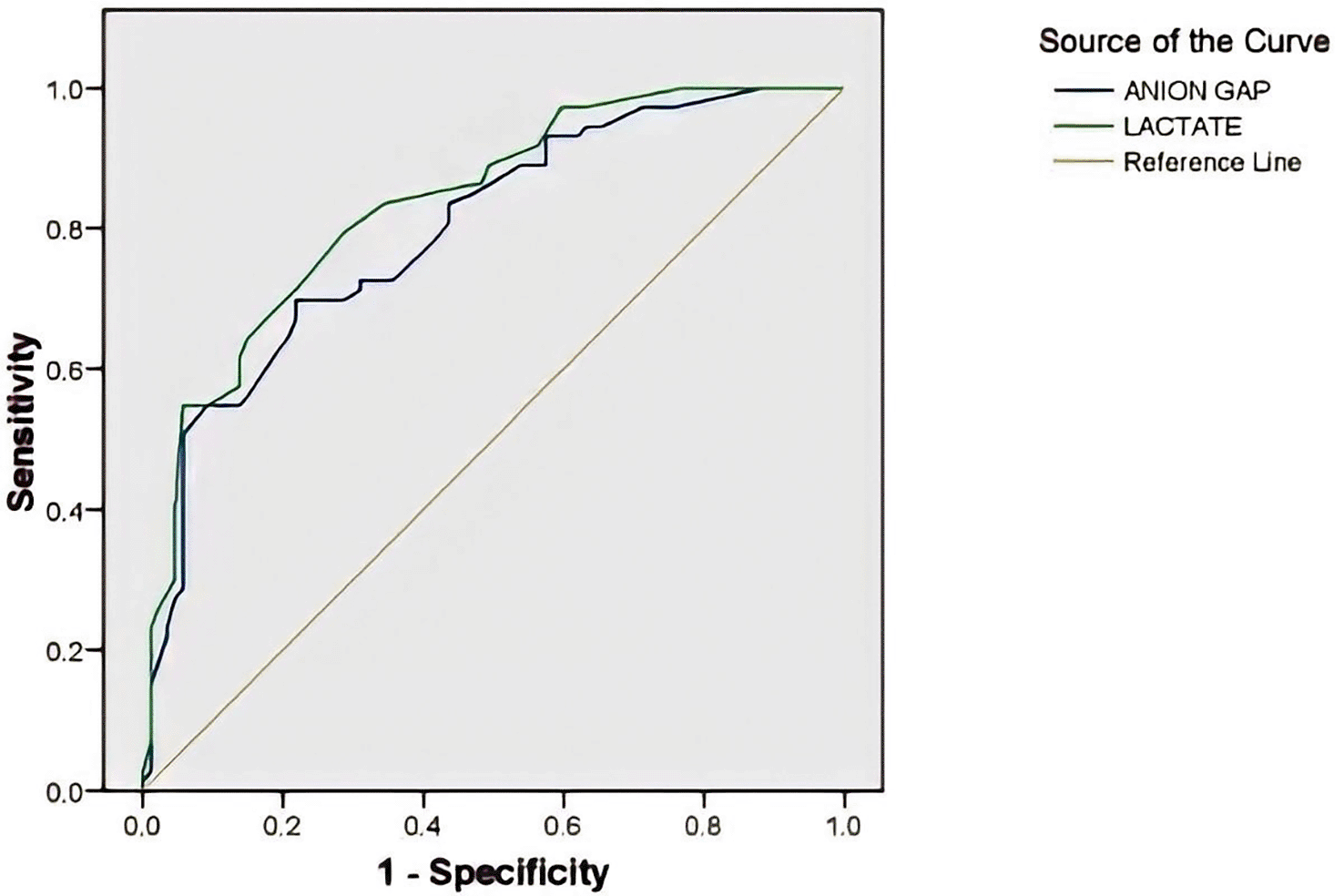
As shown in Table 8 the AUC of ROC in predicting mortality was significant for both serum lactate (AUC 0.797, p<0.0001) and AG (AUC 0.835, p<0.0001).
| Test variable | Area under curve | 95% confidence interval | P value | |
|---|---|---|---|---|
| Anion gap | 0.797 | 0.728 | 0.865 | <0.0001 |
| Serum lactate | 0.835 | 0.773 | 0.896 | <0.0001 |
Among various other parameters, significant predictors of mortality were elevated HR, RR, SOFA score and lower levels of SBP, O2 saturation, PiO2/FiO2 ratio. Also, need of ventilator and inotropes predicted mortality. Hospital stay was shorter in non-survivors than survivors.
In this study, we included 160 patients with sepsis with SOFA score >2.
Mean age of the population was 53.1±17.0 and 61.9% were males. Hypertension (41.3%) and diabetes (30%) were major comorbidities. On admission, mean HR was 123.1±20.8 bpm, mean RR was 29.8±9.2 per min, mean SBP was 82.3±28.7 mmHg, and O2 saturation level was 84±12%. The mean SOFA score was 9.5±5.3. 53.8%, patients had alerted mental status. 58.8% required ventilator whereas 63.1% needed inotrope support. Among laboratory parameters, mean Hb was 10.5±2.8 gm/dl, mean serum albumin was 3.0±0.8 mg/dl and mean serum creatinine was 2.6±2.6 mg/dl.
Mean serum lactate was 5.1±1.2 mmol/L and it was ≥4 mmol/L in 90% of patients.
Mean AG was 14.0±3.9 and it was ≥12 in 75.6% of patients. During a mean hospital stay of 10.6±8.8 days, mortality occurred in 45.6% of patients. Mean lactate level was significantly higher in patients with high AG (5.6±1.1 vs. 4.1±0.8 mmol/L, p<0.0001). Similarly, the proportion of patients who had lactate levels ≥4 mmol/L was higher in those with AG ≥12 than AG <12 (95.9% vs. 71.8%, p<0.0001). Among non-survivors, serum lactate level was significantly higher than survivors (5.8±1.1 vs. 4.5±0.9 mmol/L, odds ratio 4.55 (95% CI 2.68-7.70), p<0.0001). All non-survivors had lactate levels ≥4 mmol/L. Among non-survivors, mean AG was significantly higher than survivors (15.8±3.7 vs. 11.9±3.4, odds ratio 1.39 (95% CI 1.23-1.56) p<0.0001). AG ≥12 was seen in significantly higher proportion non-survivors than survivors (89% vs. 64.4%, p<0.0001). The area under curve of receiver operating characteristic (AUC of ROC) in predicting mortality was significant for both serum lactate (AUC 0.797, p<0.0001) and AG (AUC 0.835, p<0.0001).
Among various other parameters, significant predictors of mortality were elevated HR, RR, SOFA score and lower levels of SBP, O2 saturation, PiO2/FiO2 ratio. Also, need of ventilator and inotropes predicted mortality. Hospital stay was shorter in non-survivors than survivors.
Sepsis is one the commonly encountered diagnoses in ICU setting. In an Indian setting, beside the Gram-negative bacterial infections, tropical illnesses like dengue, malaria, leptospirosis, enteric fever, and tuberculosis are also significant causes of severe sepsis/septic shock.5 In the sepsis diagnosis and predicting the outcome of sepsis, multiple scores and biomarkers are used across the world. CRP, procalcitonin, proinflammatory cytokines, complement system biomarkers, and organ dysfunction biomarkers are employed.23 Elevation of serum lactate levels has been identified as important marker for predicting the outcome of sepsis.24 An elevated corrected AG usually reflects the presence of metabolic acidosis caused by the overproduction or decreased excretion of organic acids. In addition, elevated AG has been reported as a predictor of mortality in critically ill patients.25 We estimated the serum lactate levels and AG in sepsis patients and evaluated their role in predicting the in-hospital outcome (death or discharge). Below we discuss the findings from our study.
Age and gender
In our study, mean age was 53.1±17.0 years and 38.1% were in age group pf 41 to 60 years and 36.9% were above 60 years.
A recent Sepsis in India Prevalence Study (SIPS) from Hammond et al. reported median age of 60 years among patients admitted in ICU.26
Chatterjee et al. reported a mean age of 59.4±17.9 years in their study.27
A study from Mohamed et al. observed 44% of patients of sepsis being over the age of 60 years. Thus, sepsis is more common in middle-age to elderly population than younger population.28
Males were affected with slightly greater frequency. In our study, the proportion of males was 61.9%. Chatterjee et al. reported proportion of males affected to be 56.8%.27
Mohamed et al. also reported 71.25% of males in their study diagnosed with sepsis. These data indicate males are a slightly higher risk of sepsis than female.28
Comorbidities
Hypertension (41.3%) and diabetes (30%) were major comorbidities in our study.
Hammond et al. in their SIPS study reported diabetes (44.0%) and chronic renal failure (11.6%) as common comorbidities.26
Mohamed et al. reported type 2 diabetes mellitus and systemic hypertension 46.25% of patients each. This is similar to our observations.28
Sepsis etiology
We observed respiratory etiologies as most common cause of sepsis in our study followed by gastrointestinal and urinary etiologies.
Similar to our findings, Mohamed et al. reported respiratory tract as the suspected source of sepsis in 66.25% cases.28
Another study from Chatterjee et al reported similar results with respiratory tract (53.3%) as the most frequent site of infection followed by abdomen (14.9%), blood stream (14.3%) and urinary tract (12.9%).27
A study from Abu-Humaidan et al. from Jordan identified gastrointestinal cause (37.8%) as most frequent one leading to sepsis followed by respiratory causes (24.4%), genitourinary etiology (24.4%), skin and soft tissue infections (13.3%) and others (17.8%). These results indicate that respiratory, GI and urinary infections are the most frequent causes of sepsis.29
In-hospital outcome
In our study, during a median hospital stay of 9 days, mortality occurred in 45.6% of cases. A study from West Bengal by Chatterjee et al. reported in-hospital mortality rate of 63.6% whereas the ICU and 28-day mortality rates were 56% and 62.8% respectively.27 A study in Brazilian cohort of patients with sepsis by Sogayar et al. reported mortality rate of 49.1% and 36.7% in public and private hospitals.30 Another study from North India by Nasa et al. reported ICU mortality ranging 45.6% to 60.7% and 78.9% among younger (<60 years), old (60 to 80 years) and very old (>80 years) patients who were diagnosed with sepsis.31
These data support our observation and indicate that sepsis is associated with significantly higher risk of mortality.
Serum lactate levels and anion gap estimates
Mean serum lactate levels in our study were 5.1±1.2 mg/dL and 90% had lactate levels of 4 mmol/L. A study from Mikkelsen et al. reported median serum lactate levels of 2.9 mmol/L (interquartile range: 2.0 – 4.4). The proportion of patients who had serum lactate levels >4 mmol/L was 24.7% and 50% among patients who presented without and with septic shock respectively.32 Another study from Shapiro et al. reported serum lactate levels of >4 mmol/L in 10.5% of cases.33 A study from Singh et al reported mean serum lactate level at admission to be 2.87 ± 1.25 mmol/L.34 The relatively higher on admission lactate levels in our study population can be due to majority of patient presenting in septic shock as was observed by Mikkelsen et al.32
In our study, mean AG was 14.0±3.9. High AG (AG ≥12) was seen in 75.6% of patients. A study from Berkman et al. involving 1419 adult patients with sepsis, the mean AG was 11.8±3.6 whereas the mean lactate level was 2.1±1.3 mmol/L.22 A study from Mitra et al. involved 441 patients with sepsis. The initial median AG was 15.2 (13.8–17.4) and the median lactate level was 2.1 (1.5–2.9) mml/L.35 L-lactic acidosis and ketoacidosis are the 2 most prevalent endogenous causes of high-AG metabolic acidosis (HAGMA). Type A and Type B L-lactic acidosis are the two subtypes into which L-lactic acidosis is typically subdivided. Type A L-lactic acidosis, which can be brought on by hypovolemia, heart failure, sepsis, acute severe anemia, convulsions, or cardiopulmonary arrest, is the outcome of marked tissue hypoperfusion. In patients with shock brought on by sepsis, the initial serum lactate level and the rate at which lactate levels recover are indicators of survival. If tissue perfusion cannot be quickly restored, the prognosis is typically quite poor.36 The high anion gap in our study indicates significant acidosis induced by hyperlactatemia.
Serum lactate levels and anion gap association with mortality
We first studied the association of lactate levels with AG. The mean serum lactate level was significantly higher in patients with high AG (p<0.0001). Also, 95.9% of patients with high AG (≥12) had serum lactate levels ≥4 mmol/L. Berkman et al. reported 80% sensitivity and 69% specificity of elevated AG (>12) that predicted lactate levels >4 mmol/L. They also observed a 7.3-fold increased risk of having a lactate >4 mmol/L for patients who had high AG in the emergency department. It indicates that sepsis patients may have anion gap abnormalities that is related to increased lactic acid levels. It is therefore advisable that high anion gap should be considered in the assessment of sepsis.22
The lactate level was significantly elevated in non-survivors compared to survivors (p<0.0001) and all non-survivors had elevated lactic acid levels. Similarly, the AG was significantly higher in non-survivors than survivors (p<0.0001). High AG (≥12) was seen in 89% of non-survivors than 64.4% of survivors (p<0.0001). Serum lactate levels had odds ratio of 4.55 and AG had odds ratio of 1.39 suggesting higher risk of mortality with both markers. Multiple studies have reported similar outcomes. Shapiro et al. reported higher mortality with serum lactate levels of >4 mmol/L (4.9%) compared to 2.5 mmol/L (9.0%) and 2.5 to 3.9 mmol/L (28.4%).33 In a retrospective cohort study of severe sepsis patients, Mikkelsen et al. found that in hemodynamically stable individuals with intermediate lactate levels (2.0-4.0 mmol/L) were also at substantial risk, as opposed to patients with levels <2.0 mmol/L. They used lactate cut-off of 4.0 mmol/L as a screening tool for sepsis.32 Another retrospective cohort study from Wacharasint et al. has confirmed this finding, by observing a significantly higher mortality in those with lactate levels >4 mmol/L than among patients with high normal lactate levels (1.5–2.3 mmol/L) and intermediate lactate levels (2.3–4.0 mmol/L).37 A study from Khosravani et al. reported 1.94–10.89-fold higher fatality rate with on admission serum lactate levels >2 mmol/L when compared to values below 2 mmol/L.38 Claridge et al. reported that not only the admission lactate levels but also the persistent hyperlactatemia predicts in-hospital mortality.39 Mitra et al. reported mortality rates with elevated lactate and AG levels. They reported in-hospital mortality rate of 11.7% among 231 patients with lactate >2 mmol/L and 14.9% among 221 patients with elevated AG (>16).35 Berkman et al. reported significantly higher mortality with elevated AG (>12) than lower AG (<12) (9.5% vs. 3.8%, relative risk 2.50). They also stated that compared to AG threshold of >16 and >20, AG>12 performed the best in predicting mortality.22 Adams et al. in a retrospective cohort analysis identified lactic acidosis with a sensitivity of 58.2%, specificity of 81.0%, and negative predictive value of 89.7% by using AG cutoff 12. Thus, it can be concluded that AG > 12 might be considered as one of the important predictors of in-hospital mortality in sepsis. This is further substantiated by our observations of AUC of ROC being 0.797 (95% confidence interval 0.728-0.865, p<0.0001) for AG and 0.835 (95% confidence interval 0.773 – 0.896, p<0.0001) for lactate in predicting mortality.40 Shapiro et al. reported that ROC AUC for lactate level as a predictor of death was 0.670. This finding is nearly similar to our results.33 Also, Mitra et al. reported AUC of 0.630 and 0.6800 for serum lactate and AG for predicting in-hospital mortality. Thus, both parameters have good sensitivity in predicting the mortality in sepsis.35
Other predictors of mortality
We observed that higher heart rate, higher respiratory rate, lower systolic BP, lower O2 saturation, need of invasive ventilation, inotrope requirements, higher SOFA scores, lower PaO2/FiO2 and lower hospital stay were associated with in-hospital mortality in sepsis patients. A study from Kerala by Mohamed et al. reported elevated CRP (>100), APACHE II score >25, and need of invasive ventilation as predictors of mortality in adult patients with severe sepsis. A higher heart rate and lower mean arterial pressure at the time of admission to the ICU were also predictors of mortality.28 Vincent et al. reported significantly higher mortality in patients undergoing mechanical ventilation. This imply that need of invasive mechanical ventilation could be a predictor of mortality in severe sepsis.41 A systematic review by Minne et al. identified SOFA score in patients at presentation and at 48 hours are significant predictors of mortality.42 Similarly, various other studies have identified predictors of mortality. Bale et al. reported SOFA score, Shrestha et al. reported anemia, SOFA score, SAPS II and III scores and Mohan et al. reported SAPS III, anemia, and SOFA scores as predictors of mortality.43–45 Beside these, thrombocytopenia, and acute renal failure are also identified as predictors of mortality in other studies.46,47
This was one of the few studies that evaluated AG as parameter for predicting mortality in sepsis. Higher AG can be associated with increased mortality. However, our study has certain limitations. We did not assess other commonly used biomarkers such as CRP, procalcitonin. Comparison of lactate and AG with these parameters might provide greater insights as to which parameters can be best predictor of mortality. We did not assess APACHE II score to determine the severity of illness as APACHE II score has been identified as predictor of mortality. Also, we assessed in-hospital mortality only. Assessing 28-day or 90-day mortality can provide different picture in associating AG with such short-term mortality figures.
In this, of adult patients with sepsis, the in-hospital mortality rate was 45.6% during a mean hospital stay of 10.6±8.8 days. We observed that serum lactate was associated with significantly higher mortality with increased odds by 4.5 times for mortality in hospital. Similarly, anion gap was associated with 1.3 times higher risk of mortality. High AG (>12) was significantly associated with mortality. The association between serum lactate and AG was significant with 95.9% of patients with high AG having serum lactate levels ≥4 mmol/L. Both parameters had good predictive ability with AUC in ROC being higher than 0.7 for both parameters. Along with other parameters predicting mortality, serum lactate and AG also act as important predictors of mortality in sepsis patients. We conclude that on admission serum lactate ≥4 mmol/L and AG ≥12 can be used in predicting short-term mortality in patients with sepsis. Comparison of lactate and AG with these parameters might provide greater insights as to which parameters can be best predictor of mortality. We did not assess APACHE II score to determine the severity of illness as APACHE II score has been identified as predictor of mortality. Also, we assessed in-hospital mortality only. Assessing 28-day or 90-day mortality can provide different picture in associating AG with such short-term mortality figures.
| Views | Downloads | |
|---|---|---|
| F1000Research | - | - |
|
PubMed Central
Data from PMC are received and updated monthly.
|
- | - |
Provide sufficient details of any financial or non-financial competing interests to enable users to assess whether your comments might lead a reasonable person to question your impartiality. Consider the following examples, but note that this is not an exhaustive list:
Sign up for content alerts and receive a weekly or monthly email with all newly published articles
Already registered? Sign in
The email address should be the one you originally registered with F1000.
You registered with F1000 via Google, so we cannot reset your password.
To sign in, please click here.
If you still need help with your Google account password, please click here.
You registered with F1000 via Facebook, so we cannot reset your password.
To sign in, please click here.
If you still need help with your Facebook account password, please click here.
If your email address is registered with us, we will email you instructions to reset your password.
If you think you should have received this email but it has not arrived, please check your spam filters and/or contact for further assistance.
Comments on this article Comments (0)