Keywords
Hydroxyapatite, green mussel shell, bone substitute, bone healing, alkaline phosphatase
Hydroxyapatite, green mussel shell, bone substitute, bone healing, alkaline phosphatase
The incidence of traffic accidents has increased in Indonesia, which often cause disability in the form of fractures.1,2 Based on data recorded by the Directorate of Traffic of the Regional Police of Central Java in 2018, the prevalence of fractures due to traffic injury is 5.5% in Indonesia.3 Data recorded by the World Health Organization (WHO) shows that there was an increase in the number of fractures from 2008 to 2009, from 13 million cases to 18 million cases along with a prevalence rate of 2.7% to 5.5%.4 If these conditions are not handled, the decline in quality of life and activity limitations cannot be avoided.
Fracture is a condition where there is a discontinuity of bone.5 Meanwhile, bones have the ability to heal.1 Fracture healing necessitates a combination of mechanical stability from appropriate fixation, adequate bone vascularization, osteoprogenitors and growth factors from bone cells, and interaction between shattered bone fragments. Non-union fractures can occur if a combination of these conditions is not met.6,7 As a result, bone grafts with osteogenesis, osteoinduction, and osteoconduction characteristics are required to assist in the healing of acute fractures and non-union fractures.
A substance called hydroxyapatite (HA) is frequently utilized in the development of bone substitutes. This is because HA makes up 50% of the mineral components of bone. Bone consists of 69% mineral components, 22% organic matrix, and 9% water. HA is a key component needed in the process of bone regeneration and healing.8 Virgin clam shells and green mussel shells are just two examples of wastes that could serve as a source of HA for bone substitutes.9,10 The virgin clam shell (Anadara granosa) has been suggested as a viable material for the synthesis of HA.10 On the other hand, no recent studies related to green mussel shells have been conducted. Therefore, the potential of green mussel shells as a material for HA synthesis will be examined in this research.11
According to Sangwaranatee (2016), green mussel shells contain two polymorphs of calcium carbonate (CaCO3), namely calcite and aragonite. Most organic compounds can be found among the crystallites (intercrystalline), but some organic molecules (intercrystalline) are also intercalated in the crystal lattice. More specifically, green mussel shells consist of 95-99% CaCO3 (calcite, aragonite, or vaterite) with lesser amounts of MgCO3, Al (Fe2O3), SiO2, Ca3P2O8, CaSO4, proteins, and mucopolysaccharides. This CaCO3 content will then be processed into the hydroxyapatite.12
Indonesia is able to produce 140 – 210 tons per hectare of green mussel shell waste every year.13 Green mussel shells consist of 95.69% HA, so 133.97 – 287.07 tons per hectare of HA can be produced annually.14 Therefore, green mussel shells have the potential to be an alternative material in the production of HA.
Alkaline phosphatase (ALP), an enzyme that contributes to the process of bone mineralization, is produced by osteoblasts with increased activity in the third stage. As one of the elements of a complete blood count, ALP can therefore serve as a biomarker that is frequently evaluated and easily accessed to detect bone healing.15
In this research, serum ALP levels will be measured in the weeks 2, 4, and 6 to see how the distribution of green mussel shell HA affects the ALP levels. This retrieval was timed in accordance with findings from Rathwa et al. (2021), who noted that peak serum ALP levels in fracture patients occurred in the sixth week.16 This research aims to observe the effectiveness of green mussel shell HA as a bone substitute material and to provide knowledge for further research.
Ethical clearance was issued by the Medical and Health Research Ethics Commission Faculty of Medicine, University of Diponegoro (Komite Etik Penelitian Kesehatan Fakultas Kedokteran Universitas Diponegoro), with serial number 03/EC/H/FK-UNDIP/I/2022 approved on January 11th 2021. All methods and protocol, including the research question, key design features, and analysis plan, were performed in accordance with the relevant guidelines and regulations and the study is reported in accordance with ARRIVE guidelines. All efforts were made to ameliorate any suffering of animals. All the efforts aim for acclimatization to account for their diverse origins; each rabbit was kept separately in polycarbonate cages (0.90 0.60 0.60 m) for a week on a 12-hour light/dark cycle at a constant temperature of 25°C and humidity of 50%. Animals were routinely observed for food consumption and fecal characteristics while being fed a conventional pellet diet and drinking tap water at will.
This research was carried out at the Animal Test Laboratory in the Lembah Kalipancur Tourism Village, Semarang, and the Healthy Animal Clinic Laboratory in Malang. This research was carried out for four months from September to December 2021. This study used a true experimental study with a post-test-only control group design.
The experimental animals used in this study were New Zealand guinea rabbits (Oryctolagus cuniculus) obtained from rabbit breeders in Ambarawa, Semarang Regency. The inclusion criteria in this study were skeletally matured, male New Zealand rabbits (Oryctolagus cuniculus), aged 6-12 months, with a weight of 2.5 – 3 kg. The exclusion criteria in this study were there being anatomical abnormalities, signs of infection, and rabbits that died during treatment. The required sample size was calculated using a resource equation. The equation showed that each group (9 groups) should consist of 3 rabbits with 1 additional rabbit to account for drop out (10%). Thus, 36 rabbits were used in this study.
Sampling was conducted by simple random sampling to avoid bias due to variations in age and weight. Rabbits that met the inclusion and exclusion criteria were assigned randomly after being determined to be homogeneous using a simple randomization method. Then the 4 stages of blinding were conducted which included blinding during allocation, during the experiment, during the outcome assessment, and during the data analysis. After a week of adaptation, samples were obtained randomly from the group of rabbits (Oryctolagus cuniculus). Then the 36 rabbits were divided into 9 groups (4 rabbits for each group) as shown in Figure 1.
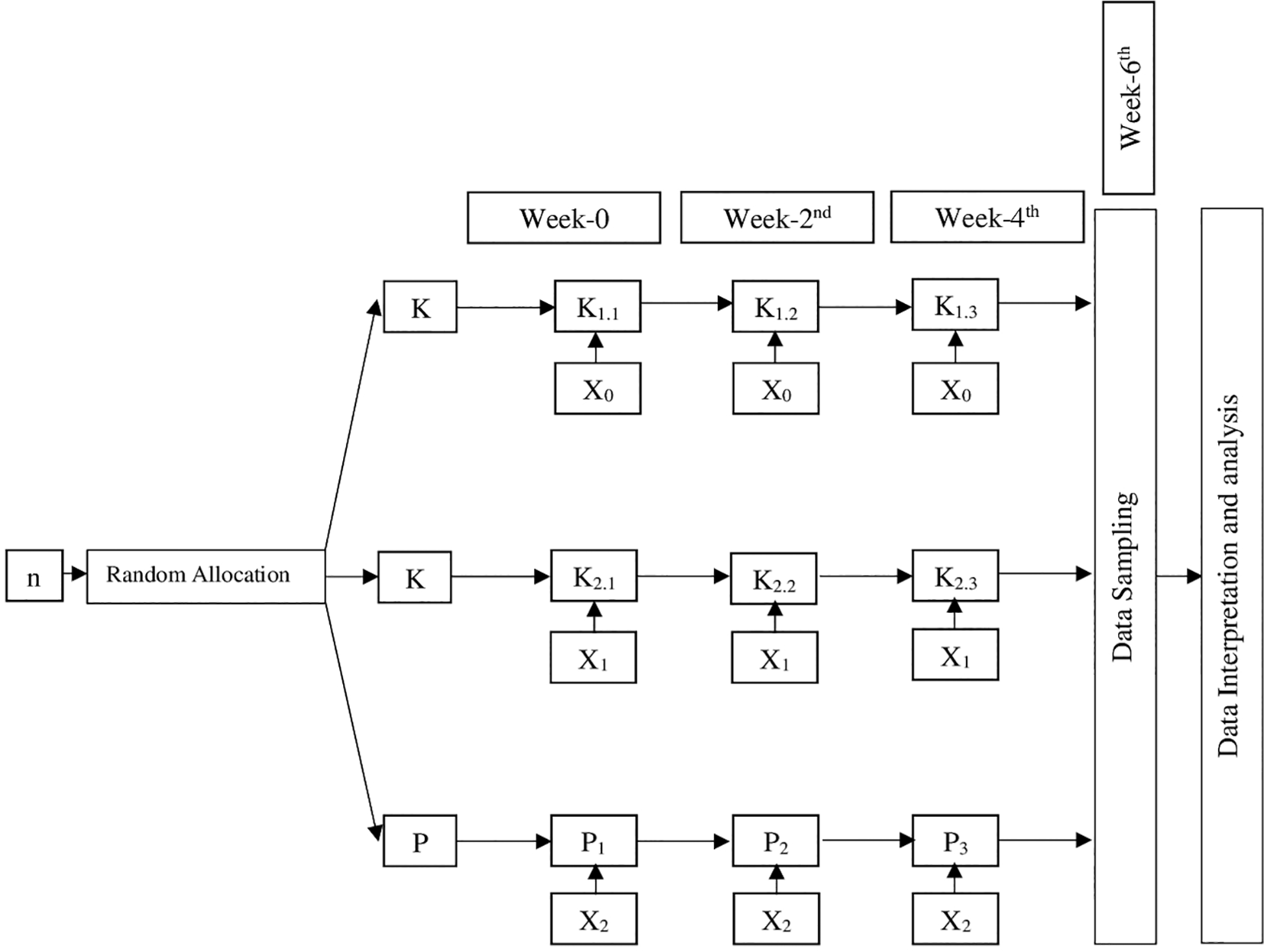
Description:
n: Sample
K1: Negative control
K1.1: Negative control observed sixth week after intervention
K1.2: Negative control observed fourth week after intervention
K1.3: Negative control observed second week after intervention
K2: Positive control
K2.1: Positive control observed sixth week after intervention
K2.2: Positive control observed fourth week after intervention
K2.3: Positive control observed second week after intervention
P: Intervention group
P1: Intervention group observed sixth week after intervention
P2: Intervention group observed fourth week after intervention
P3: Intervention group observed second week after intervention
X0: No Intervention
X1: Bovine HA implantation in rabbit femur
X2: Green mussel shell HA implantation in rabbit femur
Seven rabbits from seven different groups (P1, K1.1, P2, K2.2, P3, K2.3, K1.3) were found dead during daily monitoring several days after the surgery with no specific cause of death. None of the humane endpoint criteria (Table 1) were noted prior to their death nor found on examination after death. Rabbits from P1 and K1.1 were found dead two days after the surgery, rabbits from P2 and K2.2 were found dead three days after the surgery, and rabbits from P3, K2.3, and K1.3 were found dead five days after the surgery. The final data does not include their information. However, the data is still credible since each group achieved the minimum sample of three rabbits for each of these groups.
The rabbits were selected according to the inclusion criteria after each rabbit was weighed. A total of 36 rabbits that met the inclusion criteria were then adapted, given food and drink as necessary for a week. During the adaptation period, rabbits were given vitamins A, D, E (0.1 mL/kg BW IM), vitamin B complex (0.1 mL/kg BW IM), and ivermectin (0.4 mg/kg BW IM) for disease prevention. After a week-long adaptation period, the rabbits were separated each into one cage according to their group. The rabbits were monitored for their humane endpoint conditions before and after the intervention for 2 hours per day (16:00 – 18:00). Those rabbits that reached their humane endpoint will be terminated by the administration of ≥100 mg/kg of pentobarbital (lethal dose) intravascularly (IV). The humane endpoint criteria can be seen in this table below, from Montgomery (1990).
The research data obtained were primary data from the observation of blood serum alkaline phosphatase levels as a bone healing biomarker on rabbit femur bones from the treatment group compared to the negative and positive control groups. The collection of blood serum alkaline phosphatase levels as biomarkers of bone healing was carried out in week 2, week 4, and week 6 after surgery.
The green mussel shell HA used in this study is obtained from the production led by Rifky Ismail et al. at the Center of Biomechanics, Biomaterials, Biomechatronics, and Biosignal Processing (CBIOM3S).17 Then the obtained HA was powdered into a mortar and then the particle size was reduced using a stamper. In order to prevent infection, UV sterilization for ±3 hours was conducted. Before the implantation process, HA should be measured to prevent bias. The required amount of HA is calculated by the following calculation:
From the calculation, the mass of HA required for implantation of a tubular defect with a diameter of 5 mm and a depth of 5 mm is 300 mg.
There are two location options for making bone lesions in order to apply bone grafts, the metaphysis and the diaphysis. Both locations have advantages and disadvantages. There is a cushioning effect that prevents the risk of fracture due to the presence of layers of pars spongiosa and pars compacta which is an advantage of the metaphyseal section, but this section also has the disadvantage of being close to several large arteries and so there is risk of bleeding. In addition, the metaphysis has a lower volume and density than the diaphysis, making it easier to fracture.18,19 The diaphysis location was chosen as the surgery site because it is far from the great artery and it is not easily fractured.
The surgical procedure began by shaving the hair at the incision area followed by injection of enrofloxacin 5 mg/kg IV in the rabbit’s ear through the v. auricularis lateralis as prophylactic antibiotics, and ketamine 10-40 mg/kg and acepromazine 3-5 mg/kg intramuscularly in m. longissimus dorsi caudal as the anesthetic. After that, an incision was made in the lateral area of the distal diaphysis of the femur with blade no. 22 by incising the fascia and periosteum of the rabbit femur according to the treatment group.
The procedure was conducted according to each of the experimental groups. K1.1, K1.2, K1.3: make a defect in the lateral body of the femoris using a drill with a diameter of 5 mm and a depth of 5 mm. K2.1, K2.2, K2.3: make a defect in lateral corpus femoris using a drill with a diameter of 5 mm and a depth of 5 mm and fill the defect with bovine HA. P1, P2, P3: make a defect in the lateral part of the corpus femoris using a drill with a diameter of 5 mm and a depth of 5 mm and fill the defect with green mussel shell HA.
The fascia was sutured using absorbable catgut sutures, and the skin was sutured using non-absorbable silk. After that, the rabbits were examined until the time of data collection to know whether the rabbit showed any sign of infection, anatomical abnormalities, or died, so we could determine whether they met the exclusion criteria or not. The rabbits that showed exclusion criteria would be dropped out of the study.
The rabbits were taken care of properly by giving them food and drink as necessary until the time of blood serum data collection. The blood serum samples were taken two, four, and six weeks after surgery, depending on the intervention group of the rabbit. The intervention group is fully described in Figure 1. During this time, they were also given analgesic carbogen 2 mg/kg orally and antibiotic enrofloxacin 35mg/kg orally once per three days to keep their quality of life up.
The blood sampling was conducted by inserting needle parallel to the lateral marginal auricular vein. Once the needle reached the vein, blood will flow into the catheter. The vacutainer tube was pushed so that blood can enter the 1-2 ml tube. After collecting the required sample, the needle was immediately removed from the rabbit’s lateral marginal vein. The injected area was pressed with an alcohol swab at the end of this process.20
After the blood samples were collected, the rabbits were killed by the administration of ≥100 mg/kg of pentobarbital (lethal dose) intravascularly (IV). Then, they were monitored until a lack of heartbeat was noted for >60 seconds prior to carcass disposal.
Examination of ALP is selected due to its availability in most health centers. Detection of the ALP level from blood serum was carried out using the Mindray BC2800Vet series and blood chemistry analyzer with the brand Ubio-iChem-535 at the Animal Clinic Laboratory of Healthy Animals in Malang. In a previous study, this procedure was carried out based on measuring serum ALP levels in rabbit blood through the marginal vein. Then, the alkaline phosphatase is tested using 4-nitrophenol phosphate as a substrate and 2-amino-2-methyl-1-propanol (AMP) or diethanolamine (DEA) as a buffer, with DEA yielding higher ALP results. ALP isoenzymes can be differentiated using electrophoresis, HPLC, and other techniques.21
The data obtained from the biochemical assessment were described by the ratio of serum alkaline phosphatase (ALP) levels.
The Shapiro-Wilk test analyzed the normality of the data distribution. This test was chosen because the sample size in this study was <50. The data of this study were normally distributed with a p-value ≥0.05. The analysis was carried out using a one-way ANOVA test to see differences between groups because the data were normally distributed. A post hoc LSD test followed the results of the ANOVA test to find out more conclusively the relationship between variables that caused the data to be significant at the p-value <0.05. The comparison between the negative control group and the intervention group only shows a significant difference in the week 6.
In addition, data analysis was also carried out to compare the data for the week 2 and week 4; week 2 and week 6; and week 4 and week 6 using one-way ANOVA. The data of this study were considered insignificant at the p-value < 0.05.
In this study, a total of 36 New Zealand rabbits (Oryctolagus cuniculus) were chosen and split into 9 groups. Each group consisted of four rabbits and were treated according to their group. The alkaline phosphatase blood serum level data was collected from the remaining 29 rabbits after 7 died before samples could be taken. The timing of data collection for rabbits was carried out according to the group, namely in the week 2, week 4, and week 6 after the intervention.
Table 2 shows the serum alkaline phosphatase (ALP) levels in rabbits in each treatment group. Differences in ALP levels were found vertically at weeks 2, 4, and 6. Horizontally, differences in ALP levels can be seen between the positive control, treatment, and negative control groups.
| Positive control (bovine) | Negative control | Treatment (green mussel shell) | |
|---|---|---|---|
| Week-2nd | 44.33 (K2.3) | 72.00 (K1.3) | 60.50 (P3) |
| Week-4th | 60.40 (K2.2) | 57.70 (K1.2) | 57.33 (P2) |
| Week-6th | 42.17 (K2.1) | 62.53 (K1.1) | 18.65 (P1) |
Table 3 shows that the results of the one-way ANOVA test, which reported a p-value = 0.483 and a Levene value = 0.244. Because the p-value was > 0.05, this analysis test showed no significant difference in serum ALP levels at week 2, week 4, and week 6 in the bovine group (K2.3, K2.2, and K2.1).
| Bovine | Mean ± SD | p | Levene |
|---|---|---|---|
| Week-2nd (K2.3) | 44.33 ± 19.00 | 0.483 | 0.244 |
| Week-4th (K2.2) | 60.40 ± 7.24 | ||
| Week-6th (K2.1) | 42.17 ± 25.91 |
Table 4 shows the results of the one-way ANOVA test which reported a p-value = 0.052 and a Levene value = 0.034. Because the p-value was > 0.05, this analysis test showed no significant difference in serum ALP levels at week 2, week 4, and week 6 of the green mussel shell group (P3, P2, and P1).
| Green mussel | Mean ± SD | p | Levene |
|---|---|---|---|
| Week-2nd (P3) | 60.50 ± 34.88 | 0.052 | 0.034 |
| Week-4th (P2) | 57.33 ± 14.72 | ||
| Week-6th (P1) | 18.65 ± 5.40 |
Table 5 shows the results of the one-way ANOVA test reported a p-value = 0.510 and the Levene value = 0.772. Because the p-value was > 0.05, it can be concluded that the serum ALP levels in the negative control group (K1.3, K1.2, and K1.1) were not significantly different.
| Control | Mean ± SD | p | Levene |
|---|---|---|---|
| Week-2nd (K1.3) | 72.00 ± 11.47 | 0.510 | 0.772 |
| Week-4th (K1.2) | 57.70 ± 17.23 | ||
| Week-6th (K1.4) | 62.53 ± 16.02 |
Table 6 shows that from the one-way ANOVA test results, the p-value = 0.416 and the Levene value = 0.192. Because the p-value was > 0.05, it can be concluded that the serum ALP levels in the week 2 were not significantly different.
| Week-2nd | Mean ± SD | p | Levene |
|---|---|---|---|
| Positive control (bovine) (K2.3) | 44.33 ± 19.00 | 0.416 | 0.192 |
| Intervention (green mussel) (P3) | 60.50 ± 34.88 | ||
| Negative control (K1.3) | 72.00 ± 11.47 |
Table 7 shows that the results of the one-way ANOVA test reported a p-value = 0.959 and the Levene value = 0.454. Because the p-value was > 0.05, it can be concluded that the serum ALP levels in the week 4 were not significantly different.
| Week-4th | Mean ± SD | p | Levene |
|---|---|---|---|
| Positive control (bovine) (K2.2) | 60.40 ± 7.24 | 0.959 | 0.454 |
| Intervention (green mussel) (P2) | 57.33 ± 14.72 | ||
| Negative control (K1.2) | 57.70 ± 17.23 |
Table 8 shows that from the one-way ANOVA test results, the p-value = 0.030 and the Levene value = 0.087. Because the p-value was < 0.05 and a Levene value > 0.05, it can be concluded that the serum levels of ALP in the positive control, treatment, and negative control groups at week 6 had significant differences and were homogeneous. The test was continued with the LSD post hoc test to find out the differences between the treatment groups.
| Week 6th | Mean ± SD | p | Levene |
|---|---|---|---|
| Positive control (bovine) (K2.1) | 42.17 ± 25.91 | 0.030 | 0.087 |
| Intervention (green mussel) (P1) | 18.65 ± 5.40 | ||
| Negative control (K1.1) | 62.53 ± 16.02 |
Table 9 above shows that the results of the post hoc LSD test reported the positive control group (bovine) (K2.1) against the treatment group (green mussel shells) (P1) and the negative control group (K1.1) were not significantly different, while the treatment group (green mussel shells) (P1) against the negative control group (K1.1) had significant results.
The osteoconductive matrix is one of the elements that influences the bone healing process. It serves as a scaffold or mineralizing agent for bone and helps in the adherence of osteoconductive and osteogenic cells to the fracture site. Hydroxyapatite is one form of an osteoconductive matrix (HA). Hydroxyapatite is a substance made up of calcium (Ca2+), hydroxy (OH-), and phosphate (PO43-) ions that form a crystal structure with the chemical formula Ca5(PO4)3(OH). The body will naturally generate hydroxyapatite from Ca2+ ions in the blood and inorganic phosphate compounds (Pi or PO43-). Compound Pi is generated by dephosphorylating organic phosphate compounds (PPi), which are phosphates that happen at the ALP dephosphorylation process. As a result, the presence of a hydroxyapatite scaffold reduces ALP activity since the body no longer needs to synthesize hydroxyapatite from PPi.16,22
Because of that, ALP levels in a bone healing union will be normal or slightly elevated since the hydroxyapatite in green mussel shells functions well as a scaffold. In contrast to non-union fractures, the bone healing process takes longer, and more phosphate is required to compensate for the failure of bone healing, causing ALP levels to grow higher than in union fractures. This condition serves as the study’s measurement basis.16,22 However, ALP does not only play a role in the bone healing process, but also plays a role in other organ systems, such as hepatobiliary, genitourinary, and gastrointestinal.22,23 As a result, it is important to ensure that these body functions are in good condition so that false negatives or false positives do not occur.
This study aims to determine the effect of green mussel shell hydroxyapatite on blood alkaline phosphatase (ALP) levels in bone grafting procedures in New Zealand rabbits. The total sample was 36 New Zealand rabbits which were then divided into 9 groups. Following this, observations were made in the groups of negative control, positive control groups, and intervention groups in the weeks 2, 4, and 6. Using a surgical procedure and drilling with a width and depth of 5 mm, all groups produced lesions on the femoral diaphysis. The treatment group received green mussel shell hydroxyapatite (HA), while the negative control group did not get any HA as a follow-up intervention. The positive control group received bovine hydroxyapatite (HA). Drill lesions were made to develop the femoral diaphysis to replicate a fracture and to provide a suitable area to apply HA to the cavity formed by the drilling process. In this experiment, an effective dose of bone healing was up to 300 mg of the bone graft.10
This experiment had a number of methodological limitations, including fractures in the rabbits and post-interventional infections that were not equally exposed to all of the rabbits. This may impact the trial outcomes by increasing the time it takes for bones to recover, which could prevent all treatment groups from showing significant results between the weeks 2, 4, and 6. Even though all groups received an appropriate dose of 300 mg at the area of the lesion, the prolonged bone healing time meant that the test between the positive control group and the negative control group did not produce significant results in the week 6.
According to data analysis, the only test that produced significant results with a p-value less than 0.05 on the post-hoc LSD test was the comparison between the week 6 negative control group (K1.1) and the week 6 treatment group (P1). This demonstrates the effectiveness of green mussel shell HA in accelerating and maximizing the healing of bone fractures. however, it could not detect a significant difference between the week 2 and 4. This shows that the week 6, when ALP activity is at its peak, is the optimal week to observe how HA treatment affects the bone-healing process in femoral fractures as compared to weeks 2 and 4.16
Additionally, even though there were no statistically significant differences between the week 6 positive control group (K2.1) and the week 6 negative control group (K1.1) in the test, K2.1’s ALP level was lower than K1’s. This demonstrates that bovine HA can speed up bone repair, but its effectiveness is lower than that of HA from green mussel shells.
A comparison test of the serum ALP levels at the weeks 2, 4, and 6 in each treatment group’s results did not reveal any significant differences. This indicates that while ALP activity varied at weeks 2, 4, and 6, the differences were not statistically significant. This means that the effect of giving HA works evenly in the weeks 2, 4, and 6.
| Views | Downloads | |
|---|---|---|
| F1000Research | - | - |
|
PubMed Central
Data from PMC are received and updated monthly.
|
- | - |
Provide sufficient details of any financial or non-financial competing interests to enable users to assess whether your comments might lead a reasonable person to question your impartiality. Consider the following examples, but note that this is not an exhaustive list:
Sign up for content alerts and receive a weekly or monthly email with all newly published articles
Already registered? Sign in
The email address should be the one you originally registered with F1000.
You registered with F1000 via Google, so we cannot reset your password.
To sign in, please click here.
If you still need help with your Google account password, please click here.
You registered with F1000 via Facebook, so we cannot reset your password.
To sign in, please click here.
If you still need help with your Facebook account password, please click here.
If your email address is registered with us, we will email you instructions to reset your password.
If you think you should have received this email but it has not arrived, please check your spam filters and/or contact for further assistance.
Comments on this article Comments (0)