Keywords
Mucinous adenocarcinoma, partial urethrectomy, urethral neoplasm, radiotherapy
This article is included in the Oncology gateway.
Mucinous adenocarcinoma, partial urethrectomy, urethral neoplasm, radiotherapy
Urethral adenocarcinoma is a rare neoplasm that makes up for 0.02% of malignant tumors in females.1 There are only a few reports about this neoplasm in Indonesia. Mucinous adenocarcinomas are an extremely rare and poorly understood kind of cancer. These tumors have traits in common with a different class of tumors known as signet ring cell adenocarcinomas, which express mucin inside their cells. Compared to other adenocarcinomas, urethral adenocarcinoma frequently has a poor prognosis.2 The objective of this study was to share our experiences managing a female patient with mucinous adenocarcinoma of the urethra who had been monitored for four years.
A 67-year-old, retired single female patient from Indonesia presented with blood and clot gushing out from her urethra. She came with stranguria and hematuria. There was no previous history of urinary retention. The patient had a hysterectomy due to fibroids in 1991 and cholecystectomy in 2017. There was no previous history of cervical cancer in her family. She often consumed red meat and drank coffee. Her menarche history began at 12 years old and menopause was at 51 years old.
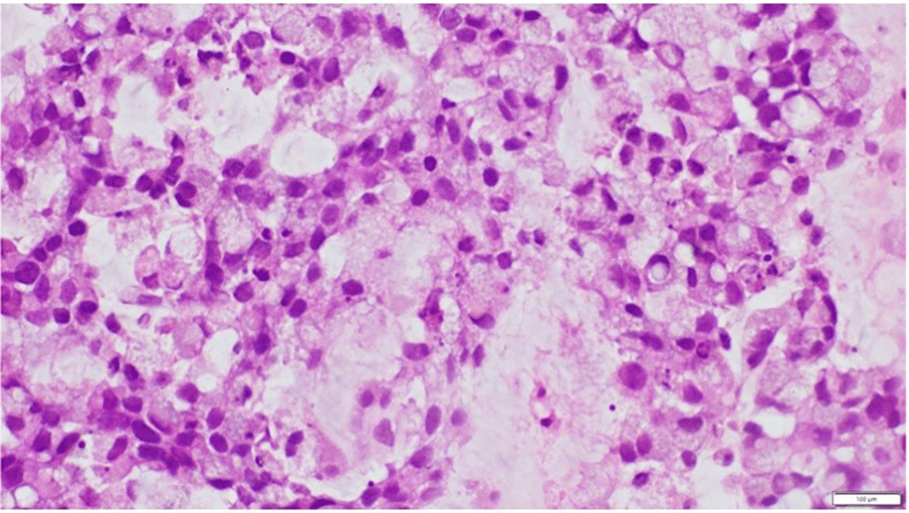
We performed a computerised tomography (CT) scan of the abdomen and suspected a urethral tumor, which was followed by a biopsy. We saw a tissue partially lined with locally hyperplastic transitional epithelial cells showing hyperplastic growth, forming a tumor mass with infiltrative growth from our histological examination. Tumor cells are arranged in small clusters and single cells are scattered among extracellular mucin, some of which form glandular structures. Tumor cells with round-oval nuclei, pleomorphic, hyperchromatic, vesicular, some with nucleolus, eosinophilic cytoplasm, some vacuolated. Tumor cells appear with a “signet ring cell” picture (Figure 1). The stroma is filled with mild to moderate acute and chronic inflammatory cells, with areas of bleeding. No tumor emboli were found in the vessels. It is consistent with mucinous adenocarcinoma.
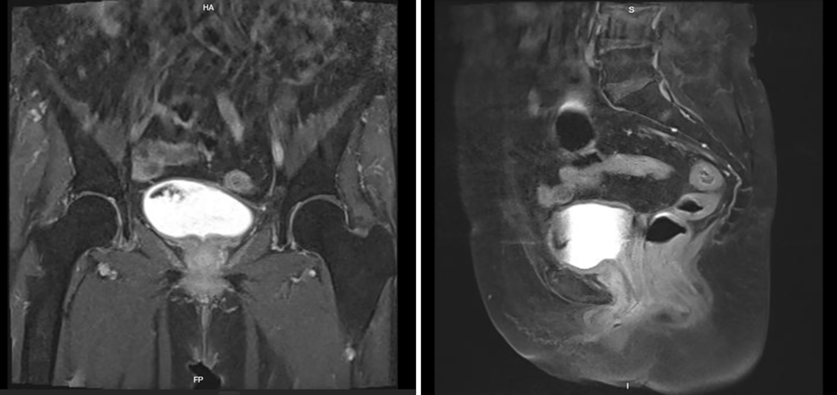
Subsequently we performed an urethrocystoscopy examination. From our examination we can distinguish between healthy tissue and tumor border. We decided to perform a partial urethrectomy. From the resected tumor, it was found that the surgical resection margins were negative from neoplastic involvement, no lymphovascular invasion was identified. After the procedure the patient had been in remission. However, after 18 months, from Magnetic Resonance Imaging (MRI) examination, recurrence had appeared. From our physical examination, it showed a mass in the urethral area, The patient did not feel any symptoms. Then the patient underwent radiotherapy for 33 sessions. Currently, she remains free of recurrence for 22 months of follow-up (Figure 2). The MRI examination showed no residual mass was seen in the urethral area. No paraaortic, parailiac, and obturator lymphadenopathy was seen. No abnormalities were seen in other intra-abdominal and pelvic organs (Figure 3).

Written informed consent was obtained from the patient for the use and publication of this clinical image.
Female urethral carcinoma is a condition with a modest prevalence, accounting for 5% of female urological tumor cases and 0.02% of all instances of malignant tumors in women.1,3 Mucinous adenocarcinomas are an extremely rare and poorly understood kind of cancer. These tumors resemble a different class of tumors known as signet ring cell adenocarcinomas, which exhibit mucin inside their cells and nearly always have a poorer prognosis than ordinary adenocarcinomas.2 Our case had the chief complaint of blood coming out of the urethra. Other symptoms that are found in urethral cancer include dysuria, dyspareunia, haematuria, perineal pain, urine retention, overflow incontinence, urethral mass, or a projecting meatal mass. It expands locally into the periurethral tissue, vagina, and vulva before spreading proximally to the bladder neck.3 Patients may present with a wide range of symptoms, the majority of which include dysuria, urine frequency, and a palpable mass. These three symptoms account for 50% of all presenting symptoms.4 Because of the shorter length of the female urethra, local spread tends to be more destructive.4
According to research by Neto et al., their patient was hospitalized with stranguria, urine retention, and sero-bloody discharge with stoppers of mucopurulent material in addition to discomfort and burning in the urethra. The patient received surgery therapy such as extended radical vulvectomy and urethrectomy, progressing with the use of indwelling urinary catheter, as well as two sessions of radiotherapy.3 In this case, partial urethrectomy seems adequate not only because of the clear ability to distinguish healthy tissue and tumor tissue, but also to maintain the integrity and function of the lower urinary tract. Another study from Satyanarayan et al. reported a case with mucinous urethral adenocarcinoma. They performed a wide local excision of the tumor and underwent radical cystectomy with ileal conduit. The patient had normal postoperative physical examination results and imaging studies. But during the cystectomy she had persistent microscopic invasive adenocarcinoma in the residual urethra. A recurrence could be avoided with early detection and surgical intervention.4
One recent study from Baffigo et al. reported a patient with mucinous adenocarcinoma of the bladder. The patient underwent anterior pelvectomy. Six months after surgery, bilateral inguinal lymph node dissection was performed because of bilateral palpable masses and the patient also received external radiotherapy of the inguinal area. After 22-months the patient appears healthy.5
A more recent study from Ndiaye M et al. reported a patient with urethra adenocarcinoma underwent partial urethrectomy. After 16 month follow up, the patient was continent without local and metastatic recurrence.6
From this case, with long term follow up we assessed response of urethral adenocarcinoma treated with radiotherapy. A limitation of this study is that the primary cause of the adenocarcinoma is still uncertain. In this case, we learned that performing partial urethrectomy is possible and able to maintain continence. Recurrence had appeared in this patient, but after radiotherapy, the patient currently remains clear from tumor.
Mucinous adenocarcinoma of female urethra is rare and there is no consensus on the optimal therapy due to the scarcity of cases. Early surgical treatment with or without adjuvant radiotherapy appears to be the best option in cases of small, organ-confined disease. Partial urethrectomy can be performed in patients with mucinous urethral adenocarcinoma, which can prevent the use of permanent urinary catheters and further improve the patient's quality of life.
| Views | Downloads | |
|---|---|---|
| F1000Research | - | - |
|
PubMed Central
Data from PMC are received and updated monthly.
|
- | - |
Provide sufficient details of any financial or non-financial competing interests to enable users to assess whether your comments might lead a reasonable person to question your impartiality. Consider the following examples, but note that this is not an exhaustive list:
Sign up for content alerts and receive a weekly or monthly email with all newly published articles
Already registered? Sign in
The email address should be the one you originally registered with F1000.
You registered with F1000 via Google, so we cannot reset your password.
To sign in, please click here.
If you still need help with your Google account password, please click here.
You registered with F1000 via Facebook, so we cannot reset your password.
To sign in, please click here.
If you still need help with your Facebook account password, please click here.
If your email address is registered with us, we will email you instructions to reset your password.
If you think you should have received this email but it has not arrived, please check your spam filters and/or contact for further assistance.
Comments on this article Comments (0)