Keywords
Vitreous/Retinal Pigment Epithelium-Relative Intensity, Proliferative Vitreoretinopathy, Uveitis
This article is included in the Eye Health gateway.
Vitreous/Retinal Pigment Epithelium-Relative Intensity, Proliferative Vitreoretinopathy, Uveitis
Rhegmatogenous retinal detachment (RRD) can produce some degree of vitreous haziness that may be associated with its severity and prognosis.1,2 RRD causes neurosensory retina and retinal pigment epithelium (RPE) separation, which leads to retinal ischemia and blood-retinal barrier disruption.3,4 These two events serve as initiating inflammatory factors of the pathogenesis of proliferative vitreoretinopathy (PVR).5 Exposure of RPE cells to the vitreous results in the migration of cytokines, inflammatory cells, and growth factors into the vitreous cavity to further stimulate cellular responses that lead to a turbid vitreous. Inflammation plays a central role in stimulating the development of epiretinal membranes and retinal fibrosis, latter findings in PVR.5,6
According to the updated Retina Society Classification, PVR is classified into grades; A, B, and C. Grade A is defined as the presence of vitreous haziness and pigment clumping. Grade B is defined as surface retinal wrinkle, rolled edges of retinal breaks, retinal stiffness, and artery-venous tortuosity; and grade C is full-thickness retinal folds and/or subretinal bands.2 However, most clinicians use this classification only as prognostic hints and a guideline in which vitreous substitute is used, i.e. gas or silicone oil. It does not address the role of inflammation that generates during RRD development. Vitreous haze caused by inflammation is not specific to PVR, as it can also be present in uveitis or a retinal detachment without PVR.7,8 Furthermore, this classification does not offer any information regarding the severity of inflammation, which plays an essential role in the pathogenesis and disease severity in PVR.2,9
Optical coherence tomography (OCT) is a non-invasive diagnostic procedure that provides cross-sectional images of ocular tissue, allowing qualitative assessment of retinal diseases and automated retinal thickness measurements. Recently, OCT has been described to visualize vitreous haziness in patients with uveitis10–12 Quantitative measurements of vitreous haze or inflammation using OCT images were obtained by measuring the vitreous intensity relative to retinal pigmented epithelium intensity, which is termed VIT/RPE-relative intensity. Studies have reported that the VIT/RPE relative intensity is significantly higher in uveitis eyes than in eyes without vitreous haze; furthermore, validation analysis shows a positive correlation with Nussenblatt vitreous haze scale.10
This study aims to objectively measure vitreous haziness in PVR compared to uveitis and normal eyes by measuring vitreous intensity relative to retinal pigment epithelium intensity captured through OCT.
We obtained OCT image sets from four groups of consecutive patients in Cipto Mangunkusumo Kirana Eye Hospital between April 2021 and December 2021.
The participants were identified during their visit to Cipto Mangunkusumo Kirana Eye Hospital. Those who met the inclusion criteria and provided informed consent were included. The procedures were done during routine appointments.
The first group of data included images from eyes with RRD and PVR. The inclusion criteria for the RRD and PVR group were patients with macula-off RRD and PVR with grade A or B, age of 18 or above, and RRD onset upon examination of 7 days to 1 month. The exclusion criteria of the RRD and PVR group were patients with media opacification (i.e., cataracts, vitreous hemorrhage), a history of intraocular surgery in less than 3 months, and patients with other eye disease comorbidities (i.e., macular hole, age-related macular degeneration, intraocular tumor).
The second and third groups were obtained from uveitis patients, which were divided into two groups; (1) intermediate and posterior uveitis; (2) panuveitis. We excluded uveitis patients with vitreous hemorrhage, retinal pigment epithelial irregularity such as in age-related macular degeneration, intraocular tumor, and choroidal neovascularization, history of triamcinolone acetate intravitreal within 6 months, and vitrectomized eyes.
The fourth group was the control group, which included image sets from normal eyes that did not show signs of intraocular inflammation or any ocular disease. We only include patients in each group with goodand readable OCT imaging quality, defined as clear visualization of the vitreous cavity, neurosensory layer, and RPE layer in one OCT image.
The estimation sample size for the present study was calculated using correlation coefficient (r) based on the previous study of Keane et al.10 (r=0.566). By implementing this method, the sample size would be 29 for the uveitis group. The sample size for the PVR and control group will be adjusted with the uveitis group.
This study was conducted based on the Declaration of Helsinki and was approved by the Ethics Committee of the Faculty of Medicine, Universitas Indonesia and Cipto Mangunkusumo Hospital (KET-87/UN2.F1/ETIK/PPM.00.02/2021) on 8th February 2021. All subjects provided written informed consent.21
All participants underwent a complete ocular examination. Clinical data collected from each patient during recruitment and examination include age, sex, and best-corrected visual acuity (BCVA) using Snellen chart and converted into LogMAR. Etiology of uveitis patient was obtained from further work-ups. OCT Image was obtained by OCT Cirrus HD 4000 (Carl Zeiss Meditec, Dublin, CA) with HD-line raster scan protocol system in grayscale. This system has a 27,000 A-scans per second imaging speed and an axial resolution of 3.9 mm. Volume scans were centered on the fovea in each OCT image. The OCT images were taken by experienced staff nurses. Each patient will have 4 to 5 images taken until the best image is obtained.
VIT/RPE Relative Intensity was measured with ImageJ software version 1.53 (National Institutes of Health, Bethesda, MD, USA). The image was manually segmented by a polygonal selection tool. OCT images were measured independently by two graders (ASA and FI). The value that will be included in the correlation analysis is the highest value of VIT/RPE-Relative Intensity obtained from the measurements. All images were randomized and the OCT image graders were masked from patient data and diagnosis to reduce the risk of bias.
OCT images were obtained from OCT and imported into free access software ImageJ. This software enables manual segmentation and measurements of OCT images for the intensity of the vitreous and retinal pigment epithelium area. We measured vitreous inflammation or hazziness using the Vitreous/RPE Relative Intensity. Images were set to 8-bit type before measurement. The boundaries definition for measurements was based on the previous study by Keane et al.10 Boundaries of the vitreous area consist of (1) “vitreous top”, the upper limit of the vitreous area visible in OCT; (2) internal limiting membrane (ILM), the inner border of the neurosensory retina. Vitreous absolute intensity is defined as the intensity of the vitreous area, which is the area between the vitreous top and internal limiting membrane. Boundaries for the RPE area consisted of (1) inner-retinal pigment epithelium (RPE), the inner border of RPE; and (2) outer-RPE, the outer boundary of the RPE. The RPE intensity is described as the space between the RPE’s inner and outer boundaries.
Clinical and imaging data were analyzed using descriptive statistics. T-independent test was used for the VIT/RPE relative intensity mean value between groups. P values<0.05 were considered statistically significant. All analyses were performed using IBM SPSS, version 26 (IBM Corp), and GraphPad Prism 9.0 (GraphPad Software, Inc., San Diego, CA).
A total of 29 eyes in the uveitis group and 37 eyes in the RRD-PVR group were eligible for the study.19,20 However, there were only 28 eyes in the uveitis group and 19 eyes in the RRD-PVR group that met the criteria of good and readable OCT. A finaltotal of 19 PVR eyes, 28 uveitis eyes (12 intermediate-posterior uveitis eyes and 16 panuveitis eyes), and 28 normal healthy eyes were recruited for this study. The mean age of patients was 43±11.95 years in the PVR group with 63.16% male patients. In the intermediate-posterior uveitis group, the mean age was 36.58±12.17 years with an equal of six male and female patients, and in the panuveitis group, the mean age was 34.75±10.88 years with 62.5% female patients. Visual acuity in the three groups ranged between 0 to 2.5 LogMAR units. The clinical characteristics of each group are described in Table 1.
Characteristics of patients in the rhegmatogenous retinal detachments and proliferative vitreoretinopathy group are described in Table 2. RRD duration upon examination was mostly 0-7 days (52.6%). All patients included in the study were PVR grade B.
| Characteristics | RRD (n=19) |
|---|---|
| Duration (days) | |
| 0-7 | 10(52.6%) |
| 8-30 | 9(47.4%) |
| PVR Grade | |
| A | 0(0%) |
| B | 19(100%) |
From 28 uveitis patients that were included in the study, the most common etiologies were toxoplasma (28.6%) and sifilis (25%). The characteristics of uveitis patients were summarized in Table 3.
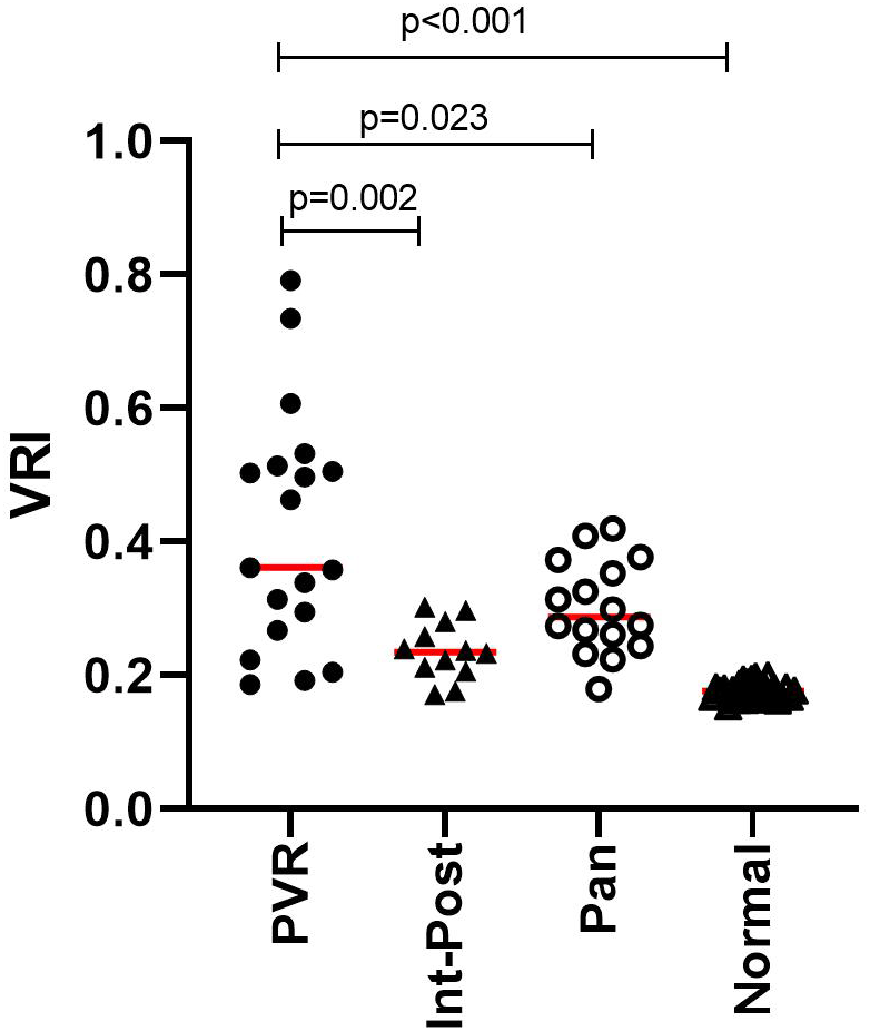
The VIT/RPE-Relative Intensity of each group (“proliferative vitreoretinopathy”, “intermediate-posterior uveitis”, “panuveitis”, and “normal eyes”) are described in Figure 1. The VIT/RPE-Relative Intensity index in the PVR eyes (0.415±0.178) was found to be significantly higher in PVR than in intermediate-posterior uveitis (0.236±0.043) (p=0.002) and in panuveitis eyes (0.30±0.07) (p=0.023). A significant difference was also found between the intermediate-posterior uveitis and panuveitis groups (p=0.008). Compared to the healthy controls, PVR and both uveitis groups have significantly higher VIT/RPE relative intensity (p≤0.001 in each group).
Vitreous haze is defined as the accumulation of inflammatory cells and protein exudate in the vitreous cavity, resulting in the decreased visibility of the retina and optic nerve. In uveitis, vitreous haziness is one of the important clinical outcomes in disease evaluation.7,13 OCT imaging enables direct visualization of vitreous haze.14 Recent studies have developed an objective, quantitative measurement of vitreous signal intensity, compared to RPE-absolute intensity. VIT/RPE-Relative Intensity is used to acquire quantitative measurements of vitreous haze in uveitis eyes, as studies reported to be significantly higher compared with normal eyes.10,11
Previous studies reported increased levels of cytokines, growth factors, and inflammatory cells in the vitreous of RRD-PVR eyes, including macrophages, transforming growth factor-β (TGF-β), platelet-derived growth factor (PDGF), vascular endothelial growth factor (VEGF), tumor necrosis factor-alpha (TNF-α), interleukin (IL)-6 and IL-8.6,15–18 These inflammatory cells, pro-inflammatory cytokines, and growth factors facilitate RPE cell migration, proliferation, and epithelial-mesenchymal transition, which leads to the development of retinal fibrosis and contraction.17
Our study showed that VIT/RPE-Relative Intensity in the PVR group is significantly higher than intermediate-posterior uveitis, panuveitis, and normal eyes. We also compared intermediate-posterior uveitis with panuveitis, with the result of the value of VIT/RPE Relative Intensity in the panuveitis group being significantly higher than the intermediate-posterior uveitis (p=0.008). Both uveitis groups also are significantly higher compared to normal eyes. These results may indicate that the inflammation and protein exudate into the vitreous cavity of PVR is higher than in uveitis cases. This study also showed similar findings for the VIT/RPE-Relative Intensity of uveitis eyes described in previous studies.10–12
These results are a preliminary finding in validating the VIT/RPE-Relative Intensity use for measuring vitreous inflammation objectively in the role of the pathogenesis of PVR and may be useful to obtain a new standard classification of PVR that corresponds to the inflammatory status of the disease. The higher value of VIT/RPE-Relative Intensity in PVR than in uveitis may indicate the need for anti-inflammatory drug use in PVR, in addition to surgery, to decrease disease severity.
We understand that this study had some limitations regarding the subject number, with only 19 subjects in the PVR group. A larger study comparing the VIT/RPE-Relative Intensity index and vitreous inflammatory cells, cytokines, and growth factors in PVR patients will be required to validate our significant findings.
| Views | Downloads | |
|---|---|---|
| F1000Research | - | - |
|
PubMed Central
Data from PMC are received and updated monthly.
|
- | - |
Provide sufficient details of any financial or non-financial competing interests to enable users to assess whether your comments might lead a reasonable person to question your impartiality. Consider the following examples, but note that this is not an exhaustive list:
Sign up for content alerts and receive a weekly or monthly email with all newly published articles
Already registered? Sign in
The email address should be the one you originally registered with F1000.
You registered with F1000 via Google, so we cannot reset your password.
To sign in, please click here.
If you still need help with your Google account password, please click here.
You registered with F1000 via Facebook, so we cannot reset your password.
To sign in, please click here.
If you still need help with your Facebook account password, please click here.
If your email address is registered with us, we will email you instructions to reset your password.
If you think you should have received this email but it has not arrived, please check your spam filters and/or contact for further assistance.
Comments on this article Comments (0)