Keywords
Abdominal Mass, Extrapulmonary TB, Mantoux Test, Ultra-sonography, Anti Tubercular Therapy
This article is included in the Datta Meghe Institute of Higher Education and Research collection.
Abdominal Mass, Extrapulmonary TB, Mantoux Test, Ultra-sonography, Anti Tubercular Therapy
Among the most common locations for extrapulmonary tuberculosis is the digestive tract.1,2 About one-eighth of all cases of TB are extrapulmonary,3,4 and abdominal TB accounts for 11 to 16 percent of cases.5,6 Up to 50% of HIV-positive people may develop extrapulmonary TB.1,6 The sixth most prevalent extrapulmonary site for tuberculosis is the gastrointestinal tract, followed by lymphatic, genitourinary, bone and joint, miliary and meningeal.7 The abdomen is either the primary site of infection or the site of secondary infection when TB spreads to it through ingested sputum, hematogenous transmission from a nearby infected organ, consumption of unpasteurized milk, or revival of a previously acquired dormant focus.2,5
A 53-year-old female patient who had been experiencing right lower abdomen pain, loss of appetite, vomiting, and weight loss for the previous two months was our patient. Abdominal pain was dull, aching, and non-radiating, located in the right lower abdomen, and it was associated with vomiting, which was projectile and non-bilious. She belonged to low socio-economic status and had no prior antitubercular therapy (ATT). She had no personal history of diabetes, hypertension, and seizure disorder. No significant family history was present. On examination, she had a blood pressure of about 126/84 mm Hg, a heart rate of about 80/min, and a temperature of about 98.9°F.
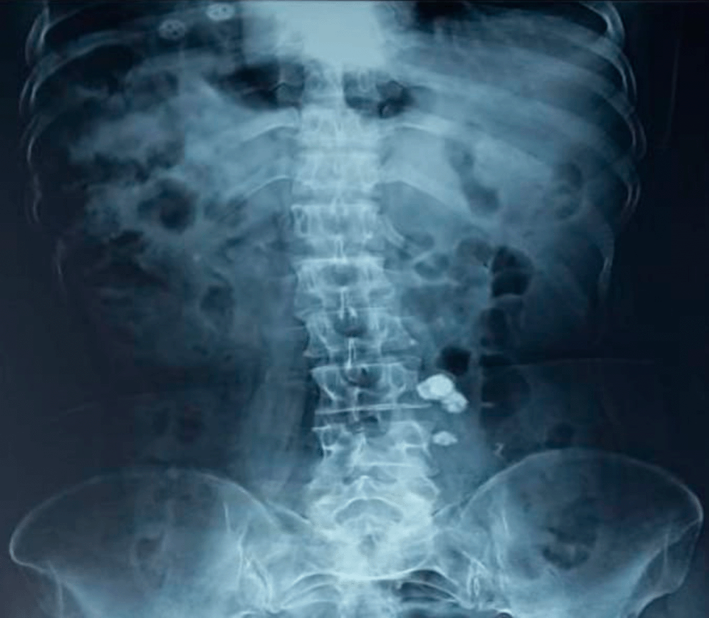
On abdominal examination, a non-tender palpable swelling of 5.0×5.0 cm was present in the right groin. Hematological investigations showed hemoglobin of 9.7 gm%, total leukocyte counts of 13100/mm3 with a differential count of polymorphs of 80%, and lymphocytes of 18%. The erythrocyte sedimentation rate was 58mm in 1st hour. Mantoux test showed an induration of 22 millimeters. Left renal calculus was found on the X-ray abdomen (Figure 1). Ultrasound whole abdomen of the patient revealed 88×51×42 mm thickened bowel loops at the ileocaecal junction. Its wall thickness was 19 mm, with evidence of a 40×23 mm cystic lesion.
The patient visited hospital in the month of January 2023 and was diagnosed with Tuberculosis of abdomen. After the diagnostic investigations done the patient was on medications of antitubercular drugs for six months.
The Diagnostic Assessment done for the patient includes the ultrasound, haematological assessment, general examination, clinical examination, sonography this all the following assessment done for the patient. The patient shows the normal temperature, normal blood pressure, slight increase in heart rate and the radiological finding shows the presence of the mass on the left side of abdomen the haemolytic assessment shoes the normal findings.
Based on these radiological findings, antitubercular treatment with rifampicin, ethambutol, isoniazid, and pyrazinamide was started. Patient symptoms gradually improved, and therapy was continued for two months. After that repeat Ultrasound abdomen revealed a decrease in mass size to 56×43× 32 mm, and bowel wall thickness was reduced with a cyst of 25×16mm. The patient continued with rifampicin, ethambutol, and isoniazid for another four months. Her ultrasonography after six months of treatment suggested a significantly reduced mass of 1.5 × 1cm size. Written informed consent was taken from the subject prior to the publication of case details and accompanying images.
Mycobacterium tuberculosis infection of the peritoneum and abdominal organs is known as abdominal TB. The signs of abdominal TB are vague and can resemble colon cancer, Crohn's disease, or lung cancer.8 They can also resemble Crohn's disease or pulmonary TB. The symptoms of abdominal tuberculosis include abdominal pain (80.4 % of cases) and weight loss. (74.65 %), appetite loss (62.67 %), fever (40.5 %), loose stools (16.44%), and abnormal bowel habits (25.35 %).9 No respiratory issues or prior antitubercular therapy histories were present in our patient. Age, underlying illness, bacterial genotype, and immunological state are all connected to the pathogenesis of abdominal TB.10 Some of the postulated mechanisms by which the tubercle bacilli reach include direct spread from nearby organs, hematogenous spread from the primary lung focus in childhood with later reactivation, ingestion of bacilli in sputum from adjacent organs, direct spread from adjacent organs, ingestion of bacilli in sputum from infected nodes, and through lymph channels from infected node Because of the ambiguous and nonspecific clinical signs and the poor yield of mycobacterium cultures or smears, diagnosing abdominal tuberculosis is challenging. The ileocaecal region is the most frequently affected area, maybe due to the heightened physiological stasis, accelerated rate of fluid and electrolyte absorption, low digestive activity, and profusion of lymphoid tissue in this location. The implicated bowel's gross morphological appearance was initially divided into ulcerative, ulcerohyperplastic, and hyperplastic variants by Hoon et al.11 Imaging tests like the Mantoux test and barium X-rays, CT scans, and ultrasounds only provide supportive information. The diagnosis can occasionally be due to the patient's response to antitubercular medication therapy trials. In our patient, the diagnosis was made on clinical and radiological grounds. Most importantly patient didn't require surgical intervention and improved on antitubercular therapy. Colonic TB can be treated with conservative care and anti-TB drugs, excluding surgical emergencies like perforation or obstruction. It is important to closely monitor the patient's reaction to medical care because a failure of the symptoms to go away and the lesions to shrink could indicate more serious underlying pathology, such as cancer. A high index of suspicion for abdominal TB should exist in individuals with vague symptoms and colonic thickening on imaging.
As a leading global killer and source of pain, tuberculosis (TB) significantly lowers patients' health-related quality of life (HRQoL). Patient awareness of their physical and mental wellbeing is indicated by HRQoL. As a result, it is crucial to understanding and calculating the precise effects of the disease state.12 The public health system in India continues to be seriously threatened by tuberculosis (TB). Adverse drug reactions (ADR) continue to be a problem in treatment adherence and completion despite the National Tuberculosis Elimination Programme (NTEP) offering a comprehensive variety of treatments from early diagnosis through complete treatment to minimise morbidity and death from TB.13 Skin with many lesions caused by extrapulmonary tuberculosis (ETB) is a rare manifestation of mycobacterial infection. It is uncommon to describe cutaneous tuberculosis (TB) with numerous lesions and Poncet's illness (tuberculous rheumatism).14 There is still debate over how well interferon-gamma release assays (IGRA) and pure protein derivative tuberculin skin tests (TST) predict incident active tuberculosis (TB).15 The second-leading cause of infectious disease-related death worldwide is tuberculosis (TB), and delays in the TB care cascade are one of the main obstacles to meeting the objectives of TB control programmes.16
Written informed consent for publication of their clinical details and/or clinical images was obtained from the patient/parent/guardian/relative of the patient.’
Zenodo. Abdominal tuberculosis presenting as abdominal mass. DOI: https://doi.org/10.5281/zenodo.8123081.
Data are available under the terms of the Creative Commons Attribution 4.0 International license (CC-BY 4.0).
| Views | Downloads | |
|---|---|---|
| F1000Research | - | - |
|
PubMed Central
Data from PMC are received and updated monthly.
|
- | - |
Provide sufficient details of any financial or non-financial competing interests to enable users to assess whether your comments might lead a reasonable person to question your impartiality. Consider the following examples, but note that this is not an exhaustive list:
Sign up for content alerts and receive a weekly or monthly email with all newly published articles
Already registered? Sign in
The email address should be the one you originally registered with F1000.
You registered with F1000 via Google, so we cannot reset your password.
To sign in, please click here.
If you still need help with your Google account password, please click here.
You registered with F1000 via Facebook, so we cannot reset your password.
To sign in, please click here.
If you still need help with your Facebook account password, please click here.
If your email address is registered with us, we will email you instructions to reset your password.
If you think you should have received this email but it has not arrived, please check your spam filters and/or contact for further assistance.
Comments on this article Comments (1)