Keywords
Breast Abcess; Mastitis; Tuberculous Mastitis; Tuberculosis; Breast ulcer.
This article is included in the Global Public Health gateway.
Tuberculous mastitis (TM) is a rare form of tuberculosis, occurring as a primary disease when there is no evidence of tuberculosis in other locations. There are no clear clinical features of TM, especially in the absence of a previous tuberculosis infection. Due to its unclear clinical picture, diagnosis is difficult, and it is often confused with breast carcinoma or pyogenic abscesses. The aim of this study was to report our experience and discuss the characteristics and diagnostic modalities in cases of primary TM in a tuberculosis-endemic area.
A case series study was conducted at the Arifin Achmad Regional Hospital, reporting four cases of primary tuberculous mastitis in January 2024. The patients were women aged 24-41 years.
All patients presented with complaints of breast pain for the last 2 weeks to 2 months and complained of symptoms in the form of a lump in the breast that was reddish in colour and mastalgia. One patient was diagnosed during pregnancy, and one had a history of prior breastfeeding. One patient presented with FNAB results for breast carcinoma. The other patient complained of an ulcer on her breast. Physical examination revealed axillary lymphadenopathy in all patients. Histopathological examination revealed tuberculous mastitis in all patients and 1 with tuberculous mastitis, fibrocystic changes, and Atypical Ductal Hyperplasia (ADH).
Patients with tuberculous mastitis who visited our institution had symptoms similar to those of abscesses and breast cancer. The FNAC test is the most reliable, but false-negative results can occur. Diagnosis requires teamwork between the patient’s doctor, anatomical pathologist, and radiologist. This research requires a larger scale to describe actual conditions.
Breast Abcess; Mastitis; Tuberculous Mastitis; Tuberculosis; Breast ulcer.
Tuberculosis is widely known as a disease of the lungs; however, extrapulmonary manifestations account for nearly 20% of all tuberculosis cases.1 Tuberculous Mastitis (TM) is a rare form of tuberculosis, that occurs as a primary disease when there is no evidence of tuberculosis in other locations.2 TM was first described in 1829 by Cooper as “swelling of the breast gland in young women”. Further detailed explanations of this disease were reported at the end of the 19th century by Richet and Powers.3,4 TM accounts for less than 0.1% of all breast diseases and less than 2% of all tuberculosis cases.5 Even in developed countries and tuberculosis-endemic areas, this condition is rare.6–8 Indonesia is a developing country that is endemic for tuberculosis.6 The prevalence of TM in developing countries is estimated to be 0.1% of all histologically examined breast lesions and 3-4.5% of operated patients.9 TM often resembles breast cancer and breast abscess both clinically and radiologically, leading to frequent misdiagnosis. Biopsy is necessary to confirm the diagnosis of TM.7 In patients from endemic areas, tuberculosis infection should be suspected if mastitis occurs repeatedly and is refractory to antibiotic treatment.10 Without a history of tuberculosis infection, TM has no apparent clinical manifestation. Dubbed “the great masquerader,” TM is difficult to diagnose owing to its ambiguous clinical appearance.4,11 This disease is often mistaken for breast carcinoma or pyogenic abscess.4 Doctors must understand TM presentations for a correct diagnosis.12 Our study reported our experience identified primary TM, and discussed its characteristics and diagnostic methods in tuberculosis-endemic areas.
A 25-year-old female patient presented with complaints of a lump in her right breast that had burst one day before admission to the hospital. The patient complained of a sudden discharge of pus accompanied by intermittent pain. A lump in her right breast appeared 1 month before admission. The lump initially felt small but gradually grew larger with redness. Lumps were not influenced by the menstrual cycle. The patient also complained of a palpable lump in the axilla. There was no shortness of breath, back pain, or headaches.
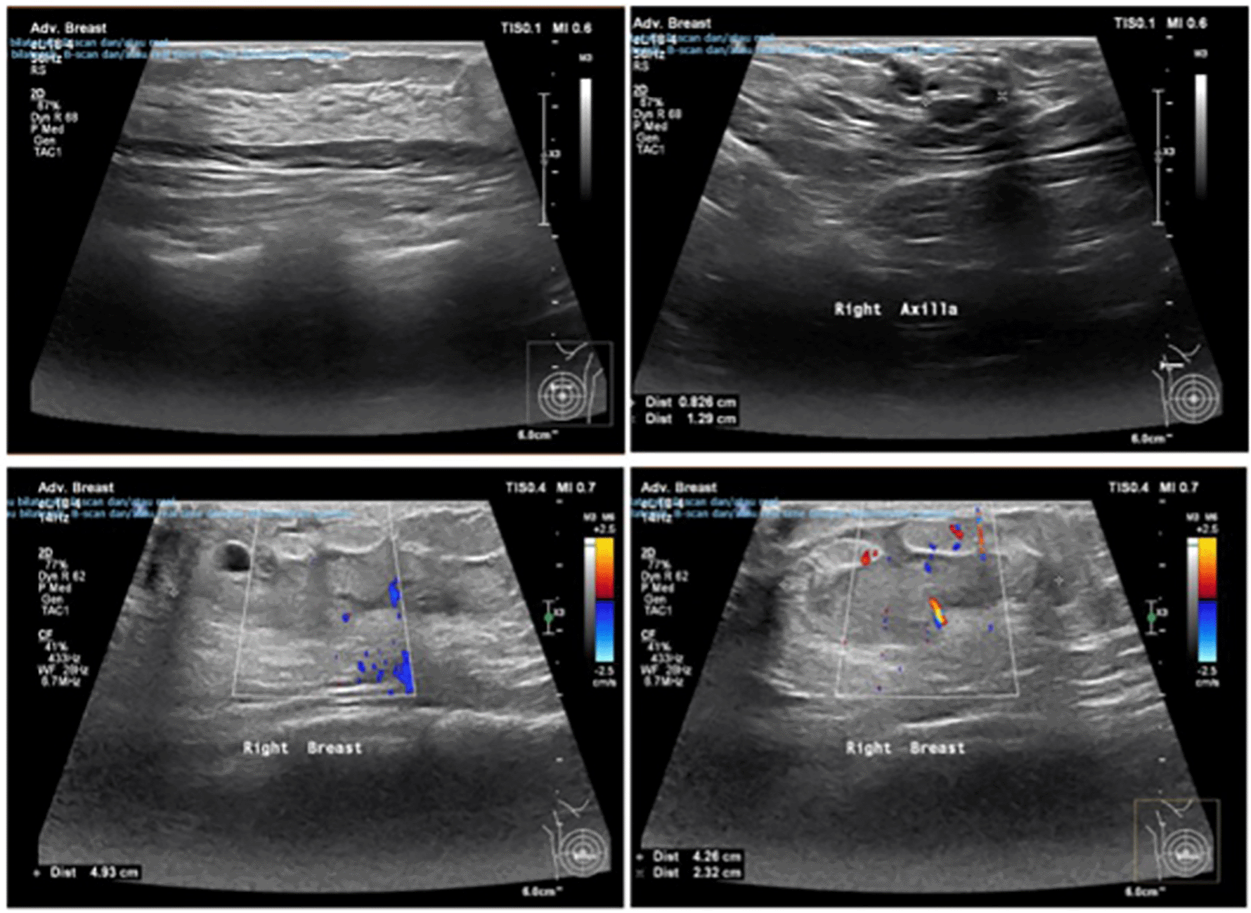
On physical examination, both breasts appeared symmetrical. The right breast had ulcers, sloughts, and pus. Upon palpation, a mass measuring 5 × 4 cm was felt, with a smooth surface, unclear borders, hard consistency, tenderness upon pressure, and warmth. No abnormalities were observed in the left breast. Upon examination of the right axillary lymph nodes, a mass with elastic consistency, clear borders, and a size of 5 × 4 cm was palpated.
Based on the results of breast ultrasound, the patient was suspected to have mastitis with an abscess in the right breast and lymphadenopathy in the right axilla. Chest radiography revealed no abnormalities.

We conducted a surgical removal of the tumor in the patient’s breast, followed by a histological analysis. The histopathological study confirmed the presence of tuberculous mastitis (TM). Subsequently, we initiated antituberculosis treatment for the patient, consisting of rifampicin 150 mg, isoniazid 75 mg, pyrazinamide 400 mg, and ethambutol Hcl 275 mg for the initial intense phase lasting 2 months. This was followed by a continuation phase of rifampicin 150 mg and isoniazid 75 mg for an additional 4 months. One month following the procedure, a fluctuating mass formed on the lateral side of the excision site (see Figure 3). The mass then bursts, spewing pus multiple times. We choose to continue antituberculosis medication for up to nine months. After completing 9 months of treatment, the patient was declared to be completely cured of TM.
A 41-year-old female patient presented with a lump in her left breast that had persisted for 2 months. The lump was the size of a quail egg, tender upon pressure, soft to touch, appeared reddish, and felt warm when palpated. The patient also complained of intermittent fever. Physical examination revealed a red nodule with a size of 3 cm × 3 cm × 1 cm on the left breast and felt fluctuant with a regular border, and consistency was soft and mobile.
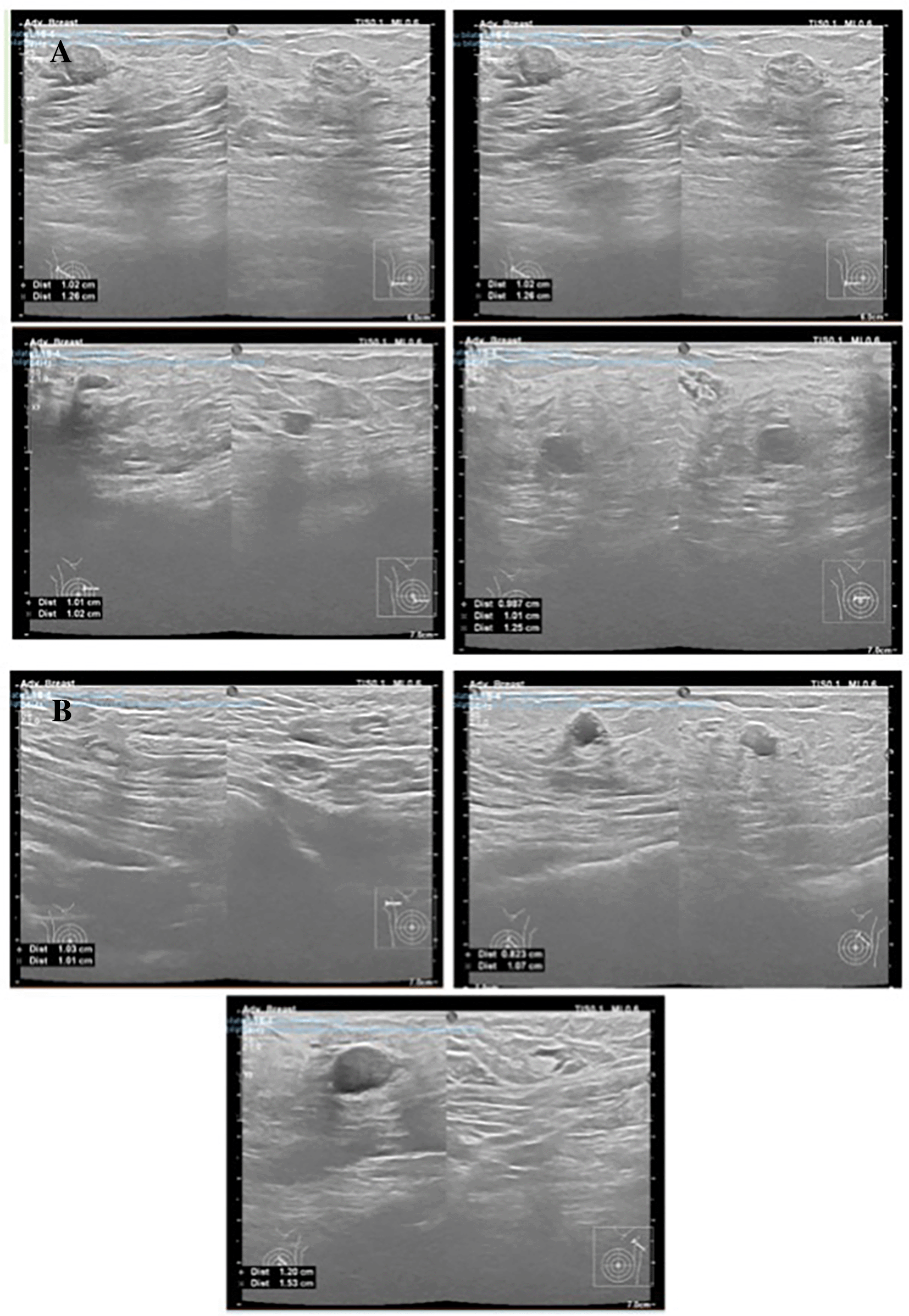
Based on the breast ultrasound results, an abscess was found in the left breast and lymphadenopathy in the left axilla. Chest radiography revealed no abnormalities.
The excision of the lump in the patient’s breast was performed, followed by histological investigation and pus culture. The histopathological analysis confirmed the presence of tuberculous mastitis (TM). The results of the pus culture indicated the absence of any bacterial growth. Following this, we initiated antituberculosis treatment for the patient, consisting of rifampicin 150 mg, isoniazid 75 mg, pyrazinamide 400 mg, and ethambutol Hcl 275 mg, during the intense phase for a duration of 2 months. Then we continued the treatment with rifampicin 150 mg and isoniazid 75 mg for an additional 4 months during the continuation phase. After completing 6 months of treatment, the patient was declared to be completely cured of TM.
A 25-year-old female patient presented with a lump in her right breast that had been present for two weeks before hospital admission. Upon physical examination, a mass measuring 4 × 3 cm was found in the right breast, with unclear borders, smooth surface, and mobility. A previous FNAB result indicated breast carcinoma.
The patient underwent ultrasound examination, which revealed a suspected benign tumor in both breasts and lymphadenopathy in both axillae.

We performed an excision procedure on the patient, followed by histopathological examination. The histopathological findings indicate tuberculous mastitis accompanied by fibrocystic changes and a focus of atypical ductal hyperplasia, which differed from the findings of prior examinations. The patient was then treated with antituberculosis medication for a duration of 6 months. During the 6th month follow-up, it was reported that the patient had fully recuperated from TM.
A 24-year-old pregnant female presented with a lump in her left breast that had persisted for 1 month. The lump started small but gradually grew larger and became more painful. Physical examination revealed a mass with a size of 5 × 5 cm, unclear border, immobility, and hard consistency.
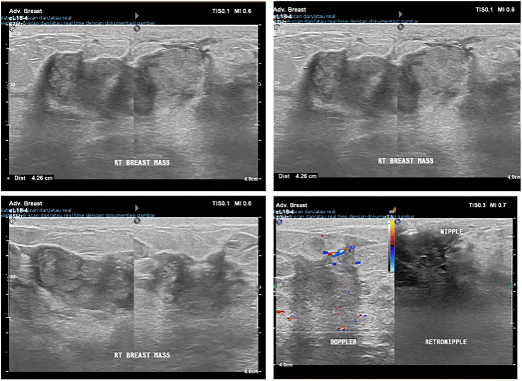
Based on the breast ultrasound results, an inhomogeneous mass was found in the left breast with an irregular border, along with subcutaneous thickening and blurring of the fibroglandular tissue around it. The ultrasound Breast Imaging Reporting and Data System (BIRADS) score was 4, indicating suspicious malignancy with differential diagnosis of mastitis or an infectious process, and lymphadenopathy in the left axilla was suspected. Chest radiography revealed no abnormalities.

We performed an FNAB on the patient. The cytology examination results indicate tuberculous mastitis with suppurative process. Therefore, we performed an excision procedure on the breast mass of the patient, followed by histopathological examination to confirm the diagnosis. The histopathological examination yielded similar findings to the FNAB results, prompting the immediate initiation of antituberculosis therapy in the patient. After 6 months of using antituberculosis medication, the patient was declared cured.
An abscess or a painless breast lump may indicate rare extrapulmonary TB. This disorder includes both the primary and secondary subgroups. The rare primary form is breast-only tuberculosis (TB). Primary tuberculous foci such as the lungs, pleura, and lymph nodes are related to the more common secondary form. Even in endemic areas, low clinical suspicion and similarity to other diseases make TM diagnosis difficult. TM can be diagnosed by a qualified surgeon, radiologist, or pathologist. In this study, during January 2024 we had 4 women in their perimenopausal diagnosed with tuberculous mastitis. The average age of the patients was 28.75 (24–41 years). One patient was diagnosed during pregnancy, and one had a history of prior breastfeeding. The findings for all patients are summarized in Table 1.
TM usually affects women of reproductive age, likely due to the hypervascularization of breast tissue and widening of lactiferous ducts during this period.12 However, TM is also found in adolescent girls, elderly women, and men.13 Multiparity, lactation, and pregnancy are known risk factors for the development of breast tuberculosis.14,15 A case series of 32 cases reported that 12.5% of all their cases were pregnant and lactating women.12 Lactation causes breasts to be vulnerable to trauma, making it easier for pathogenic bacteria to infect the tissue, especially if Mycobacterium tuberculosis is present in the baby’s faucial tonsils, which is suspected to be a potential route of infection in mothers.12,16 Lactation makes the breast highly vascularized and ducts dilated, making newborns and multiparous women more susceptible to TB. Pregnancy suppresses the pro-inflammatory T-helper response, which may enhance the tendency toward infection, as breast TB symptom presentation usually lasts less than a year but can last months or years.17 Patients with TM often show symptoms for several weeks to months before diagnosis, with varying durations, but often less than one year.13,16 In our findings, patients began experiencing symptoms from 2 weeks to 2 months before being diagnosed with TM.
All the patients had previously received a BCG vaccine. BCG vaccination has a 37% efficacy rate in preventing all types of tuberculosis in children under 5 years of age and a 42% efficacy rate in preventing lung disease in children under 3 years of age. However, it does not offer protection to teenagers or people in intimate contact with the disease.18 It is important to enhance the immune response against M.tuberculosis in order to provide long-lasting protection, particularly after infancy.19,20 According to the World Health Organization (WHO) data, Indonesia ranks second in the world, with the highest number of TB cases. In 2021, the incidence of TB in Indonesia was 354 cases per 100,000 people.21 Initially, it was believed that 60% of TM cases were caused by primary conditions; however, it is now believed that TM is almost always caused by secondary lesions in other parts of the body. The most common spread is retrograde lymphatic spread from the lung focus through the paratracheal and internal breast lymph nodes. Hematogenous spread and direct extension from adjacent structures are other routes of infection.16 Primary TM is very rare because the breast parenchyma is not an ideal site for Mycobacterium tuberculosis.22 Based on history taking, it can be established that the patient is likely to have primary TM. A patient reported a lump in the armpit area, which was discovered after a breast complaint. Extrapulmonary tuberculosis is often asymptomatic23; hence, it is probable that some of our patients had secondary TM.
The most typical sign of breast tuberculosis is a lump, typically in the upper outer or central quadrant of the breast, due to frequent spread from the axillary lymph nodes to the breast. The lump often shows irregular and poorly defined features, along with a firm texture. It may cause pain and can move or attach to the underlying skin or muscle. Furthermore, this condition can manifest as a solitary nodule, typically without the accompanying constitutional symptoms commonly seen in tuberculosis.24,25 Nipple retraction, peau d’orange, and breast edema have also been reported.8,26,27 This lump occasionally resembles a cancer.17,25 The results showed that all patients complained of breast lumps with or without abscesses, the majority of which were painful, firm, and had unclear borders. However, the lumps in our patient were typically found in the inner area of the breast. Half of our patients were clinically suspected to have breast malignancy.
TM is often unilateral, but both breasts can be infected, although this is very rare, occurring in only approximately 3% of all cases.16,28,29 Multiple lumps have rarely been reported in cases of TM. However, a recent case series report found multiple lumps in 35.3% of the cases.12 TM was first classified by McKeon and Sir Geoffrey Wilkinson into five categories: acute miliary, nodular, disseminated, sclerosing, and obliterative.16,22 Nodular caseous inflammation is the most common type. This type presents symptoms such as non-vascular hypoechoic mass without pain, elongated shape, well-defined borders, slow growth, resembling fibroadenoma in the early stages of the disease, and usually develops into a fistula within the nipple-areolar complex or in the skin and very similar to cancer.22
Radiographic results of breast tuberculosis are vague.30 In our case, all patients underwent an ultrasound examination. Chest radiography did not reveal any abnormalities or signs of pulmonary tuberculosis infection. One patient in this case series underwent FNAB examination with a diagnosis of breast carcinoma before arriving at our hospital. However, after re-examination, a diagnosis of tuberculosis mastitis accompanied by fibrocystic changes of Atypical Ductal Hyperplasia (ADH) type was made. Figure 8a shows breast tissue infested with acute inflammatory cells and chronic inflammation accompanied by necrotic areas. Langhan-type cells are visible (white arrows) between the connective tissue and hyperemic blood vessels. No visible signs of malignancy were observed. Figure 8b shows breast tissue showing chronic inflammatory cell-infested tissue (short black arrow) accompanied by necrotic areas. Necrotic breast glands (long black arrows) and Langhan-type data cells (white arrows) are visible between connective tissue and hyperemic blood vessels. No visible signs of malignancy were observed. Variations in diagnostic interpretation and reporting practices in anatomical pathology are not unusual. The percentage of inaccurate diagnoses ranges from 3% to 9%.31 The lack of clear quality criteria for anatomical pathology laboratories as well as the objectivity of pathological anatomy specialists may have contributed to this inaccurate diagnosis. The second view lessens the need for modifications to the initial pathology reports and is a useful quality metric in anatomical pathology.32
In this series, TM presented with symptoms similar to those of breast cancer, such as ulceration, redness, a hard palpable lump with uneven borders, and an ultrasound examination that suggested a breast tumor. The gold standard examination for distinguishing it from breast cancer is histopathological investigation. Diagnosis of TM requires several specific tests. The Mantoux test does not provide a diagnosis but can confirm whether a patient has tuberculosis infection. Mammography is not always helpful for diagnosis, especially in young women, because of the density of breast tissue. However, mammography findings in elderly women cannot be differentiated from mammary carcinomas. Ultrasonography shows hypoechoic masses in 60% of patients, and this method can also detect fistulas or sinus cavities, which can be observed in tuberculosis cases. Computed tomography (CT) and Nuclear Magnetic Resonance (NMR) were used to assess the extent of the lesions outside the body, especially in the chest. The gold standard for diagnosing TM is to isolate M. tuberculosis in bacteriological cultures or to use the Ziehl-Neelsen stain. However, M. tuberculosis is present in one-fifth of TM cases. Acid-fast bacteria were found in less than 12% of patients. As a result, some authors concluded that a histological finding of granulomatosis accompanied by caseous necrosis is adequate for diagnosis. Fine-needle aspiration (FNA) is the most frequently used diagnostic procedure, with 73% specificity for identifying granulomatosis and caseous necrosis. In other investigations, FNA was frequently inconclusive, with a sensitivity of 28%, compared to 94% for histology. Acid-fast bacteria were detected in 10.3% of FNA specimens and in 29.7% of histological specimens. Some researchers discovered that FNA was able to diagnose TM in 18% of the cases. When FNA is ambiguous, obtaining a larger histologic sample may be recommended, especially if other breast illnesses such as carcinoma have to be ruled out. In addition, skin testing, interferon-gamma assays, imaging tests, biopsies, and PCR-TB can confirm diagnosis.17,24,33 Polymerase Chain Reaction (PCR) is a highly sensitive method for the diagnosis of tuberculosis. Although popular, this method is only used in patients with negative culture results or other types of granulomatosis. Histopathological lesions identified chronic granulomatous inflammation with caseous necrosis and Langhans giant cells, contributing to the diagnosis in most cases. The main differential diagnosis for TM is breast carcinoma. Other differential diagnoses include fat necrosis, plasma cell mastitis, idiopathic granulomatous mastitis, and infections such as actinomycosis and blastomycosis.13,16,28,34
All of our patients received a 6-to-9-month course of anti-tuberculosis treatment. After one month of surgical excision, one person appeared with a breast ulcer and reported the presence of a pus-discharging lesion. We proceeded with the advanced stage of treatment till the ninth month. Debridement should be performed in this patient to enhance the efficacy of the treatment. All patients were reported to have achieved a complete cure of TM at the conclusion of their treatment.
Patients with tuberculous mastitis may present with nonspecific symptoms, although their condition may resemble that of mastitis or breast cancer. Due to the atypical symptoms presented by the patient, it is imperative to conduct comprehensive screening to rule out alternative probable diagnoses. The FNAC test is often regarded as the most dependable technique; however, false-negative results can still be obtained. Establishing a diagnosis requires effective teamwork between the attending physician, anatomical pathologist, and radiologist. Comprehensive research on a larger scale is required to accurately describe the existing situation.
This study was conducted using the primary patient data. Informed consent was obtained at the beginning of the patient’s visit by signing a letter of consent for examination, taking pictures, and publication for scientific purposes, and the patient’s identity was strictly protected. Written informed consent for publication of their clinical details and clinical images was obtained from the patient. The data collected were strictly used to achieve the research objectives.
The authors are grateful to the Department of Oncology Surgery at the Arifin Achmad General Hospital for their participation in this study.
| Views | Downloads | |
|---|---|---|
| F1000Research | - | - |
|
PubMed Central
Data from PMC are received and updated monthly.
|
- | - |
Is the background of the cases’ history and progression described in sufficient detail?
Yes
Are enough details provided of any physical examination and diagnostic tests, treatment given and outcomes?
No
Is sufficient discussion included of the importance of the findings and their relevance to future understanding of disease processes, diagnosis or treatment?
Partly
Is the conclusion balanced and justified on the basis of the findings?
Partly
Competing Interests: No competing interests were disclosed.
Reviewer Expertise: Breast physcian and Oncosurgeon
Alongside their report, reviewers assign a status to the article:
| Invited Reviewers | |
|---|---|
| 1 | |
|
Version 1 10 Sep 24 |
read |
Provide sufficient details of any financial or non-financial competing interests to enable users to assess whether your comments might lead a reasonable person to question your impartiality. Consider the following examples, but note that this is not an exhaustive list:
Sign up for content alerts and receive a weekly or monthly email with all newly published articles
Already registered? Sign in
The email address should be the one you originally registered with F1000.
You registered with F1000 via Google, so we cannot reset your password.
To sign in, please click here.
If you still need help with your Google account password, please click here.
You registered with F1000 via Facebook, so we cannot reset your password.
To sign in, please click here.
If you still need help with your Facebook account password, please click here.
If your email address is registered with us, we will email you instructions to reset your password.
If you think you should have received this email but it has not arrived, please check your spam filters and/or contact for further assistance.
Comments on this article Comments (0)