Keywords
Allergic bronchopulmonary Aspergillus(ABPA), Aspergillus fumigatus , Hypersensitivity pneumonia, Aspergillus flavus, cystic fibrosis.
This article is included in the Datta Meghe Institute of Higher Education and Research collection.
Allergic bronchopulmonary Aspergillus(ABPA), Aspergillus fumigatus , Hypersensitivity pneumonia, Aspergillus flavus, cystic fibrosis.
In immunocompetent patients, lung infections caused by fungi are uncommon. Numerous organisms, including Aspergillus, Pneumocytis jirovecii, and Cryptococcus, are capable of causing these infections. However, these infections can harm even immunocompetent humans. In reality, they can have an impact on people who have bronchopulmonary diseases such as chronic obstructive pulmonary disease, cystic fibrosis, and asthma.1
Lung disorder is linked to radiological manifestations such as localized infiltrates, segmental lung collapse, bronchiectasis, and Aspergillus microbiological and serological findings. These taken collectively, make up the diagnostic criteria for Allergic bronchopulmonary aspergillosis.2 Internationally accessible statistics show that up to 6% of asthmatic individuals may have ABPA.3 Aspergillus spores, especially Aspergillus fumigatus, cause airway colonization in susceptible hosts and trigger an allergic reaction when inhaled repeatedly. Various fungi can potentially cause allergic bronchopulmonary mycoses (ABPM), but Aspergillus fumigatus is most frequently linked to these pulmonary conditions. It is a pervasive, saprophytic mold found in both indoor and outdoor air, potting soil, crawl spaces, compost piles, mulches, freshly cut grass, decomposing plants, and sewage treatment facilities.4 Elevated total blood IgE levels, the presence of aspergillus IgE antibodies, and the formation of central bronchiectasis are some specific immunologic and radiologic indicators of illness. Effective management frequently necessitates corticosteroid therapy over an extended period of time.5 There is no specific test that can be used to determine whether someone has allergic bronchopulmonary aspergillosis. The diagnosis is supported by immunological results, radiographic findings, and conventional clinical symptoms.6
A 21-year-old female medical student presented with complains of breathlessness, which was MMRC grade II in beginning and gradually progressed to MMRC grade III, and history of seasonal variation for the last 15 years. She is known case of breathlessness evaluation since 2018 on rotacap. She also complained of cough with yellowish expectoration along with cold & chest pain over the last 5 days. The patient had a history of dust allergy with biomass exposure for 11 years with no any addictions.
The patient’s vital signs, which included a value of 110/70 mmHg for blood pressure, 84 for pulse rate, 20 for respiratory rate, and 94% for peripheral oxygen saturation while breathing room air, were all within the normal range. During the chest examination, bilateral rhonchi were detected upon auscultation, indicating the presence of abnormal breath sounds suggestive of respiratory issues. Heart sounds were present and no murmurs were found during a cardiovascular (CVS) examination, indicating no severe cardiac abnormalities at the time. The patient was discovered to be conscious and oriented throughout the CNS (central nervous system) assessment, demonstrating that they were completely mindful of their surroundings and position. According to laboratory results, the patient had eosinophilia, or an elevated eosinophil count, in their peripheral blood. The patient’s present respiratory symptoms may be related to eosinophilia, which has been linked to several medical disorders, including allergic responses and several respiratory illnesses. The patient’s diagnosis and continuing medical care benefited greatly from these clinical observations and test findings.
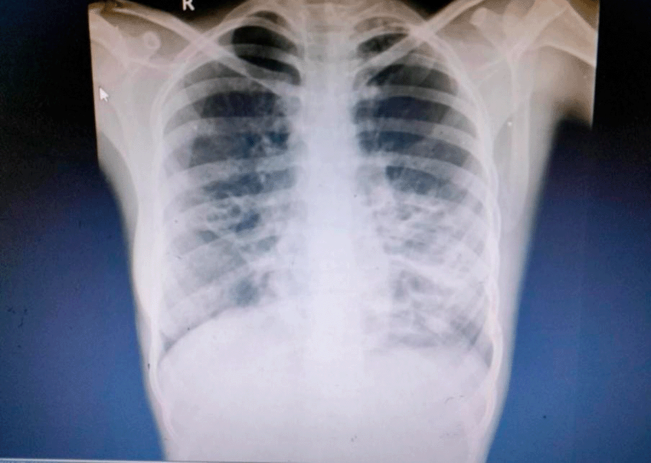
Chest X ray (Figure 1) was suggestive of Bilateral Lower Zone Bronchiectatic changes.
Pulmonary function tests demonstrated obstructive pattern with reduced FEV1 (Forced expiratory Volume in 1 second) and FEV1/FVC (Forced vital capacity ratio).
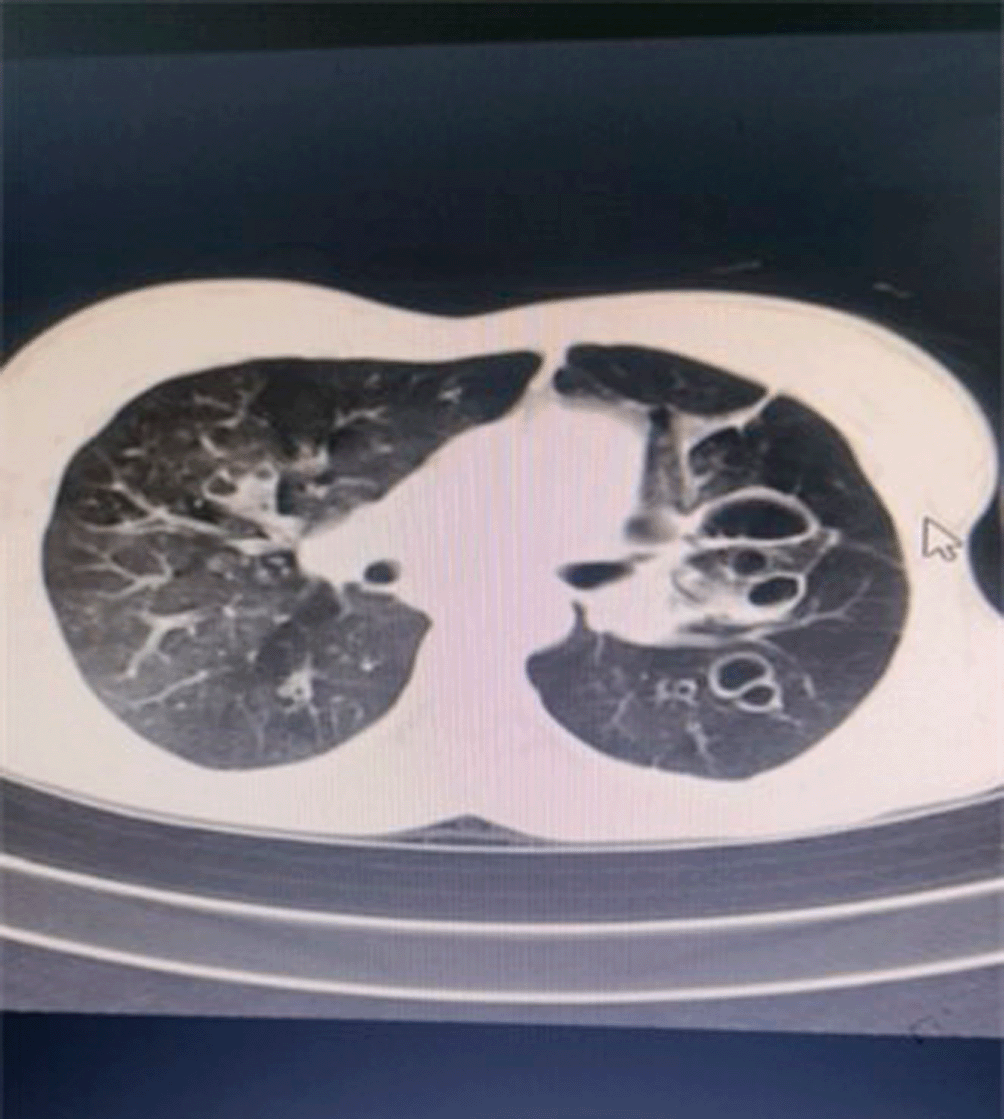
HRCT scan (Figure 2) showed cystic bronchiectatic changes in the left upper lobe, right lower lobe with fibrobronchiectatic changes in bilateral upper lobes, right middle lobe, diffuse ground glass opacities giving mosaic pattern in the bilateral lung parenchyma.
The patient’s laboratory investigations reveal several abnormal findings. First, the serum IgE level is significantly elevated at 658.1 IU/mL, well above the normal range of 52-423 IU/mL. Elevated IgE levels are often associated with allergic conditions and can be indicative of an immune response, such as the one seen in allergic bronchopulmonary aspergillosis (ABPA). The white blood cell count is also elevated at 13,300 wbcs/microliter, exceeding the normal range of 4,500-11,000 wbcs/microliter. This elevation may suggest an ongoing inflammatory or infectious process. The platelet count is elevated at 481,000/ml of blood, which is above the normal range of 150,000-450,000/ml. This could be a response to an underlying condition or inflammation in the body. The hemoglobin (Hb) level is within a slightly lower normal range at 12.5 gm/dl, with the normal range being 13-17 gm/dl. This might indicate mild anemia. Serum creatinine is slightly below the normal range at 0.5 mg/dL, which could be an indicator of kidney function. It falls below the reference range of 0.59-1.04 mg/dL. Sodium levels are lower than normal at 130 mmol/L (normal range: 136-145 mmol/L), indicating hyponatremia, which could be related to other medical conditions or fluid imbalances. The erythrocyte sedimentation rate (ESR) is markedly elevated at 46 mm/hr, well above the normal range of 0-20 mm/hr. An elevated ESR is often associated with inflammation and can be indicative of an underlying inflammatory or infectious process. HbA1c, a measure of long-term blood sugar control, is elevated at 6.2%, which is above the normal range of 4%-5.6%. This suggests poorly controlled blood sugar levels, indicative of diabetes mellitus. Fasting glucose levels are significantly elevated at 216 mg/dL, well above the normal reference range of <100 mg/dL, further confirming the presence of poorly controlled diabetes. Urine sugars and urine albumin are both reported as “Nil,” indicating that there are no detectable sugars or albumin in the urine, which is reassuring in diabetes management. Tests for AFB smear & PCR and HHH (possibly for HIV) are reported as negative, suggesting no evidence of tuberculosis (AFB) or HIV infection. Acid-fast bacilli are a group of bacteria that have a unique cell wall structure, making them resistant to decolorization by acids during laboratory staining procedures.
In summary, these laboratory findings collectively suggest a complex clinical picture with potential underlying issues related to allergic bronchopulmonary aspergillosis (elevated IgE), inflammation (elevated ESR and white blood cell count), mild anemia (slightly low Hb), hyponatremia (low sodium), and poorly controlled diabetes (elevated HbA1c and fasting glucose). Further evaluation and consultation with a healthcare provider are essential to determine the underlying causes and develop an appropriate treatment plan.
Sputum culture identified Aspergillus fumigatus as the causative agent.
The patient was admitted to the hospital’s respiratory department and received antifungal therapy, specifically oral itraconazole (200 mg) was given twice a day for 10 days in an effort to stop Aspergillus from spreading to the patient’s airways. An oral corticosteroid called prednisolone 0.5 mg/kg/days was given for 14 days on alternate days and, after discharge, 5 mg every two weeks for an additional three months to help treat the condition further. The patient was given these corticosteroids to lessen the inflammation of the eosinophils that was noticed. Nebulization therapy was used in conjunction with medication to treat the patient. The patient undertook nebulization with Budecort twice a day for 10 days and Duolin thrice a day for 10 days to focus relief and assist in managing respiratory problems.
Monitoring the patient’s progress and treatment reaction was one of the most crucial components of their care. In order to gauge the patient’s response to treatment, many follow-up appointments were set up. Lung function tests, IgE (immunoglobulin E) levels, and imaging examinations were repeated on a regular basis as part of this monitoring approach. These measurements aided medical personnel in assessing the efficacy of the therapy and making any necessary modifications to guarantee the patient’s well-being and recovery.
Outcome: The patient responded positively to the prescribed treatment regimen. Her symptoms improved significantly, and repeat HRCT scans showed improved changes. Long-term management remains essential to prevent relapses and minimize potential corticosteroid-related side effects.
A complex respiratory illness known as allergic bronchopulmonary aspergillosis (ABPA) is brought on by an overactive immunological reaction to the Aspergillus fungus, most frequently Aspergillus fumigatus. This condition primarily affects individuals with underlying lung diseases such as asthma or cystic fibrosis.7 ABPA is characterized by inflammation and allergic reactions in the airways and lung tissue due to exposure to Aspergillus spores.
Moulds called Aspergillus species have about 100 species globally, and Aspergillus fumigatus, Aspergillus niger, Aspergillus clavatus, and Aspergillus flavus are responsible for the majority of infections.8 ABPA is a complex pulmonary disorder that requires a multifaceted approach to treatment. Early diagnosis, prompt initiation of appropriate medications, and ongoing monitoring are crucial for managing the disease effectively and preventing long-term lung damage. Multidisciplinary care involving pulmonologists, allergists, and other healthcare professionals is essential for successful management.9 Various medications have demonstrated their effectiveness in the management of ABPA. The very first chemicals to be employed are glucocorticoids. A randomized trial revealed that both the medium-dose regimen & high-dose regimen are effective against ABPA, with the medium-dose treatment having fewer side effects.10 Prednisolone is administered for a total of three to five months in the medium-dose regimen as monotherapy (0.5 mg/kg/day for 2 weeks, then on alternate days for 8 weeks, then 5 mg less every 2 weeks).11 Chest radiography & total serum IgE levels should be checked on patients every 2 months until remission.12 When the baseline total IgE levels double along with clinical or radiological worsening, an exacerbation has occurred. Clinical radiographic improvement, a minimum of 25% reduction in total IgE levels, & confirmation of remission occur at least 6 months after the patient has stopped receiving all medications.13 However, it has not been proven that treating ABPA found on standard testing in asymptomatic patients with well-controlled asthma results in benefits. The prognosis for patients with ABPA over the course of time is still unclear.14 However, a positive prognosis is achieved by early disease detection and treatment recommendations. Patients who are not treated eventually develop permanent lung fibrosis & respiratory failure.15
In conclusion, a case study of ABPA provides a comprehensive view of the disease, from initial presentation and diagnostic challenges to treatment strategies and long-term management. Such case studies contribute to our understanding of this condition and help healthcare professionals provide better care for individuals with ABPA.
| Views | Downloads | |
|---|---|---|
| F1000Research | - | - |
|
PubMed Central
Data from PMC are received and updated monthly.
|
- | - |
Provide sufficient details of any financial or non-financial competing interests to enable users to assess whether your comments might lead a reasonable person to question your impartiality. Consider the following examples, but note that this is not an exhaustive list:
Sign up for content alerts and receive a weekly or monthly email with all newly published articles
Already registered? Sign in
The email address should be the one you originally registered with F1000.
You registered with F1000 via Google, so we cannot reset your password.
To sign in, please click here.
If you still need help with your Google account password, please click here.
You registered with F1000 via Facebook, so we cannot reset your password.
To sign in, please click here.
If you still need help with your Facebook account password, please click here.
If your email address is registered with us, we will email you instructions to reset your password.
If you think you should have received this email but it has not arrived, please check your spam filters and/or contact for further assistance.
Comments on this article Comments (0)