ALL Metrics
-
Views
-
Downloads
Get PDF
Get XML
Cite
Export
Track
Case Report
Revised

Case Report: Case report: Aslanger’s sign in electrocardiogram.

[version 2; peer review: 2 approved]
PUBLISHED 27 Nov 2024
Author details Author details
OPEN PEER REVIEW
REVIEWER STATUS

Abstract

Electrocardiograms (ECGs) can be affected by various factors and technical problems. It is rare for an artefact to be the cause of ST-segment elevation, especially in asymptomatic patients. An important distinction between true ST segment elevation caused by myocardial infarction and an artefact is that the baseline elevation in an artefact may begin before or after the appearance of the QRS complex. When confronted with an abnormal ECG with suspicious waveform contours and possibly only one completely normal limb leads, the diagnosis of arterial pulse artefact should be considered. It is important to exclude subjective assessments unless they are clearly labelled as such.

Keywords

Arterial pulse–tapping artifact, electrocardiographic artifact, pseudo-myocardial infarction.

Revised Amendments from Version 1

We made some changes in the Introduction (reformulated phrase) corrections in references and some details in Discussion.

See the authors' detailed response to the review by Koji Takahashi
See the authors' detailed response to the review by Nihal Sheriff

Introduction

It should be noted that artefacts on an electrocardiogram can result from a variety of causes, both internal and external. These include muscle tremors, the use of dry electrode gel and loose leads, and electromagnetic interference. These artifacts can sometimes mimic ECG abnormalities, which can cause problems for patient care.

In this report, we describe an unusual ECG artifact that caused large and bizarre T-waves on the ECG. The observed changes are aligned with those commonly associated with primary repolarisation changes characteristic of acute coronary syndrome. The artefact in question is caused by the overlapping of the artery pulse; this can be avoided by moving the lead away from the pulsating artery. This is also known as the electromechanical association artefact.

Case report

A 68-year-old man with no medical history and no cardiovascular risk factors consulted the emergency department of a district hospital due to 48 hours of atypical and paroxysmal chest pain (tingling). His physical examination revealed no abnormalities, with SBP at 120 mmHg, DBP at 50 mmHg, HR at 99 bpm, RR at 20/min, Sat O2 at 96% on air, T° at 37°C, and finger blood sugar at 0.9 g/l.

Figure 1 shows the patient’s first ECG.

94502d21-f476-4a61-9c91-0fb24b130ba3_figure1.gif

Figure 1. The prehospital 12-lead ECG.

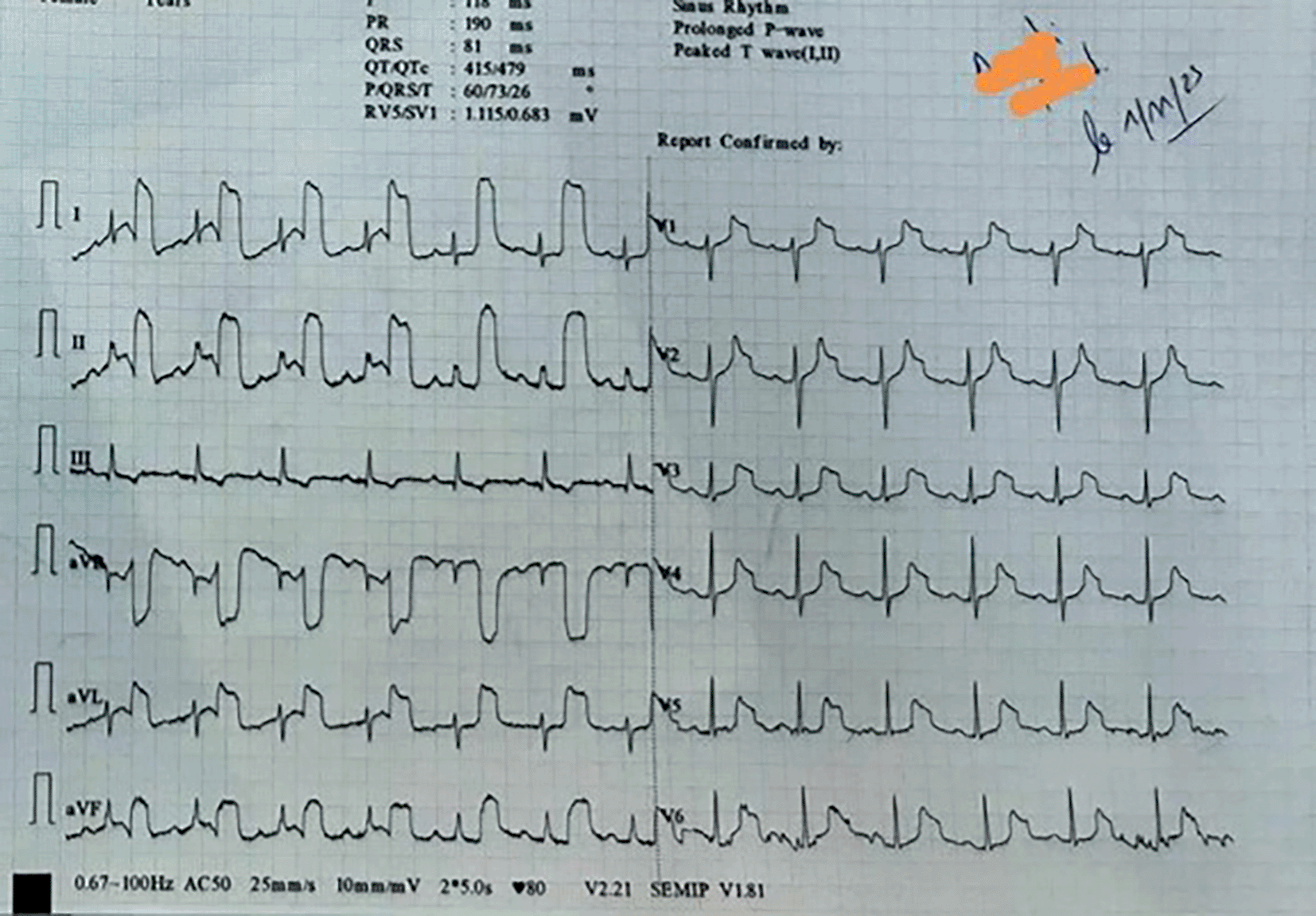
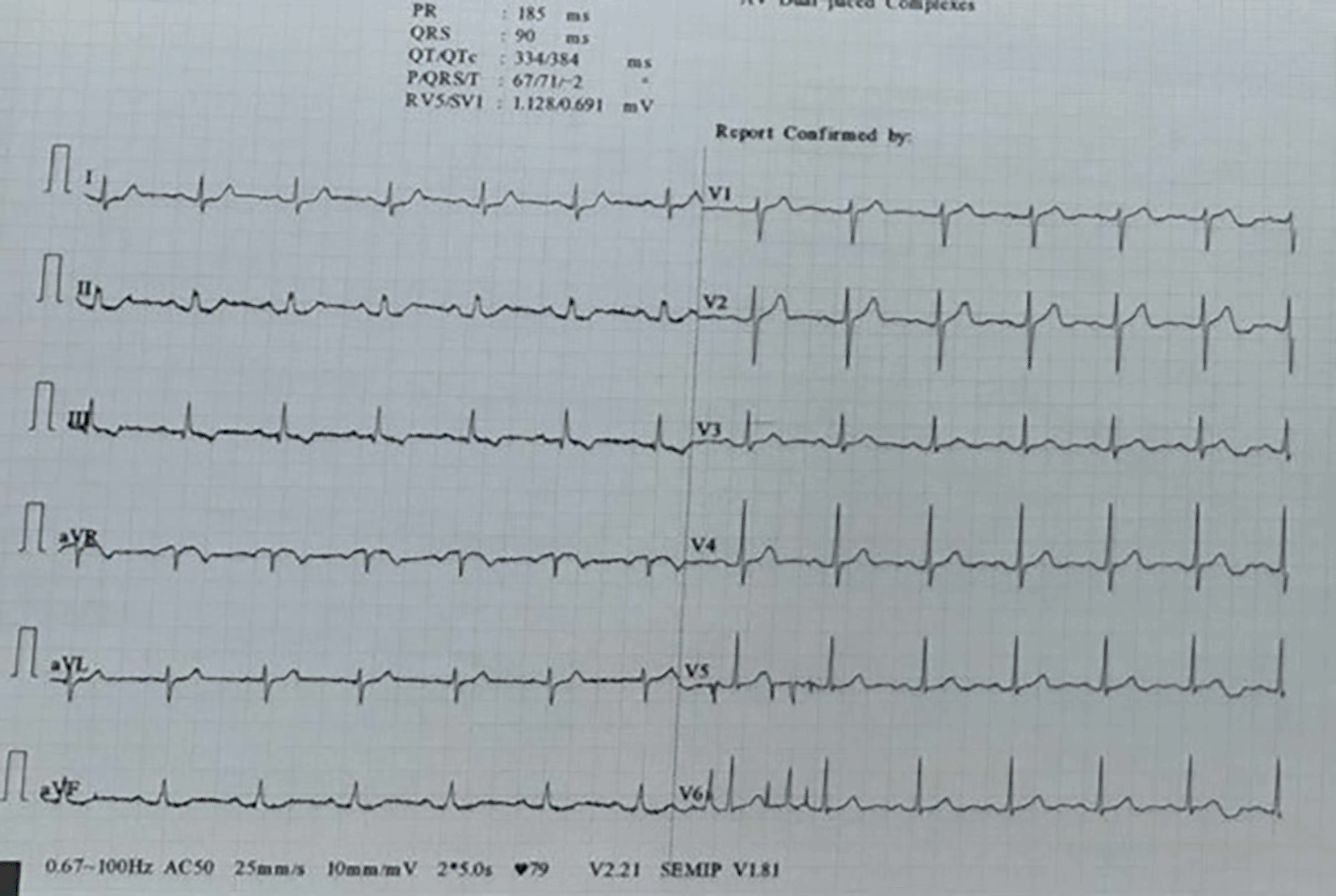
The 12-lead ECG revealed a normal sinus rhythm with a heart rate of 80 beats per minute and normal axis. Abnormal T waves with bizarre morphology were observed in leads I, II, aVL, aVR, aVF, and from V1 to V6 in precordial leads. This abnormality was also observed in all the leads except for lead III. The emergency department physician’s interpretation of the T waves was that they were indicative of an ischaemic hyperacute condition. The patient was transferred to the emergency department (ED) at the university hospital due to suspected acute coronary syndrome. However, the ECG performed in the ED was completely normal ( Figure 2), and the high-sensitivity cardiac troponin (hs-cTn) levels were twice negative.

In blood investigations no cirrhosis or severe anemia were observed. An investigation for hyperthyroidism was not done but no history of hyperthyroidism for our patient. An echocardiography was done in the emergency room without abnormality.

Therefore, the pre-hospital ECGs were recognized as artifacts, and the patient was discharged home.

94502d21-f476-4a61-9c91-0fb24b130ba3_figure2.gif

Figure 2. Normal ECG in ED.

Discussion

In this case, the electrocardiogram (ECG) showed anomalous T waves with unusual morphology in leads I, II, aVL, aVR, aVF, and V1 to V6. This distinct pattern was observed in the 12-lead ECG, with the exception of lead III. The presence of an arterial pulse-tapping artifact, also known as electromechanical association (EMA) or Aslanger’s sign, was indicated by the synchronous occurrence of this abnormality with the cardiac cycle. Aslanger initially described this phenomenon,1 which some authors refer to as Aslanger’s sign.2

The EMA artifact arises from the transmission of arterial pulsations, typically from the radial artery but also from the posterior tibial artery particularly in hyper dynamic states,3 onto the lead clips, generating aberrations in the ECG waveform. Contemporary electrocardiogram machines only record lead I and lead II, deriving the waveforms for other leads from these two. However, the majority of limb leads and augmented leads are susceptible to artifact. A consistent feature of EMA artifacts is the sparing of one lead, contingent on the limb generating the artifact. This serves as a crucial diagnostic clue, as outlined by Aslanger.4 In this case, lead III was unaffected as it represents an ECG recording between the left arm and left leg. Therefore, we concluded that the source of the artifact was the right arm. When the clip was placed proximally during a repeat ECG in the emergency department, the 12-lead ECG exhibited no artifacts.

Aslanger’s sign has been recently described and there are limited reported cases in the literature. It is important to emphasise the potential risks associated with this condition, as it can mimic symptoms of acute coronary syndrome. This can lead to unnecessary invasive investigations if not promptly recognised. The artifact, induced by the mechanical tapping of the pulse on the ECG electrode, is in synchrony with the cardiac cycle and can manifest as ST segment changes (elevation or depression) accompanied by peculiar T waves.3,5

Conclusion

This report presents a case of Aslanger sign, also known as arterial pulse-tapping ECG artifact. This anomaly presents as a primary repolarisation abnormality that is comparable to those observed in acute coronary syndrome.

It is important to be aware that an EMA artefact will almost always spare one of the limb leads, as this is a key factor in diagnosing it, which helps physicians avoid misinterpretation and unnecessary explorations.

Declarations

Participants’ consent

Informed written consent for participation in the study was obtained.

Consent to publish

Written informed consent for publication of his clinical details and/or clinical images was obtained from the patient.

Comments on this article Comments (0)

Version 2
VERSION 2 PUBLISHED 15 Oct 2024
Comment
Author details Author details
Competing interests
Grant information
Copyright
Download
 
Export To
metrics
Views Downloads
F1000Research - -
PubMed Central
Data from PMC are received and updated monthly.
- -
Citations
CITE
how to cite this article
Bradai H, Laajimi S, Mbarek R et al. Case Report: Case report: Aslanger’s sign in electrocardiogram. [version 2; peer review: 2 approved]. F1000Research 2024, 13:1233 (https://doi.org/10.12688/f1000research.156313.2)
NOTE: If applicable, it is important to ensure the information in square brackets after the title is included in all citations of this article.
track
receive updates on this article
Track an article to receive email alerts on any updates to this article.

Open Peer Review

Current Reviewer Status: ?
Key to Reviewer Statuses VIEW
ApprovedThe paper is scientifically sound in its current form and only minor, if any, improvements are suggested
Approved with reservations A number of small changes, sometimes more significant revisions are required to address specific details and improve the papers academic merit.
Not approvedFundamental flaws in the paper seriously undermine the findings and conclusions
Version 2
VERSION 2
PUBLISHED 27 Nov 2024
Revised
Views
4
Cite
Reviewer Report 09 Dec 2024
Nihal Sheriff, Madras Medical College, Chennai, India 
Approved
VIEWS 4
No further ... Continue reading
CITE
CITE
HOW TO CITE THIS REPORT
Sheriff N. Reviewer Report For: Case Report: Case report: Aslanger’s sign in electrocardiogram. [version 2; peer review: 2 approved]. F1000Research 2024, 13:1233 (https://doi.org/10.5256/f1000research.174851.r343919)
NOTE: it is important to ensure the information in square brackets after the title is included in all citations of this article.
Views
0
Cite
Reviewer Report 05 Dec 2024
Koji Takahashi, Ehime University Graduate School of Medicine, Ehime, Japan 
Approved
VIEWS 0
I have ... Continue reading
CITE
CITE
HOW TO CITE THIS REPORT
Takahashi K. Reviewer Report For: Case Report: Case report: Aslanger’s sign in electrocardiogram. [version 2; peer review: 2 approved]. F1000Research 2024, 13:1233 (https://doi.org/10.5256/f1000research.174851.r343918)
NOTE: it is important to ensure the information in square brackets after the title is included in all citations of this article.
Version 1
VERSION 1
PUBLISHED 15 Oct 2024
Views
16
Cite
Reviewer Report 06 Nov 2024
Koji Takahashi, Ehime University Graduate School of Medicine, Ehime, Japan 
Approved with Reservations
VIEWS 16
I believe that Aslanger's sign is still not well recognized, even by medical staffs working in cardiovascular and emergency medicine. Therefore, it is worthwhile to case report this typical case.

The abstract, introduction section, and case presentation ... Continue reading
CITE
CITE
HOW TO CITE THIS REPORT
Takahashi K. Reviewer Report For: Case Report: Case report: Aslanger’s sign in electrocardiogram. [version 2; peer review: 2 approved]. F1000Research 2024, 13:1233 (https://doi.org/10.5256/f1000research.171603.r336519)
NOTE: it is important to ensure the information in square brackets after the title is included in all citations of this article.
  • Author Response 27 Nov 2024
    rabeb mbarek, Department of Emergency Medical Services, Sahloul Hospital, Sousse, Tunisia
    27 Nov 2024
    Author Response
    I would like to thank the reviewer for his precious time to read critically our case and for this important corrections suggested to improve the quality of our work.
    corrections ... Continue reading
COMMENTS ON THIS REPORT
  • Author Response 27 Nov 2024
    rabeb mbarek, Department of Emergency Medical Services, Sahloul Hospital, Sousse, Tunisia
    27 Nov 2024
    Author Response
    I would like to thank the reviewer for his precious time to read critically our case and for this important corrections suggested to improve the quality of our work.
    corrections ... Continue reading
Views
16
Cite
Reviewer Report 31 Oct 2024
Nihal Sheriff, Madras Medical College, Chennai, India 
Approved with Reservations
VIEWS 16
The authors here have compiled a case report of an acute coronary syndrome mimic – “Aslanger sign” which has quite recently received attention in the field of cardiology. They have done a good job in summarising the necessary points regarding ... Continue reading
CITE
CITE
HOW TO CITE THIS REPORT
Sheriff N. Reviewer Report For: Case Report: Case report: Aslanger’s sign in electrocardiogram. [version 2; peer review: 2 approved]. F1000Research 2024, 13:1233 (https://doi.org/10.5256/f1000research.171603.r336521)
NOTE: it is important to ensure the information in square brackets after the title is included in all citations of this article.
  • Author Response 27 Nov 2024
    rabeb mbarek, Department of Emergency Medical Services, Sahloul Hospital, Sousse, Tunisia
    27 Nov 2024
    Author Response
    I would like to thank the reviewer for his precious time to read critically our case and for his important corrections suggested to improve the quality of our work. We ... Continue reading
COMMENTS ON THIS REPORT
  • Author Response 27 Nov 2024
    rabeb mbarek, Department of Emergency Medical Services, Sahloul Hospital, Sousse, Tunisia
    27 Nov 2024
    Author Response
    I would like to thank the reviewer for his precious time to read critically our case and for his important corrections suggested to improve the quality of our work. We ... Continue reading

Comments on this article Comments (0)

Version 2
VERSION 2 PUBLISHED 15 Oct 2024
Comment
Alongside their report, reviewers assign a status to the article:
Approved - the paper is scientifically sound in its current form and only minor, if any, improvements are suggested
Approved with reservations - A number of small changes, sometimes more significant revisions are required to address specific details and improve the papers academic merit.
Not approved - fundamental flaws in the paper seriously undermine the findings and conclusions
Sign In
If you've forgotten your password, please enter your email address below and we'll send you instructions on how to reset your password.

The email address should be the one you originally registered with F1000.

Email address not valid, please try again

You registered with F1000 via Google, so we cannot reset your password.

To sign in, please click here.

If you still need help with your Google account password, please click here.

You registered with F1000 via Facebook, so we cannot reset your password.

To sign in, please click here.

If you still need help with your Facebook account password, please click here.

Code not correct, please try again
Email us for further assistance.
Server error, please try again.