Keywords
E granulosus, Rib, Dorsal vertebra, Thoracic Surgery, Recurrence, Mortality
E granulosus, Rib, Dorsal vertebra, Thoracic Surgery, Recurrence, Mortality
This revised version focuses on improving clarity and completeness of the methodology, results, and references. The Methods section has been updated to provide more detailed descriptions of patient selection, inclusion and exclusion criteria, and definitions of chest wall hydatidosis, as well as clearer explanations of anatomical boundaries.
The Results section has been enriched with additional descriptive data, offering a more precise presentation of clinical characteristics, radiological findings, surgical management details, and postoperative outcomes. These improvements provide a clearer understanding of the patient population and disease patterns.
Finally, we have added new references and updated all references starting from reference number five, ensuring that the manuscript reflects the most recent and relevant literature.
See the authors' detailed response to the review by Zied Chaari
Hydatidosis or echinococcosis, also known as hydatid cyst or hydatid disease, is a cosmopolitan infectious prevalent in the Mediterranean basin, Australia, and South America. It is caused in humans, an accidental intermediate host, by the presence and development of the hydatid of a cestode of the genus “echinococcus granulosus” that resides in the small intestine of canids.1,2 Hydatid cysts represent a public health problem in endemic countries, particularly Tunisia, where the incidence is approximately 15.1 per 100,000 inhabitants,3 as well as in other countries where traditional livestock farming is still practiced, due to its frequency, severity, and socio-economic impact.
CWH refers to the presence of hydatid cysts caused by the tapeworm echinococcus granulosus within the anatomical structures of the chest wall including ribs and cartilages, sternum, thoracic vertebrae and muscles of the Chest wall (intercostal muscles, Pectoralis Major, Perctoralis Minor and Serratus anterior).
We excluded the diaphragm as it is a distinct entity that will be investigated seperately in a subsequent article deserving its own dedicated study.
The location in the chest wall presents diagnostic, therapeutic, and prognostic problems. The long symptomatic silence of this disease (5-15 years) allows the lesions to spread rapidly before they are discovered,4 thus posing a differential diagnosis problem, explaining the diagnostic delay.3,5 Chest wall hydatidosis poses unique therapeutic challenges, and recurrence remains a major concern with significant clinical implications. Bone lesions, especially vertebral ones, are extensive with a very progressive evolution and can lead to definitive neurological complications.
Our study aimed to describe chest wall surgical management and outcomes and identify possible associated recurrence and mortality risk factors.
Ethics approval
Ethical approval was waived by the local Ethics Committee of Abderrahmane Mami University Hospital under the number 20/2024 in October 2024.
Patient privacy and informed consent for publication
Written informed consent was obtained directly from the patients by the authors, regardless of their department of origin. Patients consistently provided consent before surgery as per standard hospital practice. Additionally, retrospective consent for study participation was acquired through telephone contact.
Anonymity was respected during data treatment.
Consent for publication of identifiable data
No consent for publication was required as the data had been anonymized. We confirm that such alterations have not distorted the scientific meaning.
Following the Safe Harbor methods outlined by HIPAA, all radiologic images and patient photographs used in the study were thoroughly de-identified to ensure patient privacy. This process involved removing any direct identifiers such as patient names, dates of birth, medical record numbers, and facial features that could potentially lead to the re-identification of the individuals. Additionally, any metadata embedded within the images, such as timestamps or institution-specific details, were removed. This de-identification procedure was carefully followed to comply with the HIPAA guidelines and protect patient confidentiality throughout the study.
Population source
The present study was retrospective, descriptive, and multicentric. It was over twenty-eight years: from January 1995 to December 2022.
Our study was carried out in four university hospital centers in Tunis: the thoracic surgery department of Abderrahman Mami Hospital, Ariana, the neurosurgery department of the National Institute of Neurology in Tunis, the orthopedics department of the Mohamed-Kassab Institute of Orthopedics and the orthopedics department of Charles Nicolle Hospital in Tunis. We used the admissions database of these university hospital centers to collect records of patients operated on for CWH, defined as hydatidosis affecting the osseous structures (ribs, sternum, costal cartilages), intercostal muscles, and adjacent soft tissues, excluding breast tissue and clavicle. Regarding soft tissue involvement, we considered lesions located within the muscular and subcutaneous layers of the thoracic wall, bounded:
• Superiorly by the clavicle and thoracic inlet (excluding cervical and supraclavicular regions),
• Inferiorly by the costal margin and 12th rib, without extending into the abdominal wall,
• Anteriorly up to the sternum, excluding breast tissue,
• Posteriorly to the paravertebral muscles, without extension into the vertebral canal unless explicitly associated with vertebral hydatidosis.
Inclusion criteria
We included all operated patients for hydatidosis of the chest wall, with complete records (observation, radiological findings, operative report, anatomopathological report, follow-up).
Non-inclusion criteria
We did not include the patients operated on for suspected chest wall hydatidosis (CWH) and whose diagnosis was not retained after surgery or pathological examination, patients with confirmed CWH but who were not operated on (refusal, inoperable patients with high anesthetic risks).
We applied the inclusion and non-inclusion criteria and eliminated incomplete and unusable records. We retained forty-nine patients operated on for chest wall hydatidosis.
A data collection form was drawn up in a standardized manner to collect all elements necessary to meet the objectives of our study.
For each patient, we collected information about epidemiological and anamnestic data: age, gender, geographical origin (rural or urban), the department of origin (thoracic surgery, orthopedics, or neurosurgery), notion of hydatid contagion, family history of hydatid cyst (HC), personal history of visceral hydatid cyst. We consider a history of multiple visceral hydatidosis and hydatid involvement of more than three viscera. We also examined clinical parameters such as the time to consultation (the time from the onset of symptomatology to the date of consultation), time to treatment (the time between the onset of the symptomatology and the date of surgery), the circumstances of discovery, including incidental findings, chest pain, swelling of the soft tissues of the thoracic wall presenting as a parietal tumefaction of the posterior thoracic or paravertebral wall, neurological symptoms, and respiratory involvement. Additionally, we evaluated physical examination parameters such as general examination, thoracic floor assessment, and neurological and abdominal examinations.
The radiological assessment included a chest X-ray, X-ray of the spine, coastal grid, thoracic, soft tissue, and abdominal ultrasound, CT-scan, Magnetic Resonance Imaging (MRI), and other radiological examinations such as medullary arteriography. Biological tests were carried out, including hydatid serology and a blood count to check for hypereosinophilia.
We specified the therapeutic management elements: anesthesia, surgical position, surgical approach, per-operative findings, and surgical procedure: which we have divided into three stages: surgical resection of part of the chest wall (including ribs, vertebrae, and/or muscles), surgical spinal stabilization: anterior by a bone marrow transplant or by osteosynthesis with a fixation material and conservative surgical procedure when the operation is performed only on the cystic lesion without ablating a part of the thoracic wall. We identified the used scolicidal solution (hydrogen peroxide, povidone-iodine, hypertonic saline), the protection of the operating field (use of soaked gauze in scolicidal solution to protect pleura and soft tissues for possible scolex dissemination), and we mentioned per-operative complications.
We recorded the duration of drainage, hospital stay, immediate postoperative course: simple or complicated, and late complications. Late course: was assessed by clinical examination (neurological by ASIA score to evaluate motor skills and sensitivity) and postoperative sequelae. During the follow-up period, every hydatid recurrence was recorded (time, treatment modalities, surgical treatment (approach, procedure, number of repeat surgeries, evolution after treatment of recurrences). Antiparasitic medical treatment was noted (molecule and length of treatment) and the pathological examination. We identified the mortality (frequency, causes, delay). We defined lost to follow-up patients as those who missed outpatient visits for over three consecutive years.
The data was analyzed using the IBM Statistical Package for the Social Sciences 25.0 software, accessed through the free 30-day trial available on IBM’s official website (https://www.ibm.com/products/spss-statistics#110). In our study, we proceeded to Calculate absolute and relative frequencies for categorical variables, means, medians (with minimum and maximum), and standard deviations for quantitative variables. For the quantitative variables, the distribution of the population was tested for normality. If the distribution was normal, the variables were described as mean, otherwise as median. We used the Kaplan-Meier survival curve by calculating the cumulative survival to study the survival relationships of the elected variables associated with recurrence and the elected variables associated with mortality. For the multivariate study, we used Cox regression to determine factors independently associated with recurrence and factors independently associated with mortality. All tests were two-sided, and a two-tailed probability value of 0.05 or less was considered statistically significant.
During the study period, 49 patients were operated on for CWH. The mean age of our population was 41 ± 17 years [8-76 years] with a male predominance.
At the time of diagnosis, 15 patients had associated hydatid involvement at a distance, the others had exclusive parietal involvement. Bone involvement was vertebral (n=11), only costal (n=12), costovertebral (n=21) and sternal (n=2). There was soft tissue involvement adjacent to the costovertebral involvement in the intercostal and paraspinal muscles (n=13), as well as in the soft tissue of the supraclavicular fossa (n=2) with exclusive primary involvement of the pectoralis major muscle (n=1) and the paraspinal muscle (n=1).
The diagnosis of CWH was an incidental finding in three cases. The other patients were symptomatic with a combination of functional signs: thoracic pain (n=23), parietal swelling (n=22) ( Figure 1A), neurological signs (n=22), and respiratory signs (n=7) ( Table 1).
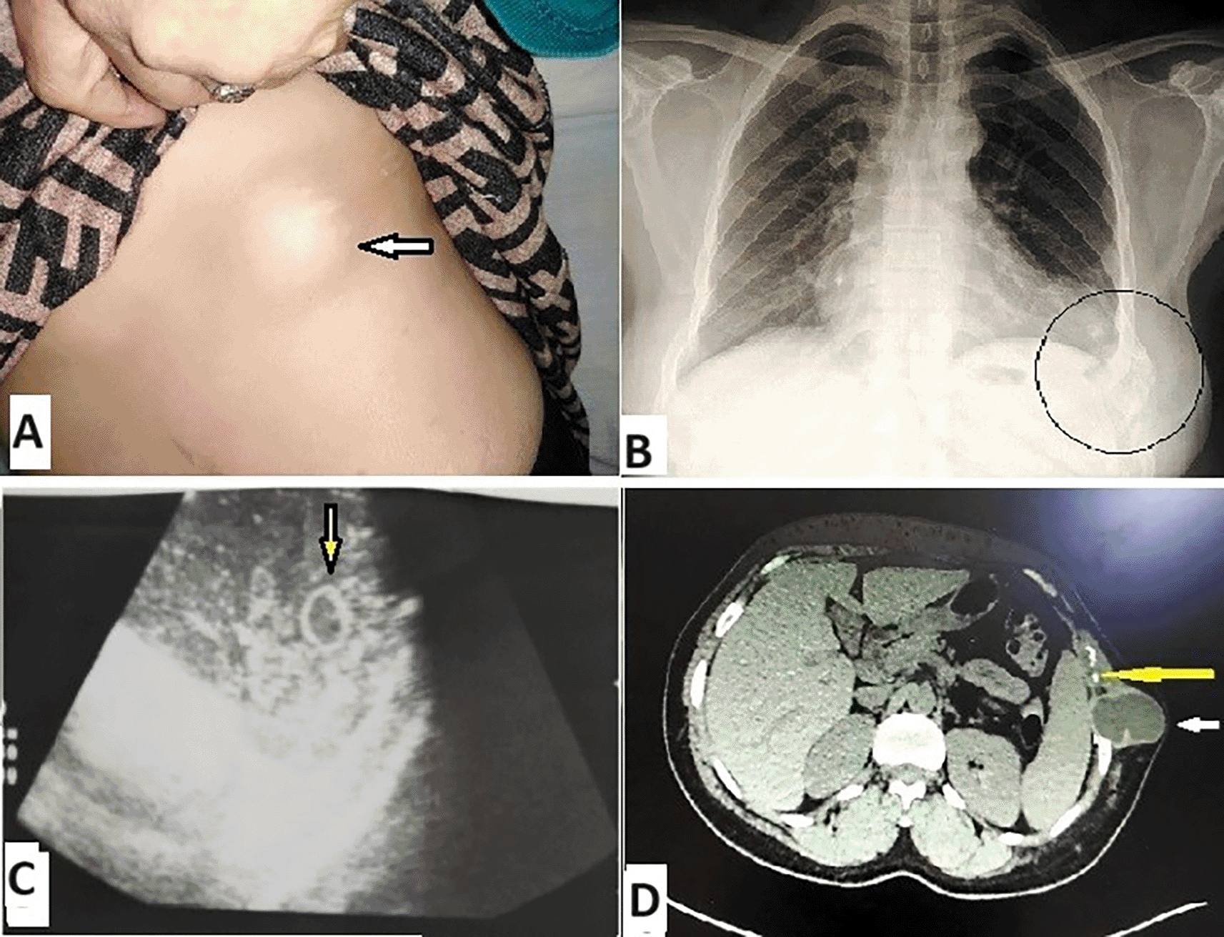
A parietal swelling (arrowhead); B Chest radiograph, circle arrowed the cyst and its effect on ribs; C echography showing hydatid vesicles (arrowhead); D CT image: cystic lesion with cortical costal lysis (arrowhead).
Patients with isolated costal or soft tissue involvement did not present with neurological symptoms, but rather with localized pain or swelling. Neurological manifestations were observed exclusively in patients with vertebral involvement.
Paraclinical investigations were carried out: chest X-ray was performed in 48 patients, i.e., 98% of cases, and mainly showed a parietal opacity in 19 cases ( Figure 1B).
Ultrasound of the soft tissues was performed in 18 cases and the most frequent aspect was a hypoechogenic multivesicular formation (n=13) which guided the diagnosis ( Figure 1C).
Abdominal ultrasound was performed in 44 cases as part of the etiological and extension assessment of the hydatid disease and showed a hepatic HC (GHARBI type IV) in nine cases.6
The thoracic CT scan was performed in 94% of cases ( Table 1). Costal damage was in the posterior arch in 25 cases (51%). A tiered involvement of more than two ribs was observed in 43% of the cases, with a predominance of lesions at the Fourth to ninth rib level. The vertebral involvement was in the vertebral body in 21 cases and the intervertebral disc in two cases.
In addition, we observed that the vertebral involvement appeared as an ossifying abscesses in the vertebral arch, pedicles, laminae, and transverse processes in 11 cases. In fact, the involvement was difficult to isolate into each component of the vertebra due to the extension and dissemination within the bone, as there was no cystic wall to limit the lesions.
In 16% of cases, the number of affected vertebrae was two or more. The most affected levels were D7-D9 (n=11) explained by the richness of the vascularization of this spine segment ( Figure 1D, Figure 2).
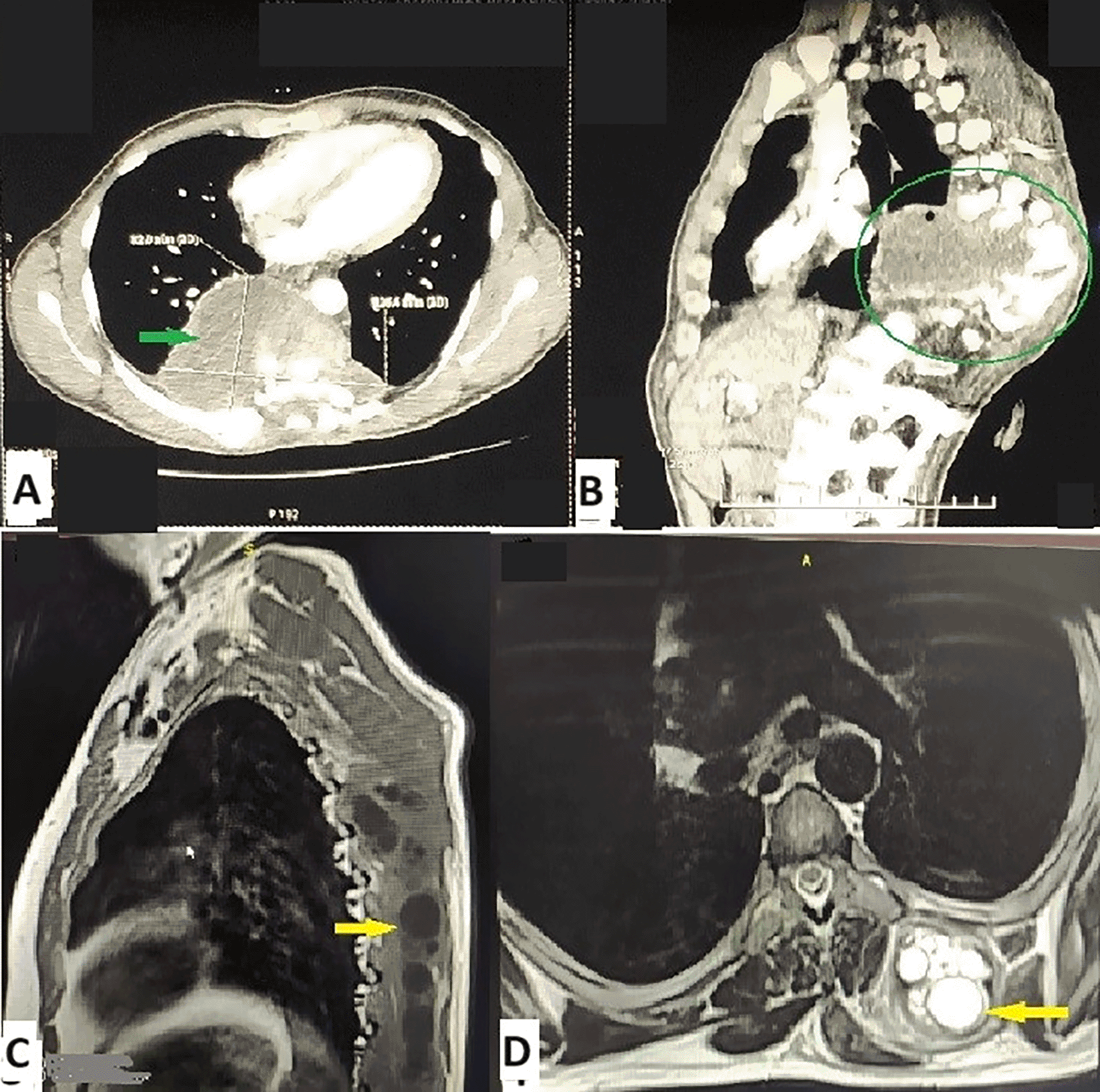
A-B Destruction of the spine and intracanal diffusion of hydatidosis (arrowhead and circle); C-D MRI showing a large multivesicular mass depending on the paravertebral muscle (arrowheads).
There was soft tissue involvement adjacent to the spinal involvement. The mean major axis of the lesion was 7.8 cm, with a size ranging from 3 to 30 cm in the major axis. Twenty-three patients had a lesion with a large diameter ranging from 5 to 10 cm.
Vertebro-medullary magnetic resonance imaging (MRI) was performed in 24 patients, i.e., 49% of cases ( Table 1).
All patients were operated on. For bone involvement, posterolateral thoracotomy (PLT) was performed in 18 cases (39% of cases), a posterior approach in 16 cases (35% of cases), and a combined approach in 3 cases (7% of cases) ( Figure 3).
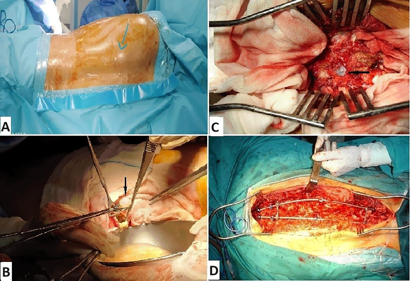
A Surgical position, arrow showing paravertebral swelling; B Flattening of hydatid abscesses; C Posterior approach revealing vertebral and intracanal hydatidosis; D Posterior spinal stabilization (osteosynthesis).
The surgical procedure was divided into three modalities: radical surgical removal, spinal stabilization, and conservative surgery without costovertebral resection ( Table 2).
| Surgical stabilization | Number (n) |
|---|---|
| No | 14 |
| Anterior spinal stabilization (by graft) | 6 |
| Posterior spinal stabilization (osteosynthesis) | 12 |
| Total | 32 |
| Conservative surgery without costovertebral resection | |
| Conservative surgery | Number (n) |
|---|---|
| Flattening hydatid abscesses | 15 |
| Cystotomy + draining + cystic wall resection | 10 |
| Fistula resection | 4 |
| Roughening of the vertebral body ± transverse process | 2 |
A primordial time is the protection of the operating field by a scolicide solution. The main solutions were hypertonic saline and hydrogen peroxide ( Figure 3).
The immediate postoperative course was simple in 94% of cases. The average length of hospitalization was 21 days [IQR: 12–32] for isolated vertebral involvement, 15 days [IQR: 9–26] for costovertebral involvement 6 days [IQR: 3–11] for isolated rib involvement, and three days [IQR: 2–4] for exclusive muscular involvement of hydatid disease. The difference between the average length of stay for the different conditions was not statistically significant (p=0.70).
Medical treatment was prescribed to all our patients even for those who had a complete resection and was taken in 63% of cases. The antiparasitic molecule prescribed was albendazole in the form of a 400 mg tablet, per day in two doses, in the form of 21-day courses separated by one week. The antiparasitic medical treatment was mainly taken postoperatively in 14 cases and after the recurrence in 11 cases. The duration of the antiparasitic medical treatment was more than one year in 17 cases, i.e., 55% of the patients who had received medical treatment.
Post-operative complications occurred in 24% of cases. There are three types of complications: neurological, infectious and decubitus. Neurological complications included paraplegia in two cases and paresis in six cases. The three main postoperative infectious complications were surgical wound infection (n=3) and pneumonia acquired by mechanical ventilation (n=3). Pressure sores were the main decubitus complication (n=4).
The postoperative evolutionary aspects were marked by a total disappearance of the initial symptomatology in 22 cases and a clinical aggravation in three cases. On the other hand, the different types of postoperative sequelae were mainly intercostal neuralgia in seven cases and paraplegia in five cases. Recurrence was a frequent complication that determines the prognosis of this rare disease and occurred in 18 cases (37%). The median time to recurrence was one year with extremes ranging from a month to 13 years. For recurrence, patients had medical and surgical treatment in most cases (n=14). For the different surgical revision procedures for recurrence, the main procedure was spinal decompression by laminectomy with partial corporectomy associated or not with spinal stabilization in eight cases. The mean number of repeat surgeries for recurrence was three times with extremes ranging from one to eleven times. Thus 61% of the patients who had a recurrence were reoperated on more than twice.
Overall survival is estimated at 80% at five years. Seven patients (70% of those who died) had vertebral involvement. Patients with vertebral hydatid involvement had a 5-year survival of 71%.
We conducted an analytical study to determine the factors associated with recurrence and mortality. The univariate study looked at several parameters, epidemiological, radio-clinical, and parameters related to the management methods, represented in Tables 3-4.
The multi-varied study of these parameters identified the factors independently associated respectively with recurrence and mortality, which are shown in Table 5.
Male gender, vertebral involvement, and antiparasitic medical treatment could be independently associated with recurrence. Associated distant hydatid cysts and postoperative infectious complications were the two factors associated with mortality.
CWH is an entity that encompasses the involvement of the soft tissues and the costovertebral and sternal bones.
The literature review revealed only case studies or articles about a few cases in the descriptive framework. Our work has added to the literature the experience of four university hospitals in a country where hydatidosis is endemic, through a descriptive and analytical study attempting to find factors associated with recurrence and factors associated with mortality that could jeopardize functional and vital prognosis.
The curative treatment for thoracic parietal echinococcosis is surgical.7,8 Results depend on the extent of the lesions, their location, and the presence of complications. It must be the subject of an exhaustive work-up and treated as a “malignant tumor” by a wide resection passing through healthy margins.9
A recent study of eight cases of primary soft tissue hydatid cysts highlighted various locations, with 25% occurring in the left posterior thoracic region, a rarely reported site.10 The management of hydatid cysts located in soft tissues remains challenging due to their rarity and potential for misdiagnosis. Complete surgical excision is the cornerstone of treatment. These findings align with the general principle that careful surgical management is critical, particularly for unusual and anatomically complex locations such as the posterior thoracic wall.
For isolated rib involvement, the main surgical procedure was resection of the anterior or posterior arch or both. This resection was extended to more than two ribs in five cases.
For sternal involvement, the procedure was a manubriectomy. For isolated soft tissue involvement, resection of the hydatid cyst was combined with partial resection of the muscle involved.
For costovertebral lesions, a multidisciplinary surgical team comprising thoracic surgeons and orthopedic surgeons or neurosurgeons is required.
Thoracic vertebral echinococcosis, particulary, of D6-D9 segment, is the most common vertebral location due to the rich vascularisation of this segment of the spine.10,11
In our series; the stages most affected D4-D6(n=10), D7-D9(n=11).
MRI is useful for checking the extent of the disease, the texture of the lesion, the degree of the spinal cord involvement and the viability of the cyst.12–14
From a technical point of view, the approach to the thoracic parietal lesion is often facilitated and guided by the ossifluent abscess, which is surgically traversed as far as the primary bony lesion, eliminating the hydatid vesicles in the process. Costovertebral lesions can be treated using a posterior approach alone, a posterolateral approach alone, or a combined approach (posterior and posterolateral). Each of these approaches has its advantages and indications, and a combined approach is recommended in most cases, particularly if a laminectomy has already been performed as part of a neurological emergency.15 Access to the vertebral bodies and ribs is difficult, and resection of hydatid material is often incomplete. The posterolateral approach has several advantages: it allows access to all the vertebral bodies of the dorsal spine and extensive excision of the spine, ribs, and muscles, with the possibility of reconstruction or osteosynthesis, and makes it possible to treat an associated pulmonary, pleural or mediastinal location. For costovertebral involvement, the data in the literature showed that most patients were operated on via the posterior approach.16,17 This was the only approach used in the series by Khazim16 and Bhavin.18 Although this approach offers the possibility of spinal decompression, excision of the hydatid process remains incomplete, with a high rate of mortality, neurological deficits, and fistulae. Other teams have used the posterior approach in addition to the posterolateral approach to search for one or more hydatid vesicles that have migrated into the spinal canal, far from the initial site of contamination, or for palliative treatment of multi-stage lesions.19 The combined posterior approach associated with PLT is recommended in most cases.11,16,17
In our study, posterolateral thoracotomy was performed most frequently. This was performed in 39% of cases of bone involvement.
Thoracic parietal resection must be carried out with multidisciplinary management. At present, it is recommended to carry out as complete an excision as possible using a “carcinological” strategy.12,20,21
Some hydatid lesions of the thoracic wall, spread over more than five vertebrae, are sometimes unresectable20 and require a conservative surgical procedure such as flattening of the hydatid abscesses, or spinal curettage, which was performed in two patients in our series, or even surgical abstention.20
Medical treatment with albendazole is the only therapeutic alternative for inoperable patients, even though it may not cure or prevent recurrence. Its use as adjuvant therapy to surgery is generally necessary to prevent systemic dissemination during vesicular rupture and to reduce and delay recurrences. We have adopted this approach in our series. However, some teams believe that albendazole does not act on infested bone without a germ membrane.22
Luan et al.23 reported a combined therapeutic strategy involving repeated surgical debridement and prolonged antiparasitic treatment with albendazole in patients with advanced vertebral hydatidosis. Although this approach aims to reduce parasitic load and limit disease progression, the authors observed a significant recurrence rate, often requiring repeated surgeries and long-term follow-up. Similarly, Kafaji et al.24 highlighted the technical challenges of spinal surgery in this context, frequently incomplete due to the infiltrative nature of hydatid cysts within vertebral structures. They also noted the limited efficacy of antiparasitic drug therapy alone, with a high postoperative recurrence rate. The earlier observations by Apt et al.25 reinforce these findings, reporting frequent recurrences even decades ago, often associated with severe neurological complications. Despite differences in clinical context and time period, these studies consistently emphasize the infiltrative and recurrent nature of vertebral hydatidosis, which continues to make curative treatment particularly challenging.
Recurrence is considered a poor prognostic factor in CWH disease, particularly when it involves the vertebrae. Operative mortality of up to 14.4% has been reported in recurrences.26
For bone involvement, the results of surgery are far from curative and recurrence is the rule.19,26,27 In our series, recurrence was noted in 37% of cases. Recurrence surgery is associated with higher morbidity and mortality than initial surgery. However, most of these operations are only palliative.28
Surgery may be repeated several times to completely eradicate echinococcosis but cysts involving the spine cannot be safely resected or excised in their entirety in most patients.26,28
There are cases of resistance that respond neither to surgery nor to medication. We report a case diagnosed as hydatidosis of the chest wall and treated with radiotherapy (RT) after medical and surgical therapy had failed. Ulger S et al report a case of hydatidosis of the sternum showing the value of RT for E. multilocularis as an alternative treatment29; however, based on the literature, we conclude that the effect of RT is not well known in this setting.
CWH, and principally the spinal location, is associated with a non-negligible mortality rate of 3 to 46%.30 The overall mortality rate was 46% in the study by Zlitni,31 and 17% in that of Ezzaouia.12 Death is, in most cases, the consequence of infectious and decubitus complications.12,21,32 In our series, ten patients died. Patients with spinal hydatid involvement had an estimated survival of 71% at five years and 29% at 10 years. There was no statistically significant difference in survival between patients with and without vertebral hydatid disease (p=0.78). This can be explained by the small sample size of our series. Seven patients, i.e., 70% of those who died, had costovertebral hydatid lesions, and four died from complications of hydatid disease, mainly from decubitus complications (n=3). The other three patients were elderly, two of whom had associated pulmonary and secondary pleural hydatidosis.
Few studies have examined survival factors, and none have explored risk factors for recurrence and mortality in this rare condition. In our multivariate analysis, we aimed to identify these factors, finding a recurrence rate of 37% and a mortality rate of 20% among our patients.
According to the literature, thoracic parietal hydatidosis is more frequent in men.13,20 In our study, we also noted a male predominance. There were 31 men and 18 women, with a sex ratio of 1.7. This may be explained by the fact that males are more likely to associate with canids in rural areas. Gender may be a factor in recurrence (P=0.006), with recidivism six times greater for males (HR=6.032).
Bone echinococcosis can affect all bones but preferentially affects the spine.10,11 This is the most common location; 42% for Dévé2 and 44% for Zlitni. The site is mainly dorsal (56%).11,31 It is the most serious because of the neurological complications and the difficulties of surgical eradication. Although hydatidosis of the bone is a parasitic disease, its prognosis should be treated as if it were a malignant lesion. In our series, vertebral involvement is a factor associated with recurrence (P=0.025) and HR=10.2. Unlike visceral sites, the rigid structure of bone prevents the formation of adventitia, allowing parasites to spread through microvesicular infiltration of bone tissue. Exogenous vesiculation is characteristic of bone due to its rigidity, and results in the release of daughter vesicles from the mother vesicle, which explains the frequency of recurrences. There is no clear dividing line between healthy and pathological tissue, which is a major difficulty during surgical removal.31
In our series, patients who had taken antiparasitic medical treatment reattract the disease earlier than those who had not (p=0.006). This seems to be unwarranted however it can be justified by the common prescription of antihelminthic treatment for extensive hydatid lesions and the absence of a standardized protocol for this treatment. Certain factors influence the efficacy of benzimidazoles. This may be also attributed to the small number of patients in our series. Todorov et al. found that larger vesicle size, the presence of daughter vesicles, and, notably, bone involvement were associated with a poor response to medical treatment. This may be attributed to the small number of patients in our series.33
In the management of recurrent spinal hydatid cysts, surgical outcomes remain variable with a significant risk of recurrence. A recent study analyzing 39 cases of recurrence identified several major prognostic factors influencing postoperative outcomes.34 Intraoperative cyst rupture was found to be significantly associated with an increased risk of recurrence (p = 0.001). Similarly, subtotal resection, often necessitated by the infiltrative nature of the lesions, correlated with a significantly higher risk of recurrence (p < 0.007). Thoracic localization of the cysts and the performance of laminectomy were also identified as independent factors associated with poorer prognosis (p < 0.04). These findings highlight the importance of complete and careful resection, as well as meticulous management of intraoperative rupture risks, to optimize clinical outcomes and reduce recurrence rates. These factors should be carefully considered in the therapeutic planning and follow-up of patients with vertebral hydatidosis.
We studied several other factors that could be associated with recurrence and that were only significant in the univariate study. These included clinico-radiological factors such as the neurological deficit, vertebral compression, scalloping of the posterior vertebral wall, vertebral lysis, paravertebral extension, intracanal extension, and involvement of more than two vertebrae. Other factors related to management methods, such as the extent of surgical removal: laminectomy alone, complete costal resection, the occurrence of neurological complications and postoperative decubitus complications, and the occurrence of postoperative sequelae.
This may be explained by the heterogeneity of our sample, with a small enough number of patients to be able to generalize.
The mortality rate of patients followed for CWH, essentially the dorsal spine is 3 to 14%.13,26,29
Hydatid involvement of the thoracic wall, and especially bone involvement, is rare, accounting for less than 2% of all hydatid cases, and is associated with a visceral cyst in 12 to 25% of cases. It is primary,2 rarely secondary, and occurs via the hematogenous route.30 In our series, patients with a visceral hydatid site at a distance from the parietal involvement at the time of diagnosis had a mortality rate 31 times higher, with a significant difference (p=0.005). This suggests that we should consider simultaneous treatment of visceral hydatid lesions if feasible.
Infectious complications can cause high mortality. In our series, mortality was 40 times higher with the occurrence of infectious complications.
We studied several other factors that could be associated with mortality, such as the onset of chest pain, exclusive parietal involvement, which is a protective factor, costovertebral involvement, and costal resection. These were significant only in the univariate study. This may be explained by the small number of patients in our series, which makes it difficult to generalize.
To validate these conclusions and identify the factors contributing to recurrence and mortality, a prospective study with a larger sample size may be necessary. However, this is challenging due to the rarity of the disease.
Surgery remains the treatment of choice for chest wall hydatidosis. If diagnosed early and treated promptly, CWH can go into long-term remission. However, detecting this disease in its early stages can be challenging. This pathology is burdened with recurrence and significant morbi mortality associated with several factors. Recurrence could be associated with male gender and vertebral involvement. Associated distant visceral hydatidosis and postoperative infectious complications could increase mortality. Larger-scale studies are needed to enhance our understanding of this rare condition, improve its management, and minimize factors that could negatively impact postoperative outcomes.
| Views | Downloads | |
|---|---|---|
| F1000Research | - | - |
|
PubMed Central
Data from PMC are received and updated monthly.
|
- | - |
Provide sufficient details of any financial or non-financial competing interests to enable users to assess whether your comments might lead a reasonable person to question your impartiality. Consider the following examples, but note that this is not an exhaustive list:
Sign up for content alerts and receive a weekly or monthly email with all newly published articles
Already registered? Sign in
The email address should be the one you originally registered with F1000.
You registered with F1000 via Google, so we cannot reset your password.
To sign in, please click here.
If you still need help with your Google account password, please click here.
You registered with F1000 via Facebook, so we cannot reset your password.
To sign in, please click here.
If you still need help with your Facebook account password, please click here.
If your email address is registered with us, we will email you instructions to reset your password.
If you think you should have received this email but it has not arrived, please check your spam filters and/or contact for further assistance.
Comments on this article Comments (0)