Keywords
spleen, hydatid cyst, echinococcosis, situs inversus, splenectomy, case report
This article is included in the Pathogens gateway.
spleen, hydatid cyst, echinococcosis, situs inversus, splenectomy, case report
This revised version of the manuscript includes improvements based on reviewer feedback.
The introduction has been updated to include a more focused thesis statement that clearly outlines the rarity, diagnostic complexity, and surgical challenges of this case. The discussion section has been reorganized for better logical flow. We have also added a detailed analysis of how this case contributes to the broader understanding of hydatid cysts in atypical locations and outlined practical strategies to address the surgical challenges posed by situs inversus. Additionally, some grammatical errors, awkward phrasing, and typographical issues have been corrected to improve clarity and readability throughout the manuscript.
See the authors' detailed response to the review by Ali Bilal Ulas
See the authors' detailed response to the review by Silvio Buscemi
See the authors' detailed response to the review by Selmy Awad
Splenic hydatic localization is extremely rare, with a worldwide incidence rate of 0.5%-4%.1 Abdominal left hypochondrium pain, mass, and fortuitous discoveries are the most frequently discovered complications.1,2 However, right hypochondrium pain due to a splenic hydatic cyst associated with situs inversus is an exceptional finding. Here, we report the case of a 50-year-old female, who underwent surgery in our department for a complicated splenic hydatic cyst with situs inversus. This case presents clinical and radiological diagnostic challenges due to the combination of a giant splenic hydatid cyst and situs inversus, as well as a therapeutic challenge posed by the presence of this congenital abnormality.
A 50-year-old female, with no medical history presented to the emergency department with right hypochondrium pain.
On physical examination, the patient was febrile at 38.4°C; anicteric, with tenderness of the right hypochondrium on abdominal examination. The hemodynamic status was stable.
Blood analysis showed a biological inflammatory syndrome. The liver test was normal.
In the face of a 50-year-old obese female who consulted for a painful and febrile syndrome of the right hypochondrium, an abdominal ultrasound was performed, which showed a complete situs inversus and a mass of the right hypochondrium with a membrane detachment, measuring 152 mm, evoking a type II splenic hydatic cyst.
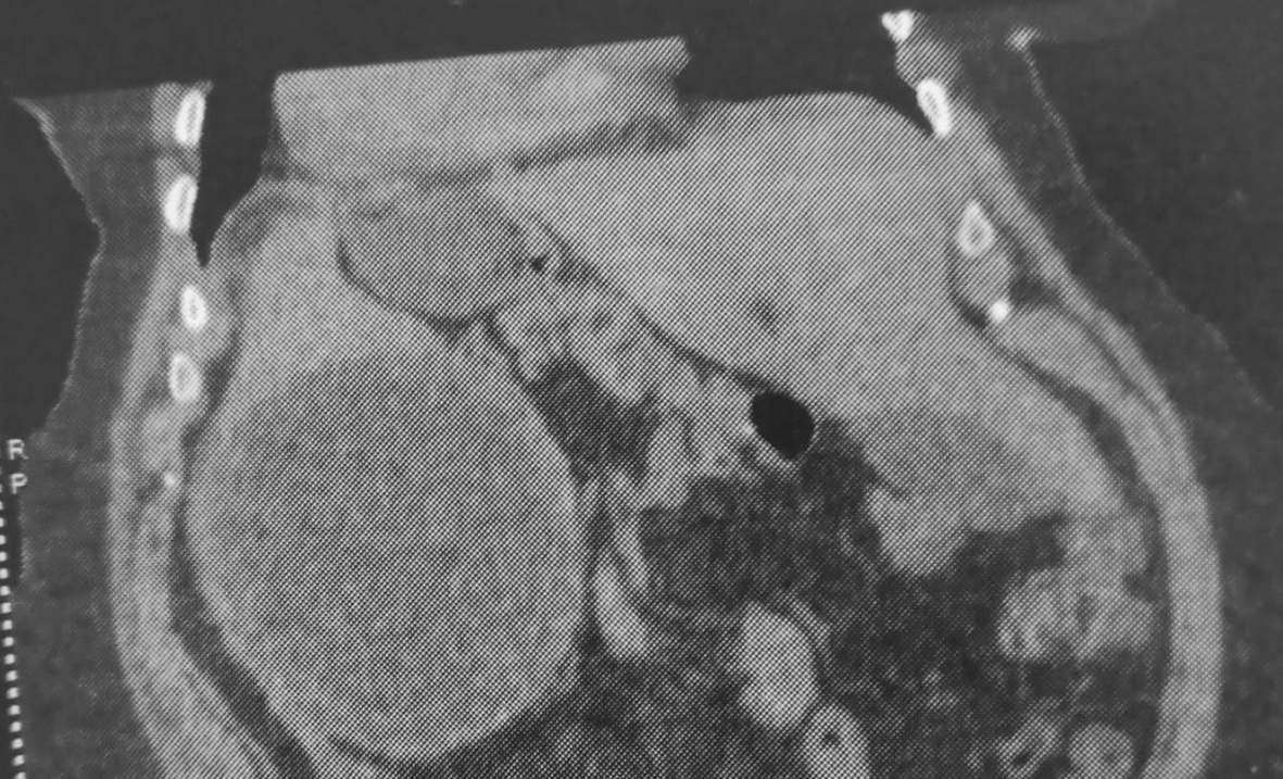
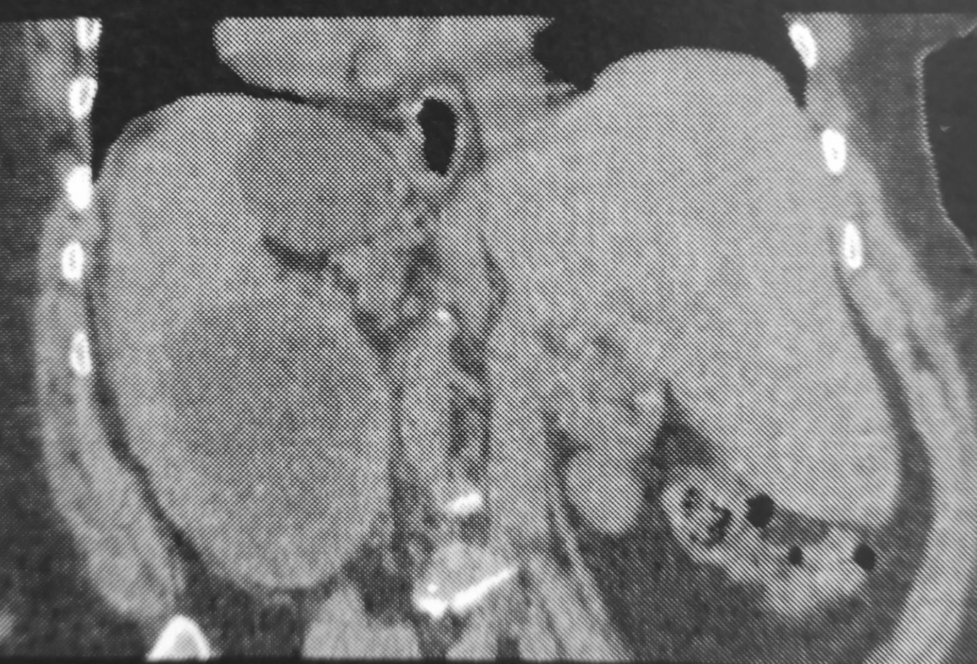
Computed tomography (CT) revealed a large cystic formation in the spleen, measuring 15 cm in its largest dimension. The cyst showed a detached internal membrane, which is typical of a type II hydatid cyst according to the Gharbi classification. Additionally, there was evidence of surrounding fat stranding and infiltration, indicative of local inflammatory changes. These findings strongly suggest a complication, specifically a hydatid cyst cracking (Figures 1, 2).
The patient underwent an emergency midline laparotomy. The exploration revealed a situs inversus, a voluminous splenic cyst occupying over 80% of the splenic volume. Exposition of the splenic pedicle is difficult. The cysto-parietal and cysto-visceral adherences, giant size of the cyst, and obesity prevented good exposure, which led to the decision to empty the cyst content after protecting the operating field with a field soaked in hypertonic serum. The cyst was infected.
Equally, the choice of the type of surgery, whether a total splenectomy or a protruding dome resection in an emergency context with complications such as cracking and surinfection, was not easy.
However, in the face of an emergency, the primary localization in the spleen, we performed a total splenectomy that allowed healing of the infested organ and avoided recurrence and surinfection of the residual cavity.
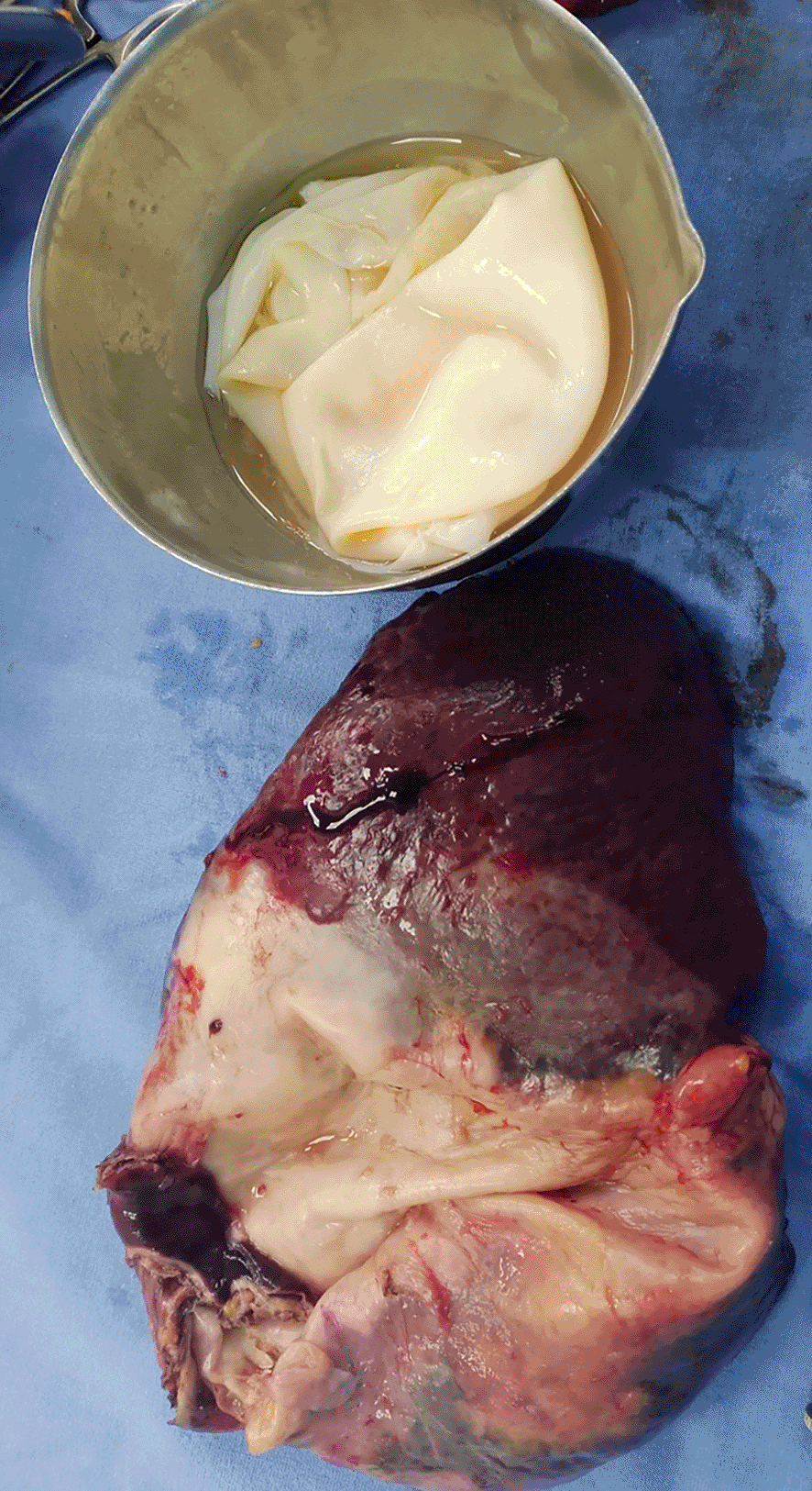
The overture of the cyst objectified the proligere membrane (Figure 3).
The post-operative period was unproblematic, and the patient was discharged with antibiotic and antiparasitic treatment and habitual vaccination.
Treatment with albendazole 400 mg twice daily was initiated from post-operative day 1, with cycles consisting of three 28-day treatments at 2-week intervals. Serum anti-Echinococcus antibody titers (hemagglutination test and ELISA) were positive in the immediate postoperative period (titers of 1/1280 and 1.9, respectively). Subsequently, the titers gradually decreased until achieving a negative serology at 2 years post-operatively. Liver function tests and complete blood counts were performed after each cycle and did not reveal any abnormalities during the treatment period. The abdominal CT scan did not reveal any recurrence of cystic echinococcosis in the thoraco-abdominal regions, particularly in the peritoneal area, with a current follow-up period of 3 years.
Hydatic cysts are a common pathology in endemic countries. The most frequent locations are the liver (70%) and lungs (20-30%).2 Splenic localization is extremely rare, with a worldwide incidence rate of 0.5%-4%.1
To our knowledge, this is the first reported case of a giant splenic hydatid cyst associated with situs inversus. In our case, the cyst was also ruptured into the abdomen, which posed both a diagnostic and therapeutic challenge.
Based on the literature of published cases of splenic primary localization, the most frequent circumstances of discovery include pain, a left hypochondrium mass, and fortuitous findings. These circumstances also occur during complications, such as infection and splenic abscess, rupture with anaphylactic shock, and dissemination to other organs.1,2
Ultrasound, computed tomography, and magnetic resonance imaging of the abdomen allow for diagnosis by objectifying membrane detachment and calcifications on the daughter vesicle wall.2,3 In the case of a complicated cyst, cross-sectional imaging, particularly CT scans, can establish the diagnosis in an emergency while also allowing for the assessment of the cyst’s location and its anatomical relationships.2,3
The treatment of splenic hydatic cysts is surgical. Total splenectomy has the advantage of avoiding recurrences. Protruuding dome resection has the advantage of being a conservative intervention of the organ and its functions and is slightly hemorrhagic at the cost of a considerable rate of residual cavity surinfection.4–6
The surgical approach depends on the localization of the splenic hydatic cyst(s) and its association with other cystic localizations.4,7 The laparoscopic approach is realizable in almost all cases, with good short-term and long-term results.6–8
Regarding complicated cysts, the treatment of a ruptured hydatid cyst typically relies on urgent surgical intervention,4–6 followed by medical therapy to prevent and manage peritoneal echinococcosis.9 However, Carola Buscemi et al.9 reported a case where prolonged treatment with albendazole was employed over 10 years for peritoneal, hepatic, and splenic hydatidosis, including a ruptured cyst. The albendazole protocol consisted of 400 mg administered twice daily for three cycles of 28 days each, with a 14-day break between cycles. This treatment was well-tolerated, and the hydatidosis showed favorable progression under this regimen.
Such cases highlight the potential of medical therapy as a complementary or alternative approach in select instances, especially when surgery carries significant risks or is incomplete. However, additional randomized prospective studies are necessary to establish standardized protocols and enhance the management of this common yet complex condition.
This case provides valuable insights into the spectrum of hydatid disease by highlighting a rare localization and an exceptional anatomical context. To our knowledge, this is the first documented instance of a giant splenic hydatid cyst associated with situs inversus, further complicated by intra-abdominal rupture. Such an association underscores the importance of maintaining a high level of suspicion for hydatid disease even in atypical locations, particularly in endemic regions. Furthermore, the presence of a ruptured cyst emphasizes the diagnostic and therapeutic urgency posed by the complications of splenic hydatidosis.
Surgical intervention in patients with situs inversus presents unique challenges due to reversed anatomy. This anomaly complicates both the orientation and execution of standard procedures, especially in minimally invasive approaches. Altered anatomical landmarks, including variations in vascular and lymphatic structures, increase the risk of intraoperative complications and may prolong operative time. To mitigate these risks, thorough preoperative imaging is essential to delineate the reversed anatomy and guide operative planning. Additionally, interdisciplinary coordination and careful intraoperative navigation are crucial. This case thus reinforces the need for meticulous planning and surgical adaptability when managing complex hydatid disease in patients with congenital anatomical variations.
Isolated splenic hydatid cysts are uncommon and present significant challenges in both diagnosis and surgical intervention. Advanced imaging techniques, particularly computed tomography (CT), play a pivotal role in accurately identifying the condition and planning the appropriate treatment strategy. In this case, preoperative imaging not only confirmed the diagnosis but also provided valuable insights into the cyst’s size, location, and relationship with adjacent structures, which were critical for minimizing intraoperative risks and guiding the surgical approach.
| Views | Downloads | |
|---|---|---|
| F1000Research | - | - |
|
PubMed Central
Data from PMC are received and updated monthly.
|
- | - |
Provide sufficient details of any financial or non-financial competing interests to enable users to assess whether your comments might lead a reasonable person to question your impartiality. Consider the following examples, but note that this is not an exhaustive list:
Sign up for content alerts and receive a weekly or monthly email with all newly published articles
Already registered? Sign in
The email address should be the one you originally registered with F1000.
You registered with F1000 via Google, so we cannot reset your password.
To sign in, please click here.
If you still need help with your Google account password, please click here.
You registered with F1000 via Facebook, so we cannot reset your password.
To sign in, please click here.
If you still need help with your Facebook account password, please click here.
If your email address is registered with us, we will email you instructions to reset your password.
If you think you should have received this email but it has not arrived, please check your spam filters and/or contact for further assistance.
Comments on this article Comments (0)