Keywords
Natural Polysaccharides, Pectin, Chitosan, Drug delivery systems, Targeted drug delivery, Implant materials.
This article is included in the QUVAE Research and Publications gateway.
Natural Polysaccharides, Pectin, Chitosan, Drug delivery systems, Targeted drug delivery, Implant materials.
The new version of this manuscript has been improved based on the reviewers' comments. Some figures have been added. Table 1 has been improved. Conclusion has been enhanced based on the comments.
See the authors' detailed response to the review by Sagar Salave
Polysaccharides such as chitosan and pectin are found in nature.1 Chitin is derived from crustaceans and insect exoskeletons, whereas pectin is derived from apple and citrus fruit peel.2 The use of pectin as a stabilizing and texturizing agent in food is safe.3 Cell walls make up approximately one third of pectin in higher plants.4 There are usually differences in the molecular structure and degree of esterification of pectins derived from different sources. Citrus peel typically contains 20-30% pectin, whereas apple pomace typically contains 10-15% pectin.5,6 In pectin, there are regions that are smooth and other regions that are hairy branched.3 The biocompatibility of pectin is generally considered to be high.7 Generally, pectin is stable between pH 2 and 4.5, although it may become unstable at extreme pH values.7 There are several combinations of pectin that exhibit distinct properties, including enhanced peptide medication-controlled release efficiency, enhanced drug loading efficiency, and improved biocompatibility.8 Due to its biodegradability, pectin is ideal for drug delivery applications because of its minimal accumulation and long-term safety.9 Furthermore, by combining pectin, the swelling size and degradation can be managed to meet drug requirements.10 Several uses for pectin have been demonstrated, including its application to obesity, diabetes, and cardiovascular disease.11
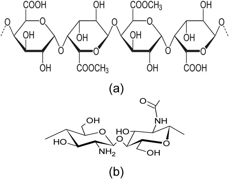
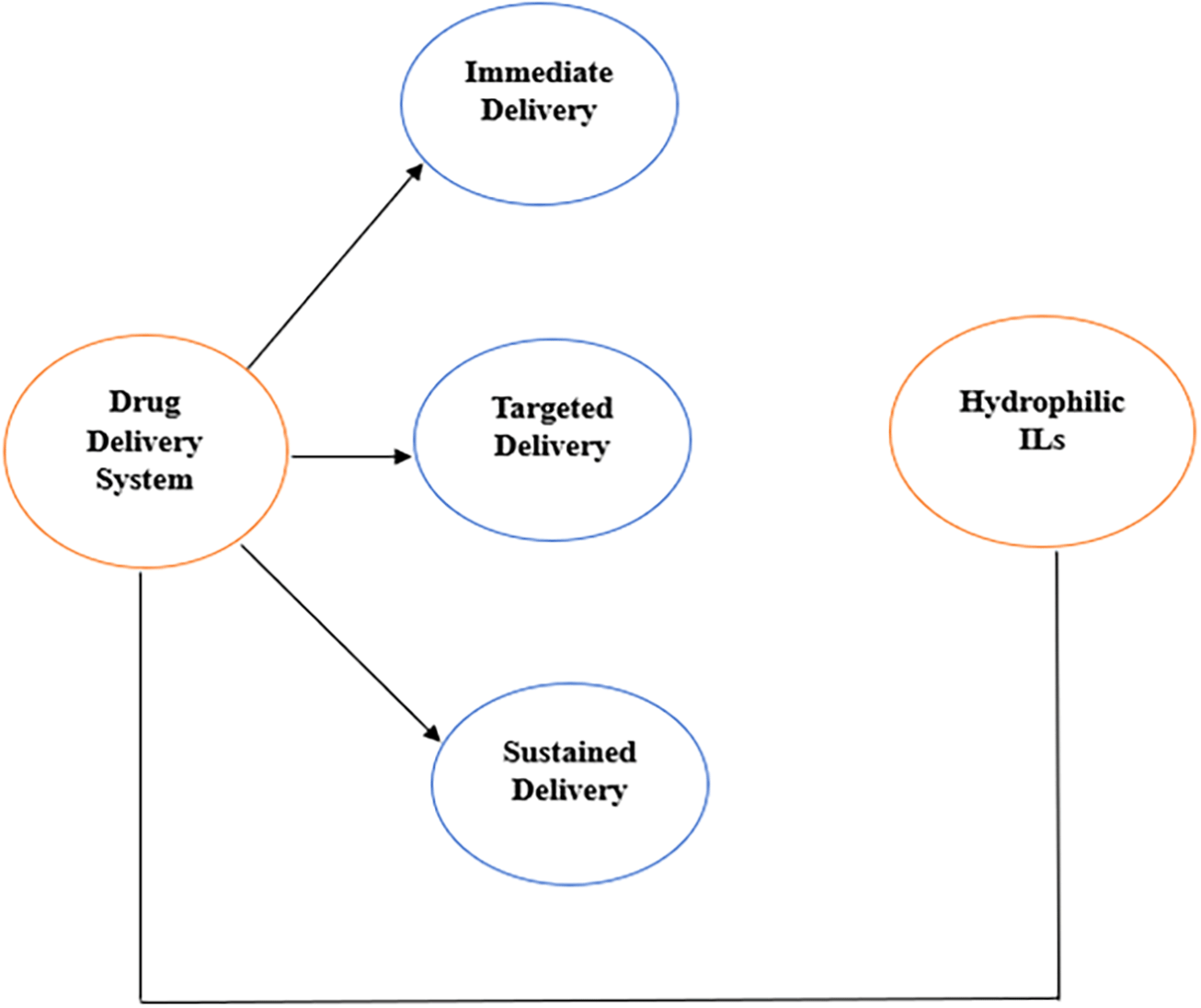
Chitosan was first discovered in 1859 by Rouget.12 Among natural biopolymers, it is the second most prevalent.13 It is estimated that 55–85% of chitin in crustacean cuticles originates from a layer of epidermal cells.14 Since chitin and cellulose share the same sugar unit and glycosidic bond type, they share several structural characteristics.15 Chitosan with a nitrogen content of approximately 6.9% is useful for chelation. Fibers, coatings, and beads made of chitosan can also be used instead of powders and solutions.16 Chitosans decompose prior to melting when heated.17 They possess strong bioadhesive properties that make them useful for adding bioadhesive properties to various drug delivery systems.18 The stability of chitosan can be challenged in highly acidic settings, which can be addressed through modifications, as it is most suitable for delivering drugs to the gastrointestinal system under neutral to acidic pH conditions.19 The inherent biodegradability of chitosan and its non-toxic degradation products make it an ideal material for biomedical applications, minimizing exposure to long-term toxic effects.20 Chitosan also possesses DNA-binding, biocompatibility, hemostatic, fungistatic, spermicidal, and anticholesteric properties and accelerates bone growth.21 Figure 1 (a & b) shows the chemical structure of pectin and chitosan. Figure 2 illustrates the overall drug delivery system, classified as immediate, targeted, and sustained delivery, which is significantly contributed by hydrophilic ILs.
In addition to increasing the delivery of specific drugs, pectin improves their absorption.3 Oral administration is widely used as the easiest and most convenient method of drug delivery. When a drug is orally administered, the dosage can be adjusted based on how quickly the digestive system absorbs the dose. Two factors that may affect oral medications administered via the gastrointestinal tract are a low stomach pH and poor stomach peristalsis. Furthermore, the enzymatic activity and digestive properties of gastric content, as well as the duration of drug residence in the stomach and intestine, are important factors.22 Different methods for encapsulating or incorporating drugs into tablets, capsules, or nanoparticles can protect them from degradation.23 Drugs can be encapsulated to mask their taste or modify their release.24 As shown in Tables 1 and 2, pectin is an effective material for the preparation of oral drug carriers.25 Over the past two decades, it has inspired academia and manufacturers to develop more effective drug delivery systems. In addition to being used as wound dressings, body fat reduction agents, and tissue engineering agents, they are also used in drug delivery systems (Table 1). Chitosan, the only biodegradable polymer with a positive charge, has distinctive properties and characteristics among the biodegradable polymers used by pharmaceutical companies. The primary amino group of chitosan is responsible for its cationic charge, which results in the ability to deliver drugs.26 Chitosan has also been shown to improve saturation, stable gelling, mucoadhesive, and efflux pump inhibitor properties. In addition, cationic interactions can be used to deliver drugs through nanoparticles based on DNA and RNA.27 Numerous studies have demonstrated that chitosan is an effective drug delivery system. The characteristics of chitosan differ depending on the salt form, degree of deacetylation, and molecular weight.27 Dornish et al. showed that high-grade and ultra-pure chitosan was safe for use in biological and physiological systems.28 Drug carriers can enhance drug bioavailability by providing mucoadhesive doses, extending release times, and maintaining drugs at absorption sites for longer durations. Chitosan has osteoinductive properties, allowing it to promote cell proliferation, adhesion, and differentiation in bone defects, in addition to biocompatibility and mechanical properties.29 Drug absorption across mucosal membranes can also be improved by chitosan depending on how it is delivered.
| Application site | Formulation of materials | In-vitro/in-vivo/Ex-vivo | Description of key features | Key findings | Drug | Date and reference |
|---|---|---|---|---|---|---|
| Colon | Eudragit: An entanglement of chitosan microcores and acrylic microspheres | In-vivo | The pH was a determining factor for Eudagrit. Following Eudagrit dissolution, the chitosan core swelled, leading to the dissolution and diffusion of drug. A molecular weight-dependent effect on drug release was found with chitosan. | Enhanced drug encapsulation, biocompatibility and stability | Sodium diclofenac | 30, 2009 |
| Crosslinked chitosan film containing citrate | In-vivo | The chitosan film was sensitive to pH. Chitosan swelling was accelerated, and drug release was faster in an acidic medium. Over the course of 24 h, 40% of the drug was released at neutral pH. | Development of self-aggregated nanoparticles | Riboflavin | 30, 2009 | |
| Chitosan succinate and chitosan phthalate | In-vivo | pH affected the dissolution of the combination, with acidic conditions causing a decrease in the release of the drug and alkaline conditions increasing dissolution. | Enhanced chitosans were utilized to encapsulate sodium diclofenac | Sodium diclofenac | 31, 1999 | |
| Chitosan capsule | In-vivo | The amount of drug directly absorbed by the cecum of rats is higher. Animals with induced colitis were treated with chitosan-based capsules which accelerated the healing process. | Chitosan was delivered to be effective in minimizing systemic absorption and enhancing concentration of local drug | 5-Aminosalicylic acid | 32, 2002 | |
| Liposomes coated with chitosan | In-vivo | In rats, coated liposomes increased alendronate oral bioavailability, and in Caco-2 cells, their absorption was also enhanced. | Liposomes coated with chitosan provided significant unique strategy for inflammatory diseases like ulcerative colitis | Alendronate | 33, 2012 | |
| Chitosan Capsules | In-vivo | The therapeutic effect was increased | Chitosan capsule was very useful dosage for the colon specific delivery of R68070 as an anti-inflammatory and ulcerative colitis drug | R68070 (thromboxane synthase inhibitor) | 34, 1999 | |
| N-(2-hydroxypropyl)-3-trimethylammonium chitosan chloride | In-vitro/Ex-vivo | It inhibited the reproduction of SARS-CoV-2 and MERS-CoV virus | HTCC provide to be more significant against both MERS-CoV ans SARS-CoV-2 | Antiviral | 35, 2021 | |
| Carboxymethyl chitosan nanoparticles cross-linked with Ca2+ ions | In-vitro | The nano film provided an extended-release pattern | This ultrasound assisted method proved to be efficient for forming carboxymethyl chitosan nanoparticles | Clindamycin HCl | 36, 2022 | |
| Crosslinked carboxymethyl chitosan nanoparticles | In-vivo | In addition to the high encapsulation efficiency (90%), its controlled release was also observed | This microfluidic procedure provides a significance for manufacturing CMCS NPs with prominent size, high dispersity | Metformin hydrochloride | 37, 2021 | |
| Chitosan capsules | In-vivo | It increased the concentration of prednisolone in the large intestinal mucosa | Encapsulation containing chitosan improves the prednisolone stability | Prednisolone | 38, 2007 | |
| Cholesterol-modified glycol chitosan | In-vivo | The compound showed prolonged circulation time in rat plasma as well as a stronger antitumor effect. | CMGC NPs Significantly synthesized. The nanoparticles possess uniform size and has favorable physicochemical composition | Doxorubicin | 39, 2009 | |
| Linoleic acid-modified glycol chitosan | In-vivo | Drug release from the nanoparticles was reduced. The nanoparticles demonstrated stronger anti-tumor efficacy in tumor bearing mice | Nanoparticles which are self-aggregated were significantly prepared using acid modified linoleic glycol chitosan | Paclitaxel | 40, 2015 | |
| Methoxy poly (ethylene glycol)-grafted-chitosan | In-vivo | In 48 h, more than 50% of the product was continuously released. | Estimation of in vitro release details of methotrexate using nanoparticles | Methotrexate | 41, 2008 | |
| N, O-carboxymethyl chitosan (NOCC) | In-vitro | NOCC nanoparticles loaded with insulin are released more slowly at higher pH levels of phosphate buffered saline | Possible enhancement in patients’ compliance and bioavailability of insulin | Insulin | 42, 2009 | |
| Folate-conjugated chitosan | In-vitro | The drug is condensed efficiently, has good tumor-targeting abilities, and has a low cytotoxicity level | Reagents and procedures utilized for the conjugation, confirming the functionalization of folic acid with chitosan | Paclitaxel | 43, 2008 | |
| pH-sensitive self-aggregated nanoparticles, amphiphilic deoxycholic acid modified carboxymethyl chitosan | In-vitro | Enhanced cellular uptake and greater retention | Chitosan provides a carrier for the hydrophobic drug DOX for effective cancer treatment against drug-resistant tumors | Doxorubicin | 44, 2012 | |
| Systemic | linoleic acid/poly (β-malic acid) double grafted chitosan | In-vivo | Nanoparticles were safe drug carriers, showed significantly potent tumor inhibition efficacy | Developed Unique chitosan parameter by using grafting linoleic acid | Paclitaxel | 45, 2009 |
| Oral | Mono- and bi-layered mucoadhesive films using CS | In-vitro | The model drug has increased tensile strength and antimicrobial activity | This study deployed films which are both monolayer and bilayer drug local loaded for treatment of the infections in the oral cavity | Cefuroxime | 46, 2019 |
| Buccal film based on Chitosan in mixture with polyvinylpyrrolidone (PVP) | In-vitro/In-vivo | The drug release at therapeutic concentrations for 6 h was continuous, but at a lower dosage than usual, because the film exhibited satisfactory mechanical and bio adhesive properties | The significant films show acceptable mechanical durability which is suitable for buccal applications in chronic periodontitis | Tenoxicam | 47, 2020 | |
| Blended CS film with PVA | Ex-vivo | The antimicrobial activity against Streptococcus mutans was effective, with a good kill time | This study expressed buccal films utilizing a set of CPC and chitosan in pediatric patients | Cetylpyridinium chloride | 48, 2020 | |
| Chitosan, sodium alginate, and ethyl cellulose | In-vivo | Presented a relative bioavailability of 246% | Film controllability and sustainability in buccal mucosal drug delivery | Zolmitriptan | 49, 2022 | |
| Chitosan, sodium alginate, and ethyl cellulose | In-vivo | Presented a relative bioavailability of 142% | Film controllability and sustainability in buccal mucosal drug delivery | Etodolac | 49, 2022 | |
| Ocular | An anionic chitosan hydrogel enhanced by disodium α-dglucose 1-phosphate | In-vivo | The drug penetrates deeply into the cornea when it is released rapidly. An exceptional level of ocular tolerance is achieved by this hydrogel. Residency in the ocular region for a prolonged period. | This is likely to proves that the Hydrogel based chitosan was significantly prepared | Levocetirizine dihydrochloride | 50, 2012 |
| The liposomes are coated with chitosan that has a low molecular weight | In-vivo | Using chitosan with a low molecular weight, the coating improved trans-corneal drug penetration. | They explore the estimation and development of coated liposomes with low molecular weight chitosan | Diclofenac sodium | 51, 2009 | |
| Nanocapsules of polycaprolactone coated with chitosan | In-vivo | Bioavailability of indomethacin in the eye was increased. | Nano capsules enhanced the bioavailability of indomethacin associated to uncoated nano capsules | Indomethacin | 52, 1997 | |
| Liver | Nanoparticles based on chitosan and poly ethylene glycol modified with glycyrrhetinic acid | In-vivo | A long-term storage of high medication concentrations is dependent on nanoparticles in the liver. Animals bearing H22 cells were not subject to tumor development when nanoparticles were applied. | These nano particles significantly delivered durgs to liver cell | Doxorubicin | 53, 2010 |
| PEG-grafted lactose conjugated chitosan micelles derived from methyl grafted chitosan | In-vivo | The drug molecules had a higher concentration of molecules and specifically targeted the liver in mice. | Characterization and development of polyion complex | Diammonium glycyrrhizinate | 54, 2009 | |
| Nanoparticles of galactosylated chitosan | In-vitro/In-vivo | The 5-Fluorouracil nanoparticles released at a sustained rate. Mouse orthotropic liver hepatic cancer cells are more effectively targeted with this drug. | It utilizes the use 5-Fu nanoparticles for the behavior of HCC | 5-Fluorouracil | 55, 2012 | |
| Conjugated chitosan nanoparticles containing glycyrrhizin (GL) | In-vivo | The loading efficiency of medication was 91.7%. Nanoparticles accumulate preferentially in hepatocytes, and cellular uptake depends on both time and dose. | Improved chitosan nanoparticles for battered drug procedure to hepatocytes | Adriamycin | 56, 2008 | |
| Kidney | Chitosan with a low molecular weight | In-vivo | There was a 13-fold increase in prednisolone dose in the kidney region when chitosan was loaded with prednisolone. | r-NAC50-LMWC provides enhanced solubility in physiological parameters | Prednisolone | 57, 2009 |
| Lung | Nanoparticles made of chitosan and poly (lactic coglycolic acid) | In-vitro | An enhanced absorption of nanoparticles by lung cancer cells. | Lung tumor-specific targeting of paclitaxel was achieved in mice by intravenous administration of chitosan-modified paclitaxel-loaded | Paclitaxel | 58, 2009 |
| Nanoparticles made of chitosan and poly (lactic coglycolic acid) | In-vivo | A negative charge on tumor cells causes nanoparticles to interact strongly with them under acidic conditions. | Enhanced electrostatic interaction between chitosan-modified NPs and acidic microenvironment of tumor cells | Paclitaxel | 59, 2009 |
| Application site | Formulation of materials | In-vitro/in-vivo/Ex-vivo | Description of key features | Drug | Date and reference |
|---|---|---|---|---|---|
| Colon | Coating of pectin at a ratio of 5:1 | In-vitro | The release of drug molecules at pH 6 is significant even in the absence of enzymes. | Indomethacin | 60, 1998 |
| The coating is made up of pectin and calcium chloride | In-vitro | The pharmacokinetics of compression-coated tablets were slower and sustained than those of core tablets. | Indomethacin | 61, 2015 | |
| Eudagrit S-100 | In-vivo | As a result of colonic enzymatic activity, drug release is delayed in the colon, and due to a high pH in the stomach, no drug release is observed. | Metronidazole | 62, 2009 | |
| Hydroxypropyl-methylcellulose Pectinate beads | In-vivo | Due to colonic pectinolytic enzymes, drug release in the colon is delayed. During the reticulation process, counterion concentration allows better control of release. | Ketoprofen | 63, 2013 | |
| Pectin with a high esterification level and a high molecular weight | In-vivo | Protection for mucosa provided by coating. Reduced production of gastric acid, reduction of pepsin secretion, and scavenging of radicals. Reduction in the reabsorption of bile acid. A reduction in cholesterol absorption and the production of volatile fatty acids. | Efficacy in lowering lipids and cholesterol | 64, 2020 | |
| Pectin with high methoxylation | In-vivo | There is a delay in drug release specific to the colon. No interference with the normal enzymatic activity of the upper gastrointestinal tract. | Ropivacaine hydrochloride monohydrate | 65, 2000 | |
| Pectin/anhydrous dibasic calcium phosphate (ADCP) matrix tablets | In-vitro | Water-soluble drugs are released into the gastrointestinal tract in a controlled manner. | Theophylline | 66, 2015 | |
| Eudragit S and Eudragit L doubly enteric-coated | In-vivo | Diminished intestinal irritation and profound drug liberation was attained | Bisacodyl | 67, 2017 | |
| Hydroxypropyl methylcellulose mixed with pectin | In-vitro/In-vivo | Sustained release inside the colon and reached the colon without disturbing in the upper gastrointestinal system | 5-fluorouracil | 68, 2020 | |
| Ocular | Nanoparticulate of thiolate pectin | Ex-vivo | Aqueous solution releases and permeates more drugs in goat corneas than normal aqueous solution. | Magnesium chloride | 69, 2012 |
| Liver | Pectin with a low esterification level and a high molecular weight | In-vivo | Harmful substances are eliminated from the body via the kidney and the intestine. The gut flora is in balance, which contributes to antioxidant properties. The harmful metal ions are removed from the system. Pectin bacterial fermentation produces short-chain fatty acids. | Hepatoprotective effects | 70, 2008 |
| Concentration of apple and orange pectin of 5% | In-vivo | Among all pectin-fed groups, hepatic cholesterol concentrations decreased significantly. The serum cholesterol level was significantly reduced only in groups that received apples. The levels of cholesterol in the liver decreased, while the levels of cholesterol in the serum and feces increased. | Cholesterol levels | 71, 1998 |
Pectin-chitosan composites have emerged as versatile materials in medicine.72 These composites possess different properties and offer various benefits in the fields of drug delivery and release, pH sensitivity, adhesion to mucosal surfaces, and enhanced stability.73 The use of pectin-chitosan composites for drug delivery offers many advantages, making them an attractive choice for drug delivery applications.74 These advantages include biocompatibility, bio-adhesiveness, and the ability to tailor drug release to ensure safe drug administration.75 In addition, their encapsulation properties are useful in protecting sensitive drugs.76 Pectin-chitosan composites for drug delivery can be formulated through various methods, providing flexibility to meet specific requirements. Common techniques include layer-by-layer assembly, gelation, cross-linking, solvent evaporation, spray drying, compression, and freeze-drying.73,77–79 The choice of method depends on the desired particle size and drug-release profile. The pectin-chitosan composite is a highly promising material for drug delivery, as it ensures biocompatibility with minimal adverse effects on living tissues. The composite shows strong bioadhesive capabilities for effective mucosal adhesion and can maintain stability within a suitable pH range.80,81 Its biodegradability and eco-friendliness reduce concerns about long-term toxicity and environmental impact, while its adaptability to a variety of drug types and ability to tailor its properties through formulation adjustments make it a versatile choice.82
The rate at which drugs are released from pectin-chitosan composites in drug delivery systems is influenced by multiple factors, necessitating the precise control of tailored drug release kinetics.83 Key factors affecting drug release include the composition of pectin and chitosan, degree of cross-linking, drug properties, composite formulation, pH, ionic strength, temperature, geometry, mechanical properties, swelling, erosion behavior, drug loading, release mechanisms, external stimuli, release mode, drug loading techniques, and degradation rate of pectin and chitosan.84 The use of pectin-chitosan composites for drug delivery is generally safe and biocompatible; however, potential side effects exist. Controlling drug release is essential for preventing overdosing or underdosing, and drug interactions should be considered.85 Systemic side effects depend on the drug and the route of administration.
Dental implants are in high demand owing to widespread tooth loss.86 In 1957, Per-Ingvar Brånemark discovered that titanium enhances bone healing properties and their regeneration.87 In the case of titanium implants, the rate of bone healing is like that following a fracture or injury to bone.88 The role of an implant and the function is to act as an osteoconductive substrate, allowing new tissue to grow around the defect.89 Blood clots, cell activation and responses to an implant wound are the three stages of wound healing.90 An implant or body surface treated with a new material elicits different biological responses. In order to improve osseointegration between implant surfaces and bone, microlevel characteristics can be applied to implant surfaces.91,92 As the implant contact surface and bone surface become more intimate, the implant acquires stronger anchorage.93 The rough texture of the surface encourages osteoblastic cell adhesion more quickly than the smooth texture.93,94 Implants with a rough surface provide a larger surface area and enhance the attachment of cells from the bone to implants. In addition to increasing attachment, biochemical interactions between the implant surface and the bone also increase. A variety of treatments and ceramic-like elements can be applied to implant surfaces to enhance osseointegration.95
According to Wennerberg, the surface roughness of implants can be classified into three levels: minimal, intermediate, and rough.96 Based on the texture, the implants were found to have concave or convex surfaces. Irregularities on the surface can be isotropic or anisotropic, giving the surface roughness a particular orientation.97 Surface roughness can be increased using various methods including blasting,98 chemical etching,99 plasma-sprayed surfaces, ion-sputtering coating,100 and anodized surfaces.100 Many materials can be used to coat the surfaces of dental implants depending on their function and intended use.101 There are two types of coating: inorganic layers (hydroxyapatite or calcium phosphate) and organic layers (growth factors).102 Among inorganic phases, ceramics dominate.103 Adding calcium phosphate to implant surfaces improves osteoconductive properties, accelerating the development of bone around the implants.104 The addition of a hydroxyapatite film to implants causes the release of fluoride ions, resulting in bone calcification.105 Hydroxyapatite also acts as a mechanical stabilizer as well as a chemical bonding agent between tissues and implant surfaces.106 As a result of adding organic phases or bioactive materials to implant surfaces, biochemical bonding capacity, hydrophilicity, and osseointegration can be enhanced, depending on the desired outcome, for example, increasing cell adhesion and growth factors or acting as local drug delivery systems.107,108
A new implant coating can enhance the properties of dental implants with the potential to improve attachment, mineralization, and protein deposition by creating a coating for osteoblasts that identify and interact with particular molecules.99 New methods of local antimicrobial prophylaxis are needed to ensure that dental prostheses are protected from infections. Implants can enhance biological reactions in situ and promote long-term bone healing. This would allow for faster recovery and a quicker return to normal activity for patients and doctors. The application of nanocoating with organic molecules can have a positive effect on osseointegration and cell behavior. In the vicinity of implants with polysaccharide nanocoating, Bone-to-implant surface contact, and bone mineral density improved.108 Biopolymers derived from marine and agricultural sources are readily available and replenishable, making them useful in the medical life sciences. Owing to their biocompatibility and biodegradability, these polymers offer ecological security and the ability to be modified by enzymes, giving them a unique set of properties.109 The anticoagulant properties of sulfated chitosan have also been demonstrated. A biopolymer membrane-covered wound showed better hemostatic properties and healed faster.110 Improvements in epithelialization and collagen deposition have been demonstrated using biopolymer-based membranes. In addition to inhibiting fibrin formation, chitosan-based dressings reduce scarring in wounds.111 Because of chitosan membranes, wounds are less likely to lose water, oxygen permeability is increased, fluid drainage is promoted, and foreign microorganism contamination is reduced.112 Chitosan and pectin are both capable of forming thin membranes that can be used to improve the delivery of medications in various ways.113 A major component of future implant technologies is the fibrin scaffold provided by biopolymers. Bone and implant surfaces can form covalent bonds with the scaffold, thereby mediating cell adhesion.107
The interaction between Fiekian diffusion and high-viscosity chitosan films has resulted in the constant release of drugs for prolonged periods of time.114 This has led to the development of antimicrobials for specific areas of periodontal therapy, addressing the disadvantages of systemic drug administration. The properties of pectin and chitosan derivatives make them ideal for local drug delivery systems because they enable easy delivery, long-term retention, and regulated medication release. These derivatives also allow for the acceptable penetration of medication into the oral cavity and sustained retention of medication. In addition to their antimicrobial properties, these compounds are highly effective and well tolerated in oral tissues. Pectin and chitosan derivatives have different chemical and physiological properties, including ionic charge, branching, various molecular weights, and enzyme modification properties, making them good candidates for biomaterial coatings.115 Chitosan and pectin with high levels of galactose and low levels of arabinose have been shown to reduce inflammation, which is vital for improving implant integration and reducing inflammation in patients with rheumatoid arthritis, periodontitis, and immunocompromised patients.116 It has been found that biopolymer derivatives inhibit a large number of gram-positive and gram-negative bacteria as well as fungi.117
Pectin and chitosan are effective and efficient because of their diverse modification capabilities. Altering the length of their side chains and removing acetyl groups from their hairy regions can change their polarity and wettability.118,119 Human tissue takes up covalently bonded pectin well in terms of immunological responses.120–122 Modifying the side chains of pectin or chitosan sugar molecules makes it easier to initiate bone cell attachment in a manner that is tailored to the human body. It would be beneficial to investigate the use of pectin or chitosan as implant nanocoating and to support their development with findings from previous studies of bone biology.
Mucoadhesive drug delivery systems using carbohydrate polymers like pectin and chitosan show potential for safer chemotherapy in colorectal cancer. Thiolation of pectin and polyelectrolyte complex formation with chitosan enhances mucoadhesion and stability. Theodore Ebenezer Leonard et al. (2023) developed a 5-fluorouracil-loaded drug delivery system combining thiolated pectin and chitosan through triple crosslinking, showing improved mucoadhesion and targeted cytotoxicity against colorectal cancer cells.123 These composites hold promise for targeted drug delivery in colorectal cancer treatment. Resveratrol, a natural polyphenol with health benefits, faces challenges due to poor solubility and low bioavailability. Sarma et al. (2022) developed core-shell nanoparticles using chitosan and pectin124 to improve their delivery. These nanoparticles showed sustained release of resveratrol for up to 30 hours, with pH-dependent efficiency. The encapsulated resveratrol exhibited enhanced antioxidant activity compared to free resveratrol. This study introduces a novel nano-based drug delivery system. A study by Nalini et al. (2022) focused on creating pectin/chitosan nanoparticle (PEC/CSNP) beads to deliver quercetin, a hydrophobic compound, using ionotropic gelation.125 The beads showed high encapsulation efficiency and sustained release of quercetin. They also exhibited improved antibacterial properties and enhanced antioxidant potency. The beads were found to be blood-compatible and showed good cell viability in vitro. Overall, the PEC/CSNP beads have potential as an effective drug delivery system, especially in low pH environments like the gastrointestinal tract. Polysaccharide-based nanoparticles like pectin/chitosan are ideal for oral drug delivery due to their solubility and biocompatibility. A study by Nidhi Mishra et al. (2023) used blending-crosslinking of pectin/chitosan polymers to create PEGylated nanoparticles for delivering phytic acid (IP6) to the colon to potentially manage colon cancer.126 Two sets of nanoparticles were prepared: one crosslinked with Glutaraldehyde (GE) and the other with sodium tripolyphosphate (TPP). The TPP- pectin/chitosan nanoparticles were smaller in size than GE - pectin/chitosan - nanoparticles. The nanoparticles showed controlled release of IP6 at different pH levels and were cytocompatible and effective in cellular uptake in cancer cell lines. The PEGylated-TPP - pectin/chitosan- nanoparticles demonstrated time-dependent uptake in cell lines. This method successfully delivered IP6 to the colon and could be applied to other drugs for colon delivery.
The efficacies of pectin and chitosan are supported by their diverse modification capabilities. It is possible to fabricate nanostructures and sizes made from these natural biopolymers for an array of innovative uses. Reviewing the application of pectin and chitosan, as well as their composites, for dental implant surface coatings is a promising endeavor because of the multifaceted benefits of the composites. These composites offer enhanced biocompatibility and can mitigate infection risks owing to their inherent antimicrobial properties. In addition, they have the potential to promote osseointegration by facilitating bone cell adhesion and growth. By enabling controlled drug delivery, these composites can release antimicrobial agents or growth factors, optimize tissue healing around implants, and minimize inflammation. The versatility of the formulation allows for tailored applications, whereas their natural sourcing contributes to eco-friendliness. This aim is to improve the safety, longevity, and success of dental implant procedures, making them an asset in dental implant technology. Studies and experimental tests should be conducted to determine the efficacy of pectin-chitosan composites as nanocoating for implants. Promising outcomes from in vitro and in vivo research may emerge in the near future. The use of pectin and chitosan in drug delivery systems has shown promising results for enhancing drug absorption and controlling drug release. These natural polysaccharides offer a wide range of applications, from wound dressings to tissue engineering agents, and hold potential for advanced biomedical implant applications. With their biocompatibility and biodegradability, pectin and chitosan composites present an exciting opportunity for the development of novel drug delivery systems and implant materials. Further research and development in this area could lead to significant advancements in the field of pharmaceutical and biomedical sciences.
| Views | Downloads | |
|---|---|---|
| F1000Research | - | - |
|
PubMed Central
Data from PMC are received and updated monthly.
|
- | - |
Provide sufficient details of any financial or non-financial competing interests to enable users to assess whether your comments might lead a reasonable person to question your impartiality. Consider the following examples, but note that this is not an exhaustive list:
Sign up for content alerts and receive a weekly or monthly email with all newly published articles
Already registered? Sign in
The email address should be the one you originally registered with F1000.
You registered with F1000 via Google, so we cannot reset your password.
To sign in, please click here.
If you still need help with your Google account password, please click here.
You registered with F1000 via Facebook, so we cannot reset your password.
To sign in, please click here.
If you still need help with your Facebook account password, please click here.
If your email address is registered with us, we will email you instructions to reset your password.
If you think you should have received this email but it has not arrived, please check your spam filters and/or contact for further assistance.
Comments on this article Comments (0)