Keywords
Torticollis, bilateral, infant, physiotherapy
Torticollis, bilateral, infant, physiotherapy
Added information about more details about how measurement was done. Measure of case I added. Information about head shape, plagiocephaly Case I.
Figure 3 and 4 added to one figure (3) to get them side by side.
Explaining that when using MFS scale infants in general are righting the trunk to some extent in addition to righting the head, especially with higher scores.
Added information about studies that report shorter treatment periods when an experienced physiotherapist performs the stretching three times a week instead of parents.
More about muscle and potential problems.
Five new references added.
See the author's detailed response to the review by Lucie Pelland
See the author's detailed response to the review by Nidhi Sharma and Parveen Kumar
See the author's detailed response to the review by Leo van Vlimmeren
See the author's detailed response to the review by Kimberly B. Castle
CMT is a common congenital musculoskeletal anomaly among infants. It results from shortening or excessive contraction of the Sterncleidoomastoid muscle (SCM). One to four weeks after birth a SMT may be visible, which consists of fibrous tissue and disappears within a few months (Cheng, Tang and Chen, 1999; Kurtycz, Logrono, Hoerl and Heatley, 2000; Tang et al., 2002; Wei, Schwartz, Weaver and Orvidas, 2001; Ohman and Beckung, 2008). The head is typically tilted towards the affected muscle, and the chin is rotated towards the other side. The true etiology of CMT has been discussed; one hypothesis is that the condition could be the sequel of an intrauterine or perinatal compartment syndrome (Davis, Wenger, Mubarak, 1993), several studies point in that direction (Bielski et al., 2006; Hardgrib, Rahbek, Möller-Madse and Maimburg, 2017; Lee et al., 2011a, 2011b; Pazonyi, Kun and Czeizel, 1982; Xiong et al., 2019). CMT can be divided into three groups:
SMT group with a clinically palpable sternomastoid tumor
MT group with tightness of the SCM muscle but no tumor
PT group with all the clinical features of torticollis, but without the tightness or tumor of the SCM muscle (Cheng et al., 2001).
For the SMT group, intrauterine or perinatal compartment syndrome is very likely to be the cause. Mandibular asymmetry is seen during examination, Fenton et al. found decreased ramal height ipsilateral to the affected SCM muscle (Fenton et al. 2018). It is possible that all three groups have the same cause, which is even more obvious in SMT and MT.
Infants with CMT have asymmetry in muscle function in the lateral flexors of the neck. The affected side is stronger than the other side (Öhman and Beckung, 2005; Ohman Mardbrink, Stensby and Beckung, 2011). Assessment of side-flexor muscle function in lateral head righting can be evaluated with the muscle function scale (MFS) (Ohman and Beckung, 2008; Kaplan, Coulter and Sargent, 2018). The MFS is found to be a reliable tool for testing infants for CMT (Ohman, Nilsson and Beckung, 2009; Seager; French and Meldrum, 2019). It gives information on asymmetry in head righting response. If the infant tilts the head without limited ROM with no difference in scores on the MFS, other causes than CMT must be considered. There are several differential diagnoses of CMT and some of them can be potentially life threating (Zvi and Thompson, 2022). If an infant has an SMT on one side and tilts to the other side with higher MFS scores on that side, bilateral torticollis must be considered.
ROM in the neck can be measured with an arthrodial protractor (Cheng et al., 2001; Ohman and Beckung, 2008; Kaplan, Coulter and Sargent, 2018).
Treatment for CMT includes manual stretching, strengthening cervical muscles through positioning, handling and exercises isolating the weaker muscle, incorporating righting reactions in different positions. Developmental exercises should be incorporated to promote symmetrical movement in weight-bearing in different positions, as well as environmental adaptions and parent/caregiver education (Kaplan, Coulter and Sargent, 2018). Kinesiology taping (KT) can be used as a complementary treatment, to give an immediate relaxing effect (Ohman, 2012; Ohman, 2015).
Bilateral CMT is rare and not often reported, Matuszewski, Pietrzyk, Kandzierski and Wilczynsk (2017) reported a case of a child with bilateral torticollis, referred to the orthopedic department at the age of 12 years. The first symptoms appeared at preschool age. This child had severe limitations in range of motion and required bilateral surgery (Matuszewski, Pietrzyk, Kandzierski and Wilczynsk, 2017). Babu et al., presented a case report of a 19-year-old girl with congenital bilateral sternocleidomastoid contracture (Babu, Lee, Mahadev and Lee, 2009).
A few articles describing case reports of bilateral torticollis have not been published in English (Chiari, 1952; Kustos and Magdics, 1993; Shi et al., 2016; Shinoda and Yamada, 1969).
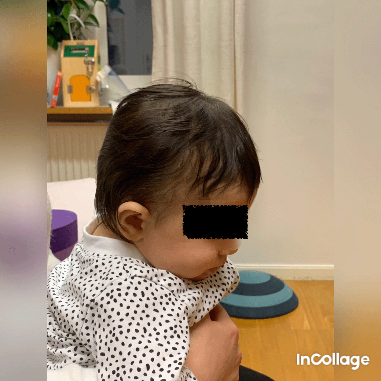
Case I is a Caucasian boy who came for a second opinion since the first examiner found it confusing, when the motion, muscle function/strength, and tilting of the head did not clearly fit left- or right-sided torticollis. On clinical examination in October 2020 when he was three and a half months of age, he had bilateral torticollis. On the right side, the patient had a SMT and some limitation in rotation towards the right side and in lateral flexion towards the left side, that is, the muscle on the right side was shortened. Case I had classification grade 3 according to Kaplan et al. CMT classification grades and decision tree for 0-12 months (Kaplan, Coulter and Sargent, 2018). When lying in the supine or prone position, he tilted his head to the right side and rotated to the left ( Figure 1), which fits well with right-sided torticollis, SMT. While sitting with support, he tilted his head to the left ( Figure 2) and was stronger in the lateral flexors on the left side ( Figure 3), which fits well with a postural left-sided torticollis (PT). Passive ROM in neck rotation was measured with an arthrodial protractor with the infant lying supine on the examination table with the shoulders stabilized. Case I had 95° towards the left side and 80° towards the right side. Lateral flexion was measured with the infant lying in supine position on a large protractor with the shoulders stabilized, 60° towards the left side and 70° towards the right side. Muscle function was evaluated using the MFS scale ( Figure 4) (Ohman and Beckung, 2008; Ohman, Nilsson and Beckung, 2009).
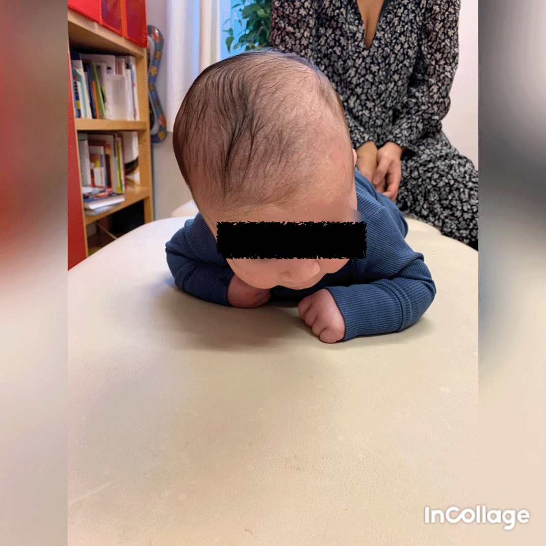
This is agreeable with right-sided torticollis. I confirm that I have obtained permission to use this image from the parents of the patient included in this presentation.

This is agreeable with left-sided torticollis. I confirm that I have obtained permission to use this image from the parents of the patient included in this presentation.
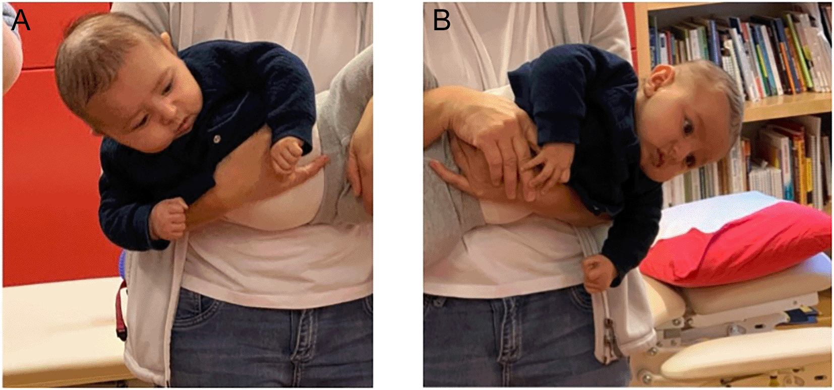
This is agreeable with left-sided torticollis Infants in general is righting the trunk to some extent in addition to righting the head, especially with higher scores. I confirm that I have obtained permission to use this image from the parents of the patient included in this presentation.
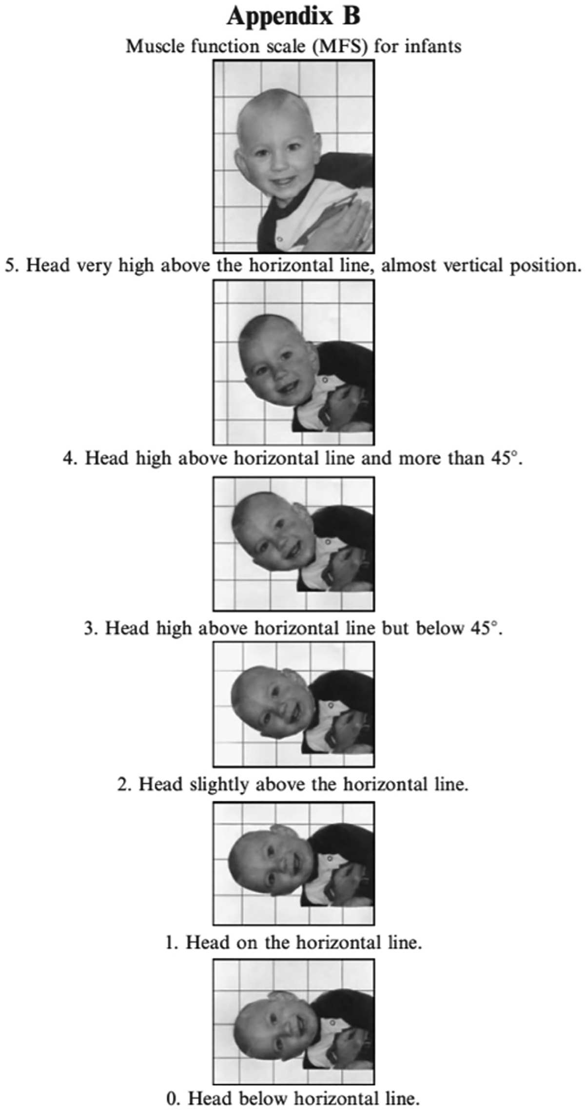
When the muscle function scale (MFS) is used, the infant is held in a vertical position and then lowered to the horizontal position in front of a mirror. The head position is observed and both sides are tested. Scores are given according to the head position in relation to the horizontal line. The infant must be observed with the head held in the same position for five seconds to obtain the score at that level. This figure has been reproduced/adapted from Ohman et al. (2009). Reprinted by permission of Informa UK Limited, trading as Taylor & Francis Group, www.tandfonline.com.
Case I had secondary features with mild plagiocefali on the left side and mild to moderate brachycefali.
Treatment involved stretching of the SCM muscle on the right side performed by the physiotherapist three times per week. Studies report shorter treatment periods when an experienced physiotherapist performs the stretching three times a week instead of parents performing daily stretching (Cheng et al., 1999, 2000, 2001; Ohman, Nilsson and Beckung, 2010). Home programmes include daily exercises done 3-5 times a day; active ROM in rotation, strengthening cervical muscles through positioning, handling and exercises isolating the weaker SCM muscle, incorporating righting reactions in different positions. The treatment at that time gave quick results, equal strength in the lateral flexors of the neck, motion in lateral flexion, and only a marginal difference in rotation. Plagiocephaly and brachycephaly were successfully treated with positioning.
At the follow-up at two years of age, he had relatively good active and passive ROM and no head tilt, but he had an indication of a discreet muscular string on the right side. At the next follow-up at two and a half years of age, he had a discreet tendency to tilt his head to the right and was slightly stronger on the right side. The muscular string is still rather discreet and felt only when stretching the muscle. He has started treatment again, and the muscular string may worsen as he grows in height. The SCM muscle grows from about 4 cm in infants to 14 cm at 13 years of age, according to measures made by Jones (1968). Case I must be followed-up for a longer time, as there is a risk that he will need surgery later in life.
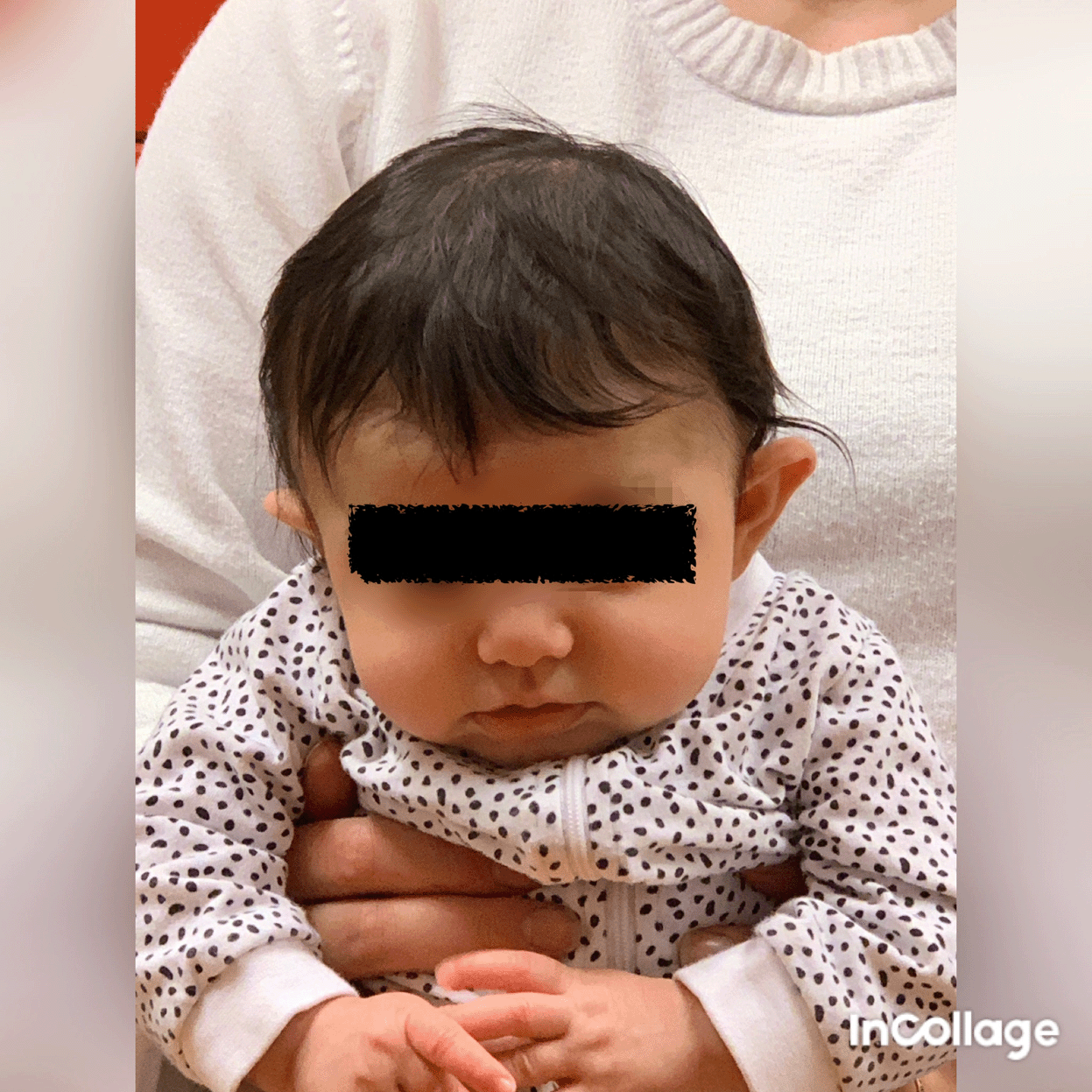
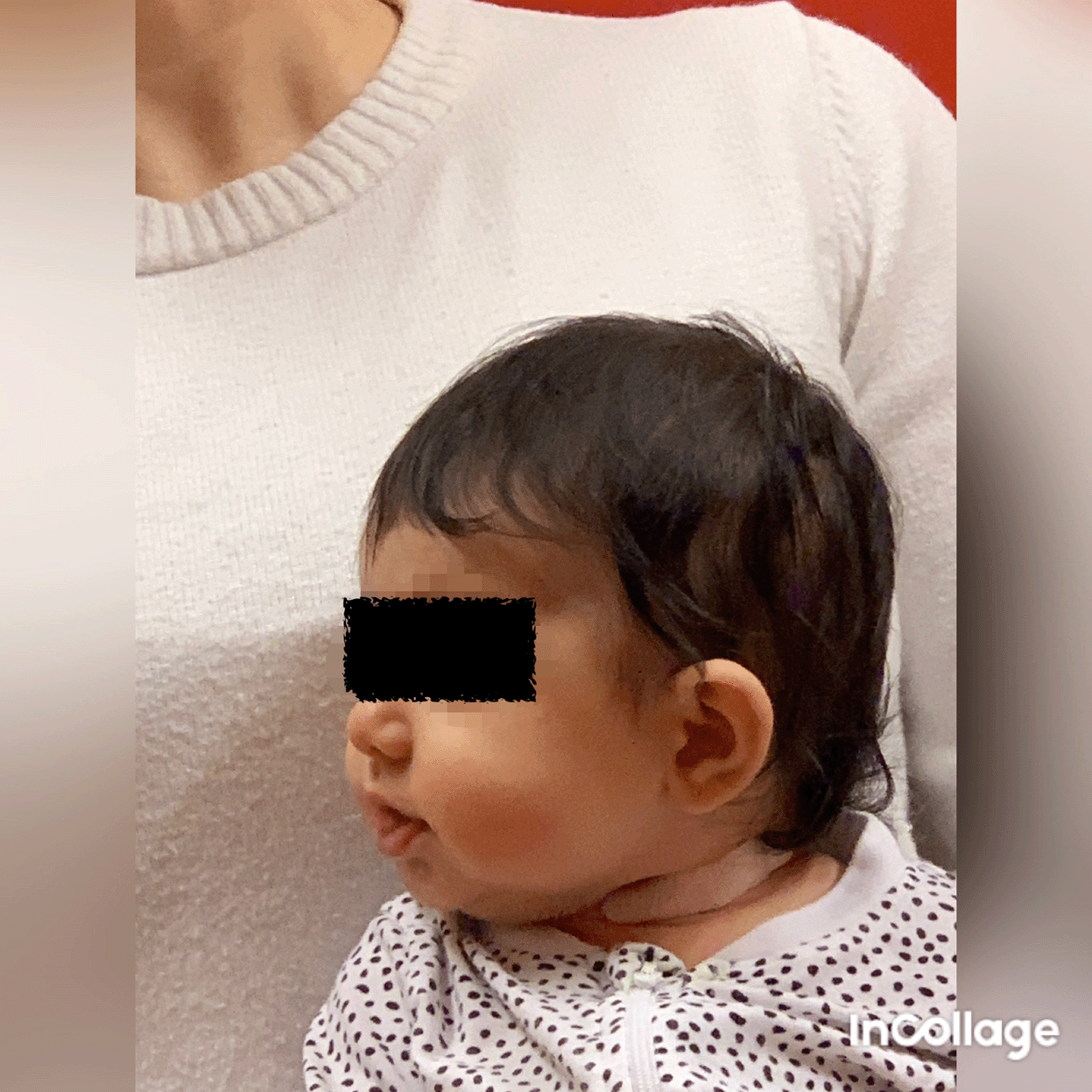
Case II is a Caucasian girl who was prenatally in breech presentation, and was turned by the healthcare provider some weeks before delivery. At the time of delivery, she was in a cephalic presentation and was fixed in the birth canal. In August 2020 at two and a half months of age, she came for clinical examination, referred for moderate brachycephaly. It was also obvious that the neck was affected, the SCM muscle was thickened bilaterally, and both active and passive rotations were affected. The head was held in flexion ( Figure 5), and active rotation was severely limited to both sides, less than 45° bilaterally ( Figure 6). Passive rotation was close to 90°, but with a clear stop, the mean normal range of rotation for infants is 110° (Ohman and Beckung, 2008). KT was used as a complement with a relaxing technique on both sides ( Figure 7), and tape was applied across the sternocleidomastoid muscle on both sides. The taping was done by the physiotherapist once a week or more often if tape came off earlier. Taping is not easy for parents without experience, infants have a short neck and a lot of skin, so taping is challenging for the inexperienced. The parents worked with the home programme several times every day with head control and exercises incorporated to promote symmetrical movement in different positions. When taped, it was easier for her to move her head with a greater range of motion. When she was about five months old, the problem was solved, and she had a good head position and active rotation of approximately 80° bilaterally and passive rotation >90° bilaterally. Her recovery was relatively rapid possibly due to intense training by the parents at home. However, after reading the case reported by Matuszewski et al. (2017) I decided to check her again.
I confirm that I have obtained permission to use this image from the parents of the patient included in this presentation.
I confirm that I have obtained permission to use this image from the parents of the patient included in this presentation.
I confirm that I have obtained permission to use this image from the parents of the patient included in this presentation.
At the age of nearly three years, she had an active rotation of 70° bilaterally and passive rotation of 90° bilaterally. Passive lateral flexion was full (i.e., ear to shoulder bilateral). Muscle function were assessed bilaterally according to the MFS scale she scored four bilaterally. The marginal flexion of the head might be within the normal span. I have decided to follow her once a year for at least some years to ensure that it stays within this span.
The combination of SMT and PT on different sides has to my knowledge, not been reported. Even though it is rare, more cases probably exist as they are easy to miss if very mild. It is important with rigorous examination and documentation to evaluate infants with torticollis, if it is bilateral CMT, the treatment must be adjusted. The MFS scale can be used in evaluating asymmetry in side-flexor muscle function in lateral head righting in PT. Healthy infants without CMT are not found to have any asymmetry on the MFS-scale (Ohman and Beckung, 2008). For example, in clinic infants with ocular torticollis show no difference on the MFS scale (author’s experience). Still photography is a reliable method for measuring habitual head deviation from midline in infants with CMT (Rahlin and Sarmiento, 2010). The examiner must follow progress attentively and when needed carefully adjust the treatment. When torticollis does not coincide with right or left CMT, it must be ensured that there is no other cause. There are several differential diagnoses, some of them more serious (Zvi and Thompson, 2022).
As the muscle grows by about 10 cm during the child’s skeletal growth, in the first 13 years, it may be important to follow up during childhood. This will ensure the early discovery of any skeletal growth problems. Dependent on how short the muscle is it will more or less limit the ability to rotate towards the affected side and make it hard to sustain a straight position of the head. Torticollis in the neck may cause secondary scolios in the back in older children and adults (Min et al., 2016; Kim, Yum and Shim, 2019; Choi et al., 2024).
In my experience, adults with neglected CMT have difficulty finding physicians or physiotherapists who understand that they have experienced CMT. Even with a clear muscular string and a typical head position, some adults do not receive a correct diagnosis and treatment. For a clinician with experience of children who need surgery for CMT, it is easy to recognize an adult with the same problem. However, bilateral torticollis is more confusing, and more knowledge of its long-term effects is needed.
KT was used as complementary treatment for Case II and worked well for her. With relaxing technique, KT has an immediate relaxing effect on the tense muscle (Ohman, 2015). However, the long-term effect of KT is uncertain and needs to be further investigated (Kaplan, Coulter and Sargent, 2018). Two studies comparing CMT treatment with and without KT came to opposite conclusions about the effect of KT (Giray et al., 2017; Hussein, Ali and El-Meniawy, 2019).
Bilateral sternocleidomastoid tumors have been reported, with or without torticollis (Dangi and Gwasikoti, 2020; Kumar, Prabhu, Chattopadhayay and Nagendhar, 2003; Tufano, Tom and Austin, 1999). However, not much information is available on long-term results. Neck problems are not uncommon among adults, and we should be aware that there could have been an earlier bilateral torticollis that contributed to this problem.
As bilateral CMT is rare and might be confusing, it needs to be further evaluated. Critical examination and documentation when evaluating CMT are important to give the right treatment when bilateral and also to make sure that a differential diagnosis is not missed by mistake. The need for longer follow-up for infants with bilateral CMT should be considered, to learn if there are any long-term problems.
| Views | Downloads | |
|---|---|---|
| F1000Research | - | - |
|
PubMed Central
Data from PMC are received and updated monthly.
|
- | - |
Provide sufficient details of any financial or non-financial competing interests to enable users to assess whether your comments might lead a reasonable person to question your impartiality. Consider the following examples, but note that this is not an exhaustive list:
Sign up for content alerts and receive a weekly or monthly email with all newly published articles
Already registered? Sign in
The email address should be the one you originally registered with F1000.
You registered with F1000 via Google, so we cannot reset your password.
To sign in, please click here.
If you still need help with your Google account password, please click here.
You registered with F1000 via Facebook, so we cannot reset your password.
To sign in, please click here.
If you still need help with your Facebook account password, please click here.
If your email address is registered with us, we will email you instructions to reset your password.
If you think you should have received this email but it has not arrived, please check your spam filters and/or contact for further assistance.
The parents did not perform passive stretching, they stimulated active rotation.
In clinic for at least the last 10-15 ... Continue reading The focus is mainly to describe how bilateral torticollis can manifest.
The parents did not perform passive stretching, they stimulated active rotation.
In clinic for at least the last 10-15 years I always perform the stretching instead of the parents and the treatment time to get acceptable ROM is about one month. In several bigger studies by Cheng et al the stretching treatment is also done by experienced physiotherapist and not by parents.
My study comparing stretching done by physiotherapist and parents showed that if the stretching is done three time each week the result is much better than when parents stretch each day several times a day (Öhman A, Nilsson S, Beckung E. Stretching treatment for infants with Congenital Muscular Torticollis physiotherapist or parents, the treatment dilemma. PM&R 2010;2(12):1073-1079). Also, earlier studies by Cheng et al. have described stretching performed by physiotherapist three times per week and not by the parents (Cheng JC, Wong MW, Tang SP, Chen TM, Shum SL, Wong EM. Clinical determinants of the outcome of manual stretching in the treatment of congenital muscular torticollis in infants. A prospective study of eight hundred and twenty-one cases J Bone Joint Surg Am. 2001 83(5):679-87.; Cheng JCY, Tang SP, Chen TMK, Wong MWN, Wong EMC The clinical presentation and outcome of treatment of congenital muscular torticollis in infants--a study of 1,086 cases Pediatr Surg 2000 Jul;35(7):1091-6).. Why we expect parents to perform the stretching treatment is another discussion that should be taken, it is likely that the parents find this more stressful than the physiotherapist observe. (Oledzka MM, Sweeney JK, Evans-Rogers DL, Coulter C, Kaplan SL.Experiences of Parents of Infants Diagnosed With Mild or Severe Grades of Congenital Muscular Torticollis Pediatr Phys Ther. 2021 Oct 1;33(4):245.) For adults that need stretching treatment in i.e. a shoulder, nobody says that “this can you partner at home do”.
Case II recovered quickly but I do not find it unexpected quick, the muscle was thick but no SMT. There was no pain.
About figure 3 and MFS scale, with higher scores the infant usually also lifts up the trunk, MFS-scores cannot be compared with measuring active lateral flexion, if the active lateral flexion was the goal with MFS scale, I agree that the body should be in line. In theory you can want the body to be completely horizontal but that is not the reality in practice. When interpreting the MFS scale it is most important to read the text to each score, it is head in relation to a horizontal line that is scored and not in relation to the body’s position. We have to be pragmatic about how it works in practice.
I have asked to get figure 3 and figure 4 side by side, but administrator says it is not possible, I do not understand why as I changed size to make them fit side by side.
Secondary abnormalities are asked for, I am aware of these, but the aim of this article is not to cover everything that can occur. The two cases described had no extreme abnormalities.
Infants that have a sternomastoid tumor are of greater risk for need of surgery later in life. As the muscle grows from 4 cm as infant to 14 cm at 13 years of age (Jones P. Torticollis in infancy and childhood. Springfield, Illinois: Charles C Thomas; 1968) some children get limited motion at an older age. What chose of surgery cannot be predicted in advance and that is also a chose by the surgeon, rarely by the physiotherapist.