Keywords
case report, salivary gland neoplasms, minor salivary gland, pleomorphic adenoma
case report, salivary gland neoplasms, minor salivary gland, pleomorphic adenoma
Salivary gland tumors are relatively uncommon, accounting for 1-3% of head and neck cancers. Pleomorphic adenoma (PA) is a benign tumor that is most frequently diagnosed not only in the major salivary glands, namely the parotid gland (85% of cases) and submandibular gland (5% of cases), but also in the minor salivary glands (10% of cases).1 The most common ectopic site of PA in the minor salivary glands is the palate, followed by the upper lip and oral mucosa.2
Salivary gland neoplasms are a heterogeneous group of tumors with variable clinical appearances and histological features.
PA, also known as “Mixed tumor, salivary gland type,” receives its name from its wide pleomorphic architectural appearance and mixed histology that consists mainly of three components: an epithelial and a myoepithelial component within a mesenchymal stroma.3 It is equipped with a fibrous capsule whose integrity must be conserved during surgical treatment to prevent recurrence.4
PA is more common in middle-aged females.
Clinically, it is known to be a slow-developing, asymptomatic lesion, typically described as firm, well-delimited, and variable in diameter. Intraoral PAs are normally located in the submucosa with a firm or rubbery consistency. The mucosal lining remains intact, but ulcerations can be observed in some cases.5
An 83-year-old man with no specific pathological history presented to the ENT Department at our hospital with a 2-year history of painless, slow-growing swelling on the right side of the upper lip.
Clinical examination revealed a well-circumscribed, mobile, firm, and non-tender submucosal mass measuring 3×1.5 cm on the right side of the upper lip. The overlying mucosa appeared intact and smooth without bleeding on palpation (Figure 1A). The lymph nodes of the head and neck were not enlarged.
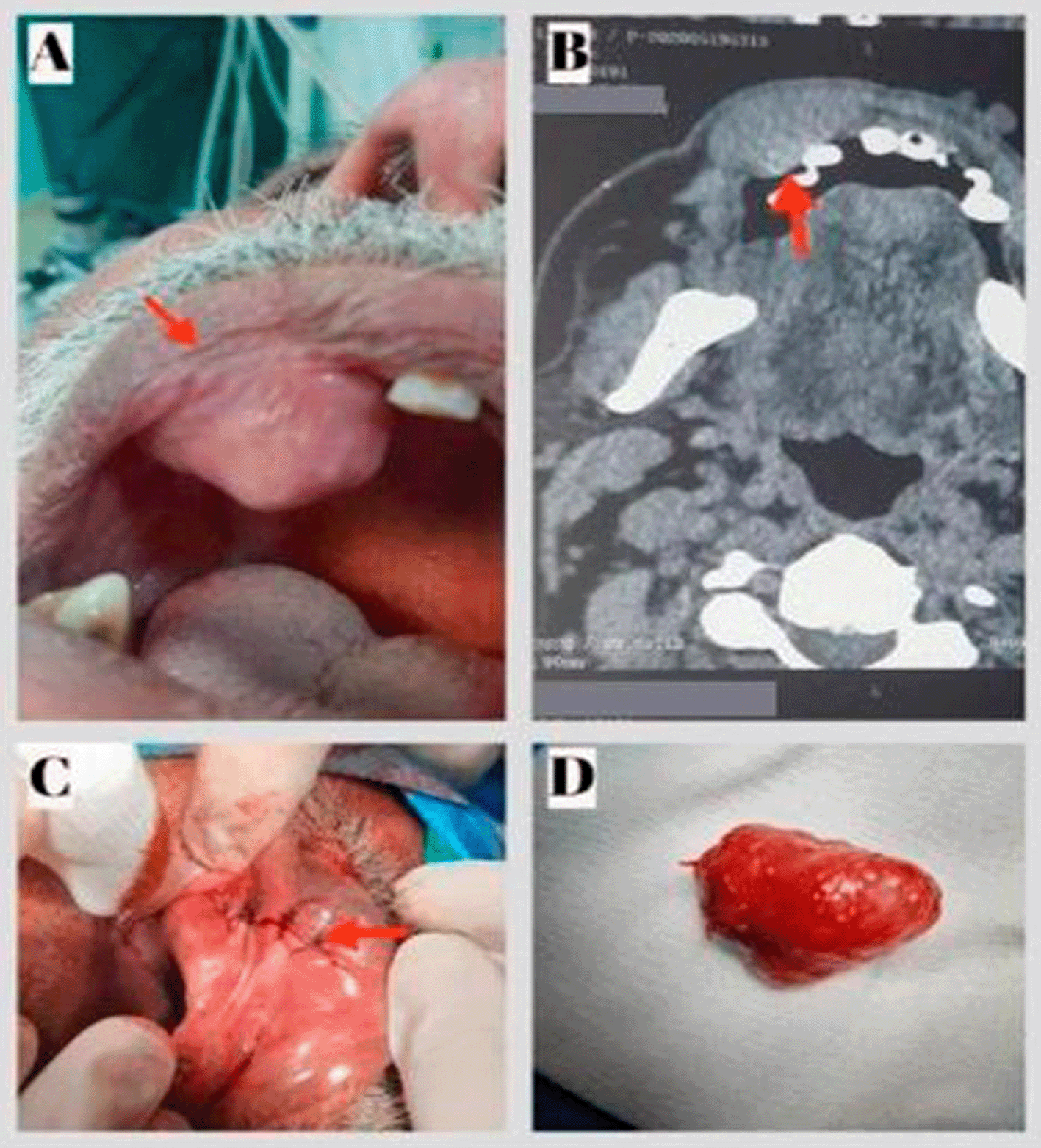
A: Preoperative view: Exposure of the mass in the palate. B: CT scan on axial section showing an oval well-defined lesion of the upper lip with homogeneous post-contrast enhancement. C: Postoperative view showing the site of the sutures. D: Excised specimen.
Computed tomography (CT) showed an oval well-defined lesion of the upper lip measuring 3.2×1.7 cm with homogeneous post-contrast enhancement (Figure 1B).
A total excision of the lesion via the sublabial approach was performed (Figure 1C). The lesion was released from the surrounding tissue, and the mass appeared to be fully encapsulated (Figure 1D). Histopathology of the resected tumor revealed the presence of a PA, a well-encapsulated soft tissue mass consisting of epithelial, myoepithelial, and stromal components (Figure 3). The follow-up 24 months after surgery showed no abnormalities and no evidence of recurrence.
A 45-year-old woman with no medical history presented to our ENT department with a slow-growing painless nodular lesion in the palate that caused difficulty swallowing. Anamnesis revealed that the mass had appeared one year previously and had rapidly increased in size over the last three months.
Intraoral examination revealed a unilocular, mobile, fibrous, endophytic nodule at the junction of the soft and hard palates, measuring approximately 4 cm in diameter. The nodule was well delimited, with a regular contour, smooth surface, and normal overlying mucosa color (Figure 2A). No lymph node involvement was observed during the physical examination.
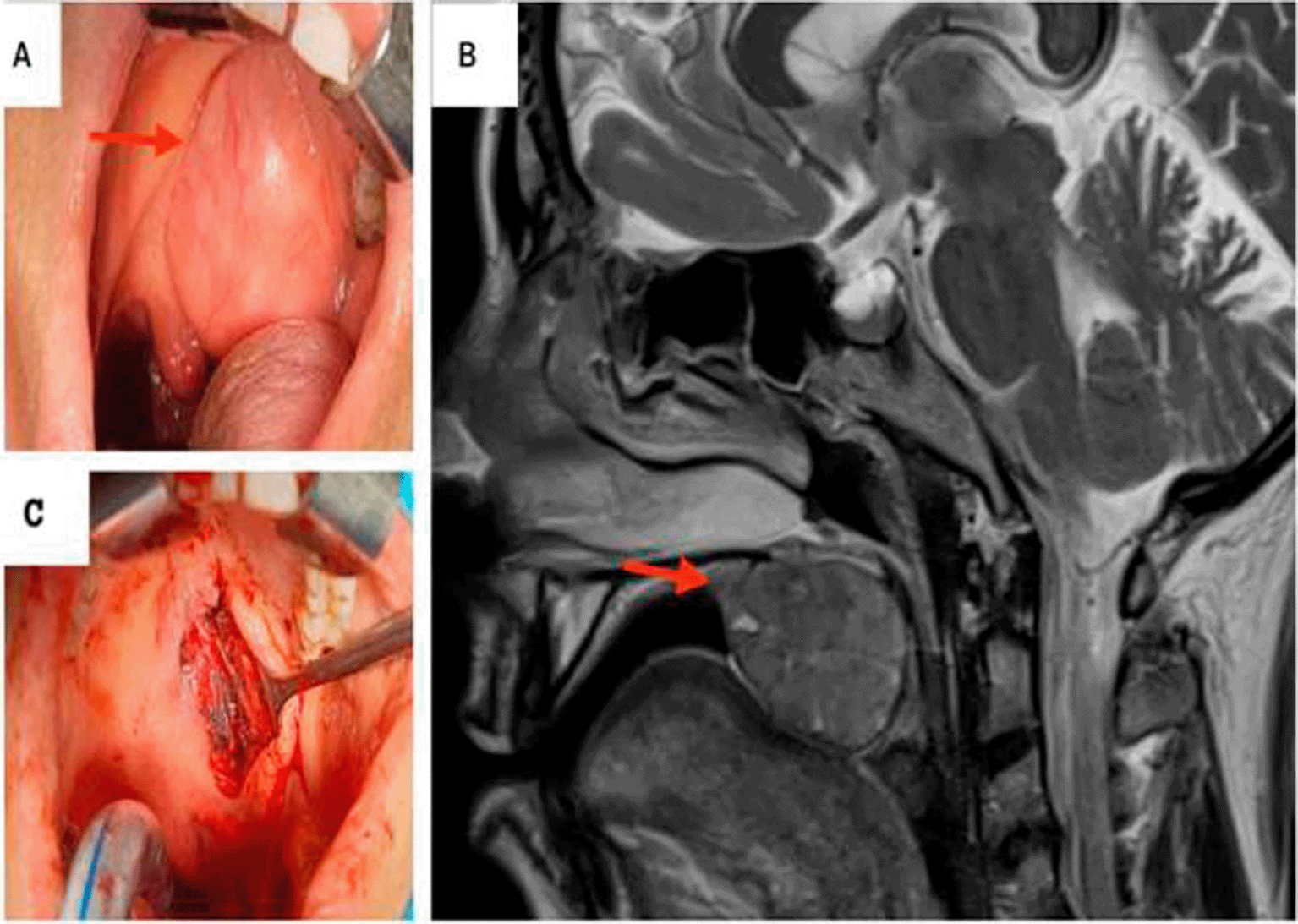
A: Preoperative view: Exposure of the mass in the palate. B: T2-weighted MRI showing a hyposignal ovoid well-defined mass within the right soft palate. C: Peroperative images: the defect post excision of the mass.
Magnetic resonance imaging (MRI) revealed an ovoid well-circumscribed encapsulated mass measuring 3.7 cm in size, within the midline of the soft palate to its right para-median side. The lesion extended backward to the oropharynx and forward in the left tonsillar pillar (Figure 2B).
The mass was completely excised with safety margins via an intraoral approach (Figure 2C).
On final histopathological examination, the report suggested a PA (Figure 3A & B).
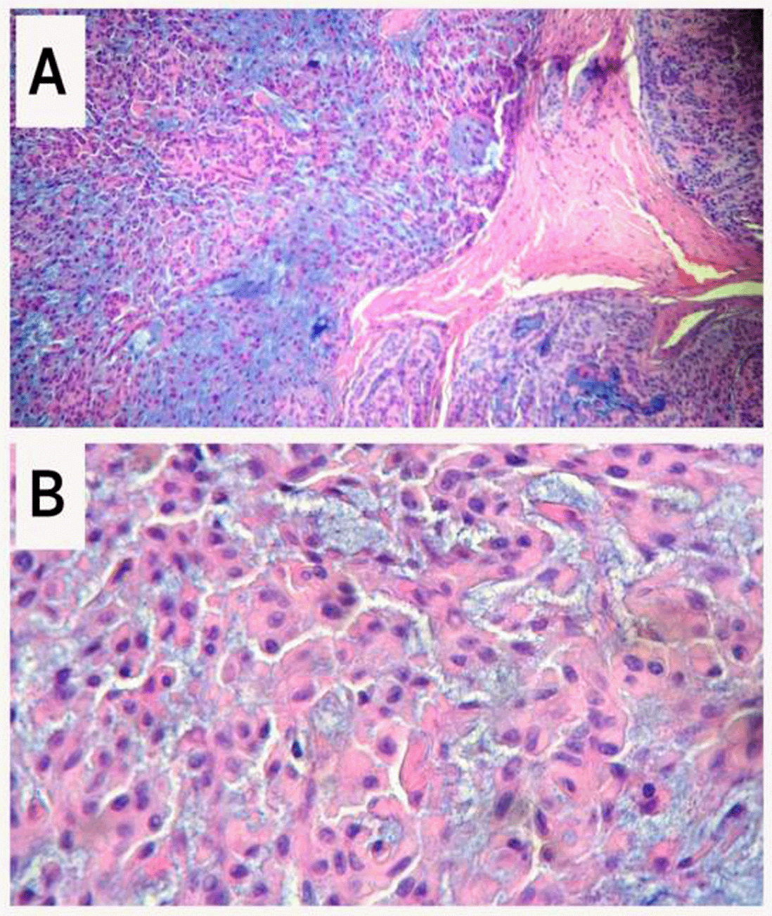
A: The tumor consists of three components: epithelial cells (right), myoepithelial cells (left), and myxochondroid tissue stroma (hematoxylin-eosin, 100×). B: Myoepithelial cells with epithelioid and plasmacytoid appearance (hematoxylin-eosin, 400×).
An 18-month follow-up after surgery showed no abnormalities or evidence of recurrence.
Although malignant lesions are mostly diagnosed in minor salivary glands, benign lesions have also been reported. PA are the most common benign salivary gland neoplasms, representing approximately 3–10% of all neoplasms of the head and neck region.6
They represent the most prevalent histopathological benign tumors diagnosed in both the major and minor salivary glands (50% of cases). Additionally, PA mainly affects the major salivary glands, particularly the parotids. The palate is the most prevalent intraoral site (42.8-68.8%), followed by the upper lip (10.1%) and cheek (5.5%).4,6
It occurs in individuals of all ages but tends to affect more women than men, especially middle-aged individuals.
The present cases of PA in the minor salivary glands of the palate and the upper lip corroborate literature data showing a relatively low prevalence at these sites.
Regarding the epidemiological features of PA, gender, and age in our present cases, one of our two patients agreed with the literature, which shows that PA in the minor salivary glands may occur in individuals of all ages but most frequently affects women in their fourth to fifth decade of life, with a relevant mean age of 40-60 years.
However, in our first case, PA was diagnosed in an 83-year-old man, which does not match the relevant literature as neither the age of occurrence nor the sex of the patient was uncommon.
The clinicopathological features of our two cases concur with those reported in previous studies. In fact, PA generally presents as a mobile, slowly developing, painless, and firm swelling that does not cause any fixation or ulceration of the overlying mucosa with no lymph node involvement.7
Histopathologically, PA is a complex mixed lesion consisting of both epithelial and myoepithelial components within a mucopolysaccharide stroma, organized predominantly in a duct-like pattern. They tend to have a fibrous capsule that separates the tumor from the surrounding tissues. The proportions of the different components can vary among individuals, parallel to changes in tumor consistency.6,8,9
The need for complementary investigations into the management of PA in minor salivary glands depends on its localization. Indeed, a biopsy may be performed for additional oral sites.
However, if the diagnosis of PA is suspected in intraoral localizations, CT, ultrasonography, and optimal MRI are useful for studying the extent of the tumor and determining eventual bone involvement.3,10
Biopsy is generally avoided because of the fear of the seedling. However, fine-needle aspiration is safe and recommended.11
According to recent studies, the treatment of choice is wide local excision of the tumor with adequate margins, followed by histopathological examination to establish the final diagnosis.8
Our therapeutic approach was consistent with the literature, as a total excision of the lesion via a sublabial approach was performed in the case of PA of the upper lip and via an intraoral approach for the patient who presented with PA of the palate.
Regular follow-up is necassary for patients with minor salivary gland neoplasms, not only due to their heightened tendency toward local recurrence but also because of their malignant potential.12,13
The diverse presentations of pleomorphic adenomas make diagnosis complicated and challenging. PAs of the minor salivary glands are rare neoplasms. While their occurrence in the minor salivary glands is uncommon, obtaining the correct diagnosis as early as possible is essential because early initiation of appropriate treatment allows for effective management and improves patient prognosis. Complete wide local surgical excision is the treatment of choice. Patients should be followed up for a longer period to detect late recurrences.
| Views | Downloads | |
|---|---|---|
| F1000Research | - | - |
|
PubMed Central
Data from PMC are received and updated monthly.
|
- | - |
Provide sufficient details of any financial or non-financial competing interests to enable users to assess whether your comments might lead a reasonable person to question your impartiality. Consider the following examples, but note that this is not an exhaustive list:
Sign up for content alerts and receive a weekly or monthly email with all newly published articles
Already registered? Sign in
The email address should be the one you originally registered with F1000.
You registered with F1000 via Google, so we cannot reset your password.
To sign in, please click here.
If you still need help with your Google account password, please click here.
You registered with F1000 via Facebook, so we cannot reset your password.
To sign in, please click here.
If you still need help with your Facebook account password, please click here.
If your email address is registered with us, we will email you instructions to reset your password.
If you think you should have received this email but it has not arrived, please check your spam filters and/or contact for further assistance.
Comments on this article Comments (0)