Keywords
Low dose, Computed tomography, Deep learning image reconstruction, Iterative reconstruction technique, Image quality
This article is included in the Manipal Academy of Higher Education gateway.
This article is included in the Artificial Intelligence and Machine Learning gateway.
Low dose, Computed tomography, Deep learning image reconstruction, Iterative reconstruction technique, Image quality
Computed tomography (CT) plays an important role in modern diagnostic radiology and assists in the identification of various complex disorders. Over the past ten years, CT scan utilization has increased significantly globally as new clinical reasons are continually identified. An estimated 375 million CT examinations are continually performed annually worldwide, with a 3-4% annual growth rate. The demands of physicians and other health care providers, as well as technology developments, have had a considerable impact on the world market for CTs. Compared to other traditional imaging modalities, CT scans offer significantly higher radiation doses (RD) despite having significant diagnostic benefits for specific patients. Adult CT scans dramatically raise cancer risk. A positive correlation between RD and cancer risks was found.1–5
A recent study reported seventeen-fold variations in high-dose CT examinations among different countries. There is a four-fold variation in effective dose [ED] for Chest and abdomen examinations with less variation for CT head in adults and suggested optimization of radiation doses.6 The most recommended practice in the CT sector is to reduce CT radiation exposure as low as reasonably achievable while maintaining the Image Quality (IQ). Reducing the exposure factors of tube voltage (kVp) and tube current (mA) reduces RD but increases image noise.7,8 Up until ten years ago, Filtered back projection (FBP) was the only technique used for image reconstruction in CT. Although this method produces high-quality images it has noise issues at low doses and is prone to artifacts. Although an iterative reconstruction (IR) method was proposed in 1970, computational power restrictions prevented its widespread use in clinical settings. The Hybrid Iterative reconstruction (HIR) method was introduced in 2009 which had low computation time and allowed it to be implemented in clinical practice. The HIR combines iteratively reconstructed images in the raw data domain with FBP images to reduce image noise (IN). The first complete model-based iterative reconstruction (MBIR) received FDA approval in 2011. Compared to the HIR technique, this reconstruction minimizes artifacts and noise. However, it requires a greater computational power demand, which results in lengthy reconstruction times. IR techniques, irrespective of type produce lower IN, artifacts, or both at lower doses than FBP.9–15 However, in general, IR at higher levels of reconstruction may result in an artificial, plastic-looking, and blotchy appearance that would eventually lower the IQ and compromise the clinician’s ability to diagnose pathologies, limiting the potential for significant RD reduction.16–18
Deep learning image reconstruction algorithms (DLIR) are the most recent developments in CT image reconstruction technology. DLIRs are increasingly replacing IR techniques due to their disadvantages such as negative image texture and nonlinear spatial resolutions. DLIR is based on deep convolution neural networks (CNN) which learn from the input data sets. It gains the ability to distinguish actual signal and IN through training using pairs of low and high-quality images. In comparison to FBP and IR, the trained CNNs can distinguish between noise and signal much better, allowing for better dose reduction while preserving the image quality. DLIR produces an image texture similar to that of FBP even at low doses and high strengths. DLIR technique may be able to detect the low contrast lesions at low doses without damaging the image texture.19–22 More research is required to determine the potential applications of DLIR in clinical settings. Our literature search showed there is no systematic review performed in head and chest CT examinations using Deep learning reconstruction algorithm for reducing RD and improving IQ in CT. Hence, the purpose of the article is to review the influence of DLIR on RD, IN, and outcomes of the studies compared with IR and FBP in CT Head and Chest examinations.
This review was carried out as per the “Preferred Reporting Items for Systematic Reviews and Meta-analysis (PRISMA)” guidelines.23
A comprehensive literature search was performed using databases such as “PubMed, Scopus, Web of Science, Cochrane Library, and Embase” to find the relevant original studies (Table 1). The MeSH terms such as “Deep Learning Image Reconstruction” “Radiation dose” “Image quality”, “Head and Chest Computed Tomography” were used (Table 2). The search was limited to the English language including both adult and paediatric populations of Head and Chest CT examinations.
| Database | Number of studies retrieved | Total |
|---|---|---|
| PubMed | 32 | 196 |
| Scopus | 52 | |
| Web of Science | 58 | |
| Cochrane Library | 8 | |
| Embase | 46 |
Articles were screened considering the Participant’s Intervention Comparison and Outcome (PICO) methodology. Case studies, case reports, conference abstracts, letters, editorial reviews, meta-analyses, or surveys were not included. The title and abstract of all the articles were independently and blindly screened by the two researchers. The articles that described a comparison of DLIR algorithms with the IR technique or FBP were included in the final review. The exclusion criteria were phantom studies, physics-based performance of DLIR, other language than English, articles with no comparison of DLIR with FBP, HIR/MBIR, and articles with no Hounsfield Unit (HU), Contrast to Noise Ratio (CNR), Signal to Noise Ratio (SNR).
Data from each article was assessed independently by two researchers and any differences were solved by the third researcher.
To evaluate the quality of all the included articles, the custom-made Quality Assessment (QA) scale was used.24 The list of all the questions for the quality assessment (underlying data). A score of 1 was given if the answer to the question was “yes” and each study was assigned a score ranging from 0 to 18. Based on the total scores obtained by each study, the studies were classified into three quality levels: Low- quality studies (score of 6 or lower), Moderate-quality studies (score between 7 and 11), High-quality studies (score of 12 or more).
Finally, 15 articles were included (Figure 1).
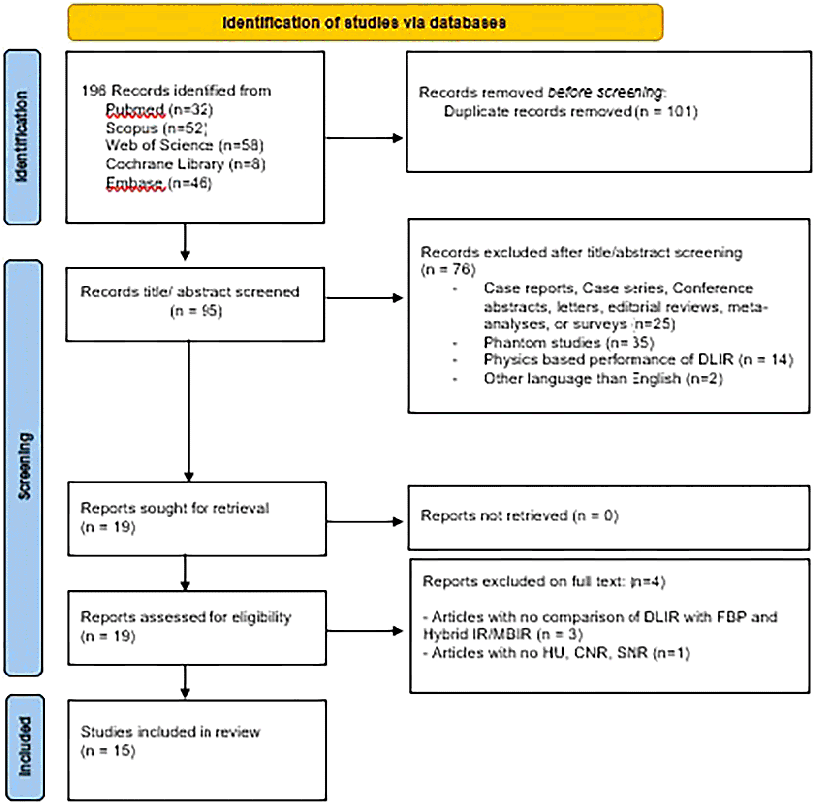
The search in PubMed, Scopus, Web of Science, Cochrane Library and Embase resulted in 196 studies. 101 duplicates were removed. The title and abstract of 95 studies were assessed and 76 studies were excluded as they did not meet the inclusion criteria. A total of 19 reports were sought for retrieval. A total of the full text of 19 articles were assessed for eligibility. Among them, 4 articles were excluded (3 studies were excluded due to no comparison of DLIR with FBP and HIR/MBIR, and 1 article were excluded due to lack of HU, CNR, and SNR). Finally, 15 articles were included in the systematic review.
CT imaging has increased recently with the advancement in CT technology. The studies included in the review covered different countries such as China (n = 8), Japan (n = 1), France (n = 1), Korea (n = 3), Netherland (n =1), Sweden (n = 1). The RD data and IQ parameters were collected from different CT vendors such as General Electric (GE) Health care (128, 256, 512-slice, and dual-energy CT), Siemens Healthineers (256-slice), Canon Medical system (320 and 640-slice). A total sample size of 1292 was collected from the included studies. 4 studies used prospective data collection, and 11 studies used retrospective data collection. The characteristics of the study and the outcomes of each study are summarized in Table 3.
| Author; Year and Country | Method | CT exam | Adult/Paediatric | Reconstruction techniques | Slice CT/Vendor | Sample | QLA | QUA & Region & Lesion detection | Outcome of the study |
|---|---|---|---|---|---|---|---|---|---|
| CT Head (Adult) | |||||||||
| Alagic et al., 2022 Sweden25 | RS | Non-contract CT Brain | Adult | AS-V (50%), DLIR-(L-H) | 512 Slice/GE | 94 | IN, brain structures, posterior fossa artifacts | CT, SD of GM, WM, ICH, SNR in the GM, WM and ICH Intracranial hemorrhage conspicuity | With substantially less non-diagnostic images, greater SNR (82.9%), and higher CNR (53.3%) compared to AS-V (50%), IQ of CT Brain with DLIR-M&H revealed dramatically increased IQ. |
| Kim I. et al., 2021 Korea26 | RS | Non contrast Brain | Adult | AS-V (30%), DLIR (L-H) | 512- slice/GE | 62 | Artefacts, IQ, IN | CT HU of GM, Image Noise, Artifact index, CNR – Basal ganglia and Centrum semiovale. SNR & SD, Reconstruction time | DLIR(M&H) exhibited reduction in IN and artifacts in Posterior fossa compared with AS-V. |
| Nagayama et al., 2023 Japan27 | RS | Non-Contrast CT Brain | Adult | LD-HIR, MBIR, DLIR | 320-Slice/CMS | 114 | Noise magnitude, IT, GM-WM differentiation, Artifact, IS, OIQ, | GM-WM differentiation, CNR, HU, SD GM, WM Lesion Conspicuity | DLIR can enhance the IQ of the CT Brain while reducing low RD and reconstruction time. |
| Oostveen et al., 2021 Netherland28 | RS | Non-Contrast CT Brain | Adult | DLIR, MBIR, HIR | 320-slice/CMS | 50 | IN, IS, natural appearance, GWM diff., artefacts, OIQ | SNR & SD of CSF, Reconstruction time | Comparing DILR to MBIR and HIR leads in reduced IN and better tissue differentiation with a little increase in reconstruction time. |
| CT Head (Pediatric) | |||||||||
| Sun J et al., 2021China29 | RS | Non contrast CT Brain | Pediatric | FBP, AS-V 50% and 100% DLIR (High) | 256-slice/GE | 50 | Clarity of cistern boundaries, GWM differentiation, Image quality | GM HU and SD, WM HU and SD, GM SNR, WM SNR,CNR | In comparison to AS-V and FBP, DLIR-H demonstrated reduced IN and increased IQ. Lesion identification is improved in 0.625 mm DLIR-H images, which also have similar image noise levels to 5 mm AS-V (50%) images. |
| CT Chest (Adult) | |||||||||
| Ferri et al., 2022 France30 | RS | Non-contrast chest CT | Adult | FBP, AS-V 70% and DLIR (L-H) | 256-Slice/GE | 54 | OIQ,IN, artefacts | HU, SD, SNR (Air, trachea, muscle). Emphysema volume | SNR was significantly raised with DLIR. Emphysema volume decrease with increase strengths of DLIR. |
| Jiang B et al., 2022 China31 | PS | Non contrast and contrast Chest | Adult | AS-V (40%, 80% and DLIR- M, H. | 256-slice/GE | 203 | Lung tissue noise, air background noise Nodule detection rate Malignant-related features | Measurement accuracy of nodule detection. | DLIR showed better enhancement of nodule detection rate and reduced image noise compared with AS-V images. For DLIR-H, 81.5% malignancy-related characteristics detections were noted. |
| Jiang J. M. et al., 2022 China32 | RS | Non contrast Chest CT | Adult | AS-V (50%) and DLIR (L-H) | 256-slice/GE | 50 | Soft tissue and lung tissue | SD, HU of (Aorta, Lung, Muscle, Liver, Vertebrae), CNR, SNR. | At the same dosage, DLIR can deliver a greater IQ, boosting the doctors DC and raising the diagnostic accuracy of LDCT. |
| Jo et al., 2023 korea33 | RS | Non contrast Chest CT | Adult | ADMIRE and DLIR (L-H) | Dual Source/Somatom Force | 100 | SN, Spatial resolution, Distortion artifact, Beam hardening artifact, OIQ | Noise, SNR, Edge rise distance Nodule detectability assessment | When compared to normal LDCT images reconstructed using IR, Quarter LD images reconstructed with DLIR demonstrated noninferior nodule detectability and IQ. |
| Kim J. H. et al., 2021 Korea34 | RS | Non-Contrast Chest CT | Adult | AS-V 30% and (DLIR M & H) | 512 Slice/GE | 58 | IC, IN & conspicuity of structures | HU, SD, SNR and CNR (Lung, Mediastinum, Liver Air) | Compared with AS-V 30% the DLIR images exhibited IN reduction in LDCT images by maintaining IQ. |
| Kim C. H. et al., 2022 China35 | RS | Non contrast chest CT | Adult | FBP, ASiR-V 30%, DLIR | 512 Slice/GE | 193 | Image contrast, OIQ | HU, SD, SNR and BRISQUE Score of lower lobes Diagnostic characterization of usual interstitial pneumonia (UIP) | DLIR images produced highest OIQ score. Comparing DLIR to AS-V and FBP, IN, SNR, and visual rating of chest LD-CT scan images all improved. For purpose of diagnosing UIP DLIR may be useful. |
| Tian et al., 2022 China36 | RS | Non contrast Chest CT | Adult | AS-V 40%, DLIR (L-H) | 256-slice/GE | 86 | IN, Image artifacts, and lesions. | HU, SD, (Lung Muscle, Fat, Aorta) | Compared with AS-V 40%, DLIR effectively reduced IN and improved IQ in LD chest CT. |
| Wang et al., H 2022 China37 | PS | Non contrast chest CT | Adult | AS- V 40%, DLIR M and H | 256-slice/GE | 48 | Nodules, Lung tissue, Artifacts and diagnostic confidence | Objective Noise, CNR, Image Signal (Aorta) | With DLIR, LD chest CT scans minimise IN, while DLIR-H provides images with a similar level of quality to SDCT AS (40%) while using just 4% of the RD. |
| Wang J et al., 2023 China38 | PS | Non contrast Chest CT | Adult | HIR, DLIR | 640-slice/CMS | 60 | OIQ | SD, HU, and SNR (Aorta, Paraspinal muscle,fat) Assessment of Pulmonary lesion Conspicuity | In comparison to HIR, LDCT-DLIR offers an excellent IQ with the exception of sub-solid nodules and reduced lung attenuation. |
| Zhao et al., 2022 China39 | PS | HRCT and Low dose Chest | Adult | HIR (AIDR3D, LDCT with DLIR | 320-slice/CMS | 70 | OIQ, SIN, artifacts | SNR Lung in ILD | Low dose CT DLIR showed better recognition of ground glass opacity and visualization of architectural distortion than HRCT-AIDR |
The results of the quality assessment are summarized in Table 4. All studies compared DLR to hybrid iterative reconstruction techniques. 5 studies compared DLIR with IR and FBP algorithms. A total of 14 studies were rated as high and 1 study as moderate quality.
| CT Head | Alagic et al.25 2022 | Kim I et al.26 2021 | Nagayama et al.27 2023 | Oostveen et al.282021 | Sun J et al.29 2021 | CT Thorax | Ferri et al.30 2022 | Jiang B et al.31 2022 | Jiang J M et al.23 2022 | Jo et al.33 2023 | Kim J. H et al. 2021 | Kim C. H et al.34 2023 | Tian et al.36 2022 | Wang H et al.37 2022 | Wang J et al.38 2023 | Zhao et al.39 2022 | |
|---|---|---|---|---|---|---|---|---|---|---|---|---|---|---|---|---|---|
| 1. | 0 | 0 | 0 | 0 | 0 | 0 | 1 | 0 | 0 | 0 | 0 | 0 | 1 | 1 | 1 | ||
| 2. | 1 | 1 | 1 | 1 | 1 | 1 | 1 | 1 | 1 | 1 | 1 | 1 | 1 | 1 | 1 | ||
| 3i. | 1 | 1 | 1 | 1 | 1 | 1 | 1 | 1 | 1 | 1 | 1 | 1 | 1 | 1 | 1 | ||
| 3ii. | 1 | 1 | 1 | 1 | 1 | 1 | 1 | 1 | 1 | 1 | 1 | 1 | 1 | 1 | 1 | ||
| 3iii. | 1 | 1 | 1 | 1 | 1 | 1 | 1 | 1 | 1 | 1 | 1 | 1 | 1 | 1 | 1 | ||
| 3iv. | 1 | 1 | 1 | 1 | 1 | 1 | 1 | 1 | 1 | 1 | 1 | 1 | 1 | 1 | 1 | ||
| 3v. | 1 | 1 | 1 | 0 | 0 | 0 | 1 | 0 | 1 | 0 | 1 | 1 | 0 | 0 | 1 | ||
| 3vi. | 0 | 0 | 1 | 0 | 0 | 0 | 1 | 0 | 0 | 0 | 1 | 0 | 0 | 0 | 0 | ||
| 4i. | 1 | 1 | 1 | 1 | 1 | 1 | 1 | 1 | 1 | 1 | 1 | 1 | 1 | 1 | 1 | ||
| 4ii. | 1 | 1 | 0 | 0 | 1 | 0 | 1 | 0 | 1 | 0 | 0 | 0 | 0 | 0 | 0 | ||
| 4iii. | 1 | 1 | 1 | 1 | 1 | 1 | 1 | 1 | 1 | 1 | 1 | 1 | 1 | 1 | 1 | ||
| 5. | 1 | 1 | 1 | 1 | 1 | 0 | 0 | 1 | 1 | 0 | 0 | 1 | 0 | 1 | 0 | ||
| 6i. | 1 | 1 | 1 | 1 | 1 | 1 | 1 | 1 | 1 | 1 | 1 | 1 | 1 | 1 | 1 | ||
| 6ii. | 1 | 1 | 1 | 1 | 1 | 1 | 1 | 1 | 1 | 1 | 1 | 1 | 1 | 1 | 1 | ||
| 7i. | 1 | 1 | 1 | 1 | 1 | 1 | 1 | 1 | 1 | 1 | 1 | 1 | 1 | 1 | 1 | ||
| 7ii. | 0 | 0 | 0 | 0 | 1 | 1 | 1 | 0 | 0 | 0 | 1 | 0 | 1 | 0 | 0 | ||
| 8i. | 0 | 0 | 0 | 1 | 0 | 0 | 0 | 0 | 0 | 0 | 0 | 0 | 0 | 0 | 0 | ||
| 8ii. | 1 | 1 | 1 | 1 | 0 | 1 | 1 | 1 | 1 | 1 | 1 | 1 | 1 | 1 | 1 | ||
| Total | 14 | 14 | 14 | 13 | 13 | 12 | 16 | 12 | 14 | 11 | 14 | 13 | 13 | 13 | 13 |
CT head examination: We summarized the percentage IN and RD reduction for various CT examinations in Table 5. A total of five studies in CT brain that used DLIR showed a reduction in IN (18-52%) compared with IR and FBP.25–29 In the brain, a study compared SD with IR (CTDIvol-70.8 mGy & EF-2.8±0.2 mSv) and LD with DLIR (CTDIvol-53.0 mGy & ED-2.1±0.1 mSv) protocol in adult CT brain and noticed a 25% reduction in RD.27 Another study was done with LD DLIR (CTDIvol-18.18 mGy, DLP- 269.43 mGy.cm) protocol of CT brain.29
| S.No | Author | CTDI vol (mGy) | DLP (mGy.cm) | SSDE (mGy) | ED (mSv) | % reduction in radiation dose | % reduction in Image noise |
|---|---|---|---|---|---|---|---|
| CT Head (Adult) | |||||||
| 1. | Alagic et al 202225 | Mean 46.96±0.49 | Mean 847.84±22.25 | - | - | - | DLIR H reduced IN by 37.2% (AS- V 50) |
| 2. | Kim et al 202126 | 35.90±3.36 | 768.38±86.61 | - | - | - | DLIR H reduced 52.25 compared to AS V 30% |
| 3. | Nagayama et al 202327 | SD-70.8 LD-53.0 | - | - | SD-2.8±0.2 LD-2.1±0.1 | 25% | LD DLIR reduced IN to 22.2% compared to LD HIR |
| 4. | Oostveen et al 202128 | 16.1-52.7 | - | - | - | - | DLIR showed reduce IN to 23.31 compared to MBIR and 11% compared to HIR |
| CT Head (Pediatric) | |||||||
| 5. | Sun J et al 202129 | LD-18.18±2.82 | LD-269.43±57.95 | - | - | - | DLIR-H showed reduced IN to 5.5% compared to AS-V 100% and 18% compared to AS-V 50% |
| CT Chest (Adult) | |||||||
| 6. | Ferri et al 202230 | LD-2.38±0.68 | LD-98.7±26.5 | - | - | - | DLIR H reduced IN by 35.1% compared with AS-V |
| 7. | Jiang B et al 202231 | CECT-4.9±0.7 ULD1-0.13 ULD2-0.27 | CECT-169.9±26.4 ULD1-5.1±0.3 ULD2-10.2±0.6 | - | CECT-2.38±0.37 ULD1-0.07 ULD2-0.14 | 94-97% | DLIR H reduced image noise by 9% compared to AV 40% |
| 8. | Jiang J M et al 202232 | LD-2.04 | LD-79.69±4.81 | LD-1.07±0.07 | - | - | DLIRH reduced IN by 11%.5 compared with AS V |
| 9. | Jo et al 202333 | QLD-0.29 LD-1.2 | QLD-11.6 LD-46.4 | - | QLD-0.16 LD-0.65 | 75% | QLD-DLIR showed reduced IN by 28.1 compared with ADMIRE |
| 10. | Kim et al 202134 | LD-1.07 | LD-53.9±2.3 | LD-0.69±0.05 | LD-0.75±0.03 | - | DLIR H reduced IN to 18.33% compared with AS V 30% |
| 11. | Kim et al 202335 | LD-1.96±0.03 | LD-70.32±5.82 | - | LD-0.98±0.08 | - | DLIR M reduced IN to 14% compared with AS V 30% |
| 12. | Tian et al (2022)36 | - | - | - | LD-1.03±0.36 | - | DLIR H reduced IN to 33.3% compared to AS-V 40% |
| 13. | Wang H et al 202237 | SD-12.46±1.16 LD-0.54 | SD-447.32±34.51 LD-19.44±1.37 | - | - | 95% | DLIR-H reduced IN to 56.29% compared to AS V 40% |
| 14. | Wang J et al 202338 | SD-4.88±1.56 (3.10-9.90) LD-0.70 | SD-146.08±46.49 (77.17-311.94) LD-20.54±2.10 (14.29-23.84) | - | SD-2.05±0.65 (1.08-4.37) LD-0.29±0.03 (0.20-0.33) | 85% | LDCT DLIR H reduced IN to 33.8% compared to LDCT HIR |
| 15. | Zhao et al 202239 | HRCT-5.38±1.49 LDCT-2.00 | 228.99±62.69 (HRCT) 87.38±6.01 (LDCT) | 7.29±1.45 (HRCT) 2.78±0.26 (LDCT) | 1.93±0.55 (HRCT) 0.72±0.07 | 61.9% | DLIR H showed reduced IN to 30.76% compared to AIDR |
CT chest examination: A total of nine studies from chest CT that used DLIR showed a reduction in IN (9-50%) compared with IR and FBP.30–39 Four studies compared LD with DLIR and SD with IR for chest CT and observed a reduction (62-97%) in RD.31,37–39 Another 4 studies done with LD chest CT with DLIR [CTDI vol (1.07-2.38 mGy.cm), DLP (79.69-08.7 mGy.cm), SSDE (0.69-1.07 mGy), ED (0.98-1.03 mSv)].30,32,34,35 One study done with Quarter low dose (QLD) CT Chest using DLIR showed 75% reduction in radiation dose compared to IR.33
This systematic review focussed on investigating the influence of DLIR on RD, IN, and outcomes of the studies compared with IR and FBP in Head and Chest CT examinations.
Our review noted that for CT Brain examination, DLIR (Medium and High) showed reduced IN (18-52%), improved IQ (GM-WM differentiation) with better detection of cerebral lesions, and reduced RD (25%). In the Pediatric CT brain, a study by Sun et al. noted that higher strength DLIR reduced image noise and noted better detection of cerebral lesions in 0.625 mm compared to 5 mm slice thickness. The thinner sections of DLIR-H were able to identify micro-hemorrhages of less than 3 mm.29 Nagayama et al. demonstrated a 25% reduction in RD with LD CT-DLIR (120 kVp, 280 mA) compared to SD -IR (120 kVp, 350 mA) and also observed that DLIR had the highest sensitivity in lesion detection (2.9±0.2) compared to MBIR (1.9±0.5) and HIR (1.2±0.4) in adult CT brain.27 Studies by Oostveen et al. and Nagayama et al. showed reduced reconstruction times 44 sec; 24±1 sec compared to MBIR (176 s & 319±17 secs) for Non-contrast CT brain.27–28 Studies by Alagic et al. and Sun et al. reported CTDIvol and DLP of 46.96±0.49 mGy; 847.84±2.25 mGy.cm and 18.18±2.82 mGy; 269.3±57.95 mGy.cm in adult and pediatric CT Head respectively.25,29
Our review noted that LDCT of the chest with DLIR showed higher image contrast and lower IN (9-56%) and reduced RD (62-97%) compared with FBP and IR. Zhao et al. compared LDCT with DLIR (120kvp, 30 mAs) and HRCT with HIR (120kVp, Automatic tube current modulation) for the patients with interstitial lung disease (ILD) and noted that LDCT DLIR showed better visualization of honeycombing and assessment of bronchiectasis.39 Kim et al. noted that DLIR-H yielded higher scores in determining the prominence of the lungs main structures of the lungs.34 Jiang et al. noted that ULD-CT with DLIR under or overestimated the long diameter and sub-solid nodules compared with CECT Thorax. DLIR-H overestimated the solid and calcified nodules while underestimating the long diameter and amount of sub-nodules.31 Wang et al. noted LDCT with DLIR provides higher scores for assessing pulmonary lesions except for sub-solid nodules or ground glass opacity nodules (GGN) compared to SD with HIR, whereas GGN greater than 4 mm can be picked up on LDCT DLIR images.38 Tian et al. reported that DLIR-H appeared to be slightly smoothed and DLIR M provides higher structures on visualization of smoother structures.36 Ferri et al. reported DLIR reconstruction series provided the smallest volume of emphysema compared with Adaptive statistical iterative reconstruction-V (ASIR-V) and FBP and also observed the increase in strength of DLIR led to a decrease in the size of emphysema.30
The study has a few limitations. Firstly, we did not include phantom studies. Secondly, we did not perform meta-analysis due to heterogeneity in terms of scanners and protocols used for head and chest examinations. The adoption of DLIR algorithms holds promise for improving IQ, reducing RD, and mitigating IN in Head and Chest CT examinations compared to traditional IR and FBP techniques. Healthcare providers may consider incorporating DLIR into their imaging protocols to enhance patient care by reducing radiation risks while maintaining diagnostic accuracy. Furthermore, future research efforts should focus on optimizing DLIR algorithms, investigating their long-term effects on patient outcomes, and evaluating cost-effectiveness compared to conventional reconstruction methods. Additional studies exploring the application of DLIR in other anatomical regions and patient populations could further expand its utility and impact on healthcare delivery.
In conclusion, DLIR is a versatile and valuable technology that consistently improves IQ, enhances lesion detection, reduces radiation exposure, and mitigates image artifacts across a wide range of medical imaging applications compared with IR and FBP. A careful selection of strengths of DLIR, slice thickness and radiation dose levels are required for evaluation of tiny lesions, which can overcome with next generation DLIR algorithms. Overall, DLIR holds promise for improving patient care and diagnostic accuracy in various clinical settings.
| Views | Downloads | |
|---|---|---|
| F1000Research | - | - |
|
PubMed Central
Data from PMC are received and updated monthly.
|
- | - |
Provide sufficient details of any financial or non-financial competing interests to enable users to assess whether your comments might lead a reasonable person to question your impartiality. Consider the following examples, but note that this is not an exhaustive list:
Sign up for content alerts and receive a weekly or monthly email with all newly published articles
Already registered? Sign in
The email address should be the one you originally registered with F1000.
You registered with F1000 via Google, so we cannot reset your password.
To sign in, please click here.
If you still need help with your Google account password, please click here.
You registered with F1000 via Facebook, so we cannot reset your password.
To sign in, please click here.
If you still need help with your Facebook account password, please click here.
If your email address is registered with us, we will email you instructions to reset your password.
If you think you should have received this email but it has not arrived, please check your spam filters and/or contact for further assistance.
Comments on this article Comments (0)