Keywords
Cellulose nanofibers, CNF, PMMA, Impact, transverse, hardness, roughness, Color stability
Cellulose nanofibers, CNF, PMMA, Impact, transverse, hardness, roughness, Color stability
Polymethyl methacrylate (PMMA) has long been used to construct partial or complete edentulous denture bases because of its good color matching with the gum, low cost of manufacture, and biocompatibility within the oral environment.1,2 In addition, PMMA is dimensionally stable, tasteless and odorless, non-irritating and non-toxic, and is insoluble in saliva.3,4 PMMA has deficiencies in certain mechanical and physical properties, including impact resistance, flexural strength, fatigue fracture,5,6 insufficient surface hardness, low strength, and brittleness upon impact.7 Dentures lose their effectiveness in a significant number of cases (63–68%) due to fractures brought on by chewing forces or impacts from dropping them on hard surfaces while they are out of the mouth.8,9 Important physical characteristics, such as color stability, must be considered when conducting research to improve the properties of PMMA denture bases via various reinforcing techniques.
Previously, in an effort to prevent fractures, strategies such as metal wires have been used to reinforce the PMMA denture base, but a lack of adhesion between the wire surface and the PMMA matrix has been a problem.10 Despite the strong impact resistance of butadiene styrene for enhancement of the denture base,11,12 the bending strength of a copolymer of polymethyl methacrylate and butadiene styrene was found to be lower than that of PMMA and standard acrylic resin.
Micro- or nano-sized fibers and fillers have been used to try to make PMMA denture base materials better in terms of their mechanical and physical properties.13
Based on recent research, incorporating natural fibers into polymers to improve their properties is an effective technique. Because natural fibers are renewable, affordable, abundant in nature, extremely biocompatible, and possess favorable mechanical properties,14 the use of natural fibers (ramie fiber or oil palm empty fruit cluster fiber) as strengtheners can enhance the flexural capabilities of heat-polymerizing PMMA denture base material.15,16
Several nanoparticles have been used to improve the mechanical and physical properties of polymer composites. Among them, cellulose nanomaterial has been dubbed “the future of materials” and has been the subject of numerous studies in the past two decades.17,18
Many researchers are interested in cellulosic nanomaterials due to their natural abundance and biodegradability, as well as their many other properties that contribute to the functionality and improvement of material performance. The creation of polymer nanocomposites utilizing nano-cellulose has grown in popularity due to the unique properties of these nanoparticles, such as their abundance of surface OH groups. Compared to other synthetic fibers, cellulose nanofibers have superior mechanical properties, high aspect ratios, and are a readily available material resource.19 Using chemical and mechanical procedures, cellulose from any source, such as wood pulp,20 is converted into cellulose nanofibers which is also known as cellulose nanofibril or nanofibrilated cellulose. CNFs have notable potential in dental applications. Their unique shape, which incorporates nano- and microscale capabilities, supports their use as a mechanical reinforcement.21
CNFs have a diameter on the nanometer scale and a length of a few micrometers.22,23 The aim of this study was to look at how mechanical and physical properties change in heat-cured acrylic resin with 0.5% and 1% cellulose nanofibers by weight added to it. As far as the authors are aware, no prior research has examined the impact of adding CNFs to heat-cured denture base material.
The null hypothesis of this study was that adding 0.5–1% by weight of CNFs would have no significant effect on the impact strength, transverse strength, surface hardness, surface roughness, and color stability of PMMA heat-cured denture base material.
Figshare: checklist for (Evaluating the effect of the addition of Nano-Cellulose fibers on certain properties of heat-cured acrylic resin denture base material), https://doi.org/10.6084/m9.figshare.25048673.v1.61
In this investigation, a conventional acrylic resin denture base material (Supracryl Plus, Czech Republic, catalogue N. 129 4328411) was used, and cellulose nanofibers (Nanografi Nano Technology Company, Germany, NG01NC0201) (CNFs; diameter: 40–80 nm; length: 2–5 μm) were added to the PMMA liquid at two different concentrations, 0.5% and 1%.
| Group | Amount of acrylic polymer (PMMA) | Amount of acrylic monomer (MMA) | Amount of CNF powder |
|---|---|---|---|
| Control | 12 g | 6 ml | 0 |
| 0.5% | 11.940g | 6 ml | 0.060 g |
| 1% | 11.880g | 6 ml | 0.120 g |
A total of 0.5, 1, 1.5, and 2 wt.% of CNFs were added to the resin, and then the transverse strength and hardness of each percentage were tested and compared with the control group (acrylic resin without CNFs). The two most appropriate percentages were determined to be 0.5 and 1% wt based on these tests. Therefore, these concentrations were utilized for the study.
A total of 150 samples were created and split evenly into three groups based on the amount of cellulose nanofibers present: 0% CNF (control), 0.5% CNF (test), and 1% CNF (test). The samples were divided into five classes based on the tests of impact and transverse strength, as well as those for surface hardness, surface roughness, and color stability.
Specimens were prepared in the shape of bars measuring 80 mm × 10 mm × 4 mm for the impact strength test,24 and bar specimens measuring 65 × 10 × 2.5 mm were prepared for the surface hardness, transverse strength, and surface roughness tests.25 A sample disc with a 20 mm diameter and a 2 mm thickness was made and used in the color change experiments. The plastic molds were cut with a laser cutter to the precise dimensions needed for each test.26 All of the samples were created following the standard practice for making removable dentures from acrylic. The molds were made by placing plastic samples in an extra-hard Type IV dental die stone. Samples of acrylic resin were packed using these molds. Control samples of heat-cured acrylic were made by combining the liquid and powder components of PMMA. To ensure that the mold was completely filled with acrylic dough, we used a hydraulic press set at a pressure of 100 Kp/cm2 to gradually squeeze the flask after joining its two halves with a polyethylene sheet. The polyethylene sheet was taken out of the flask after the pressure was released. The extra material was cut away with a razor-sharp wax knife. After removing the polyethylene sheet from the second closure, the flask was pressed (100 Kp/cm2) for five minutes. The flask was sealed and taken to a curing water bath. Following the manufacturer’s guidelines for acrylic resin, the flasks were placed clamped in a thermostatically controlled water bath at room temperature, the temperature was increased to 70 °C, the flasks were left at this temperature for 30 minutes, and finally the temperature was raised to 100 °C. The flasks were kept for 30 minutes in this temperature to complete the curing process. The polymerization flasks were left to cool, and the specimens were kept in distilled water for 48 hours before further testing.
The CNFs were surface-modified with methyl methacrylate (MMA) to create a homogenized dispersion and prevent agglomeration of the CNFs in the heat-cured acrylic resin polymer.27 CNF-incorporated samples were prepared by adding 0.5% and 1% by weight of CNFs to the liquid of heat-cured acrylic resin denture base material and mixing this mixture in a probe sonicator device (120 W, 60 KHz), (soniprep-150, England) for about 5 minutes, then adding this mixture to the acrylic polymer to manually complete the blending.
Impact strength test
Thirty samples were made in total, with ten serving as a control group and the remaining twenty being acrylic samples with varying concentrations of cellulose nanofibers added (0.5% and 1%).
The Charpy’s impact testing apparatus (Testing Machines Inc., USA) and the method specified in ISO 179-1:200024 were used to conduct the test, which involved holding the specimen horizontally at its ends and striking it with a free-swinging pendulum that could generate a force of 2 joules. A scale measured the amount of impact energy that was absorbed. In order to determine the impact energy in kilojoules per square meter, the following equation was used24:
where E is the impact energy in Joules, b is the width of the specimen in millimeters, and d is the thickness of the specimen in millimeters.Transverse strength test
Thirty samples were made in total, with ten serving as a control group and the remaining twenty being acrylic samples with varying concentrations of cellulose nanofibers added.
For this evaluation, a standard Instron device was used. The testing fixture consisted of two parallel supports spaced 50 mm apart, onto which each specimen was placed. A road positioned in the middle of the supports applied the load at a cross-head speed of 1 mm/min, resulting in deflection until fracture. The formula for determining the transverse strength was as follows:
where P is the maximum load, I is the span length, and B is the sample width.25 D denotes the sample’s thickness.Shore D surface hardness
The surface hardness was measured with a Shore D durometer (HT-5610D, China) which had been verified for use with acrylic resins. A spring-loaded indenter of 0.8 mm in diameter is the main component of this tool. The digital scale with indenter graduated from zero to one hundred. The recommended method involved making a quick, firm press on the indenter to obtain a reading. Each specimen had its center and two ends measured separately, and the average of these three readings was used.
Surface roughness test
The micro geometry of the samples was examined using a computerized profilometer (LR300, China). The apparatus was connected to a multidirectional metal stand, and this system was linked to a computer. The stylus was adjusted by the metal stand to make contact with specimens and yielded detailed measurements for each specimen. The specimen was placed on a stable and rigid surface before the stylus made contact. Denture base roughness was represented by the parameter Ra, which can be described as the mean arithmetic average of the absolute values of the roughness profile.28
Color stability test
The contrast between the test samples and the control samples was measured by acquiring digital images of both the control and experimental specimens. The digital images were taken with a Canon EOS Rebel T3i SLR camera and a Sigma 105 mm f/2.8 DG OS HSM Macro lens made in Japan.26 The digital camera was set to manual mode and placed in a perpendicular stand holder so that the shutter speed and f-stop could be adjusted to 1/60 and 5.6, respectively. These measurements were not altered during the photography process.
The digital images were transferred to a computer and saved as TIFF files. The photos were analyzed using Adobe Photoshop CS6, Version 13.0.1.1 (Adobe Systems, United States). Using mathematical modeling, values for red, green, and blue were acquired and then translated to (L-a-b) or (v-h-c).26 The color deviation (DE) was measured using the CIE (Commission Internationale de l’Éclairage) and the RGB Lab system, as shown in Figure 1.
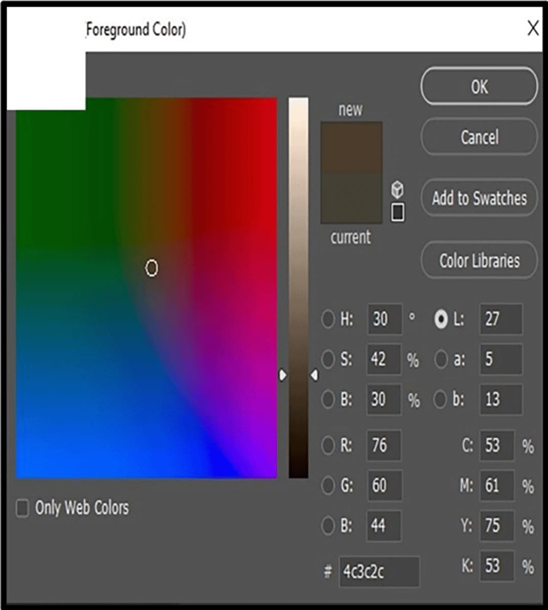
The addition of CNF to PMMA was followed by measurements of L, a, and b to determine the color shift in the samples.29 To guarantee consistent and reliable readings, the acrylic sample was attached to and detached from a stable surveyor’s table and then put parallel to the camera lens at regular intervals.30 In order to standardize the calculations, a sixty-pixel square measurement template was created in a sample’s center. The L, a, and b values for the colors were taken from the color picker’s palette window.26
The color coordinates (L, a, and b) of each sample were measured:
I. Control group: L0, a0, and b0.
II. After the incorporation of 0.5% CNFs to PMMA: L1, a1, and b1 were measured.
III. After 1% by wt. CNF addition: L2, a2, and b2 were measured.
Fourier transform infrared (FTIR) spectroscopy
Fourier transform infrared spectroscopy (Fourier-transform, 1800, Sigmadzyu, Japan) was used to determine whether PMMA heat-cured resin and CNFs exhibited any chemical interaction. The modified groups with 1% CNFs, the control group with only PMMA, and the CNF powder-only group were all analyzed.
Field-Emission Scanning Electron Microscopy (FESEM)
Four specimens were evaluated in total: one for the control group, two for the experimental groups (0.5% wt. and 1% wt. CNF), and one for the CNF powder. Using a sharp knife, square specimens (2 mm × 10 mm) were used, and 1 nm of gold was applied to the specimens’ testing surfaces. This sputter-coated film enabled uniform and profound electron dispersion throughout the specimen. A field emission scanning electron microscope (INSPECTF50, Netherlands) was used to ascertain the dispersion of CNFs in the polymethylmethacrylate (PMMA).
3.1.1 Impact strength test
The average impact strength shown a notable improvement upon incorporation of CNF at 0.5% and 1% by weight concentrations.
After 48 hours of incubation in distilled water, the reinforced groups (0.5 and 1% by wt. CNF) exhibited noticeably greater impact strength compared to the control group. Results are displayed in Figure 2(A) from the impact strength test, where the control and experimental groups were compared using the unpaired t-test.
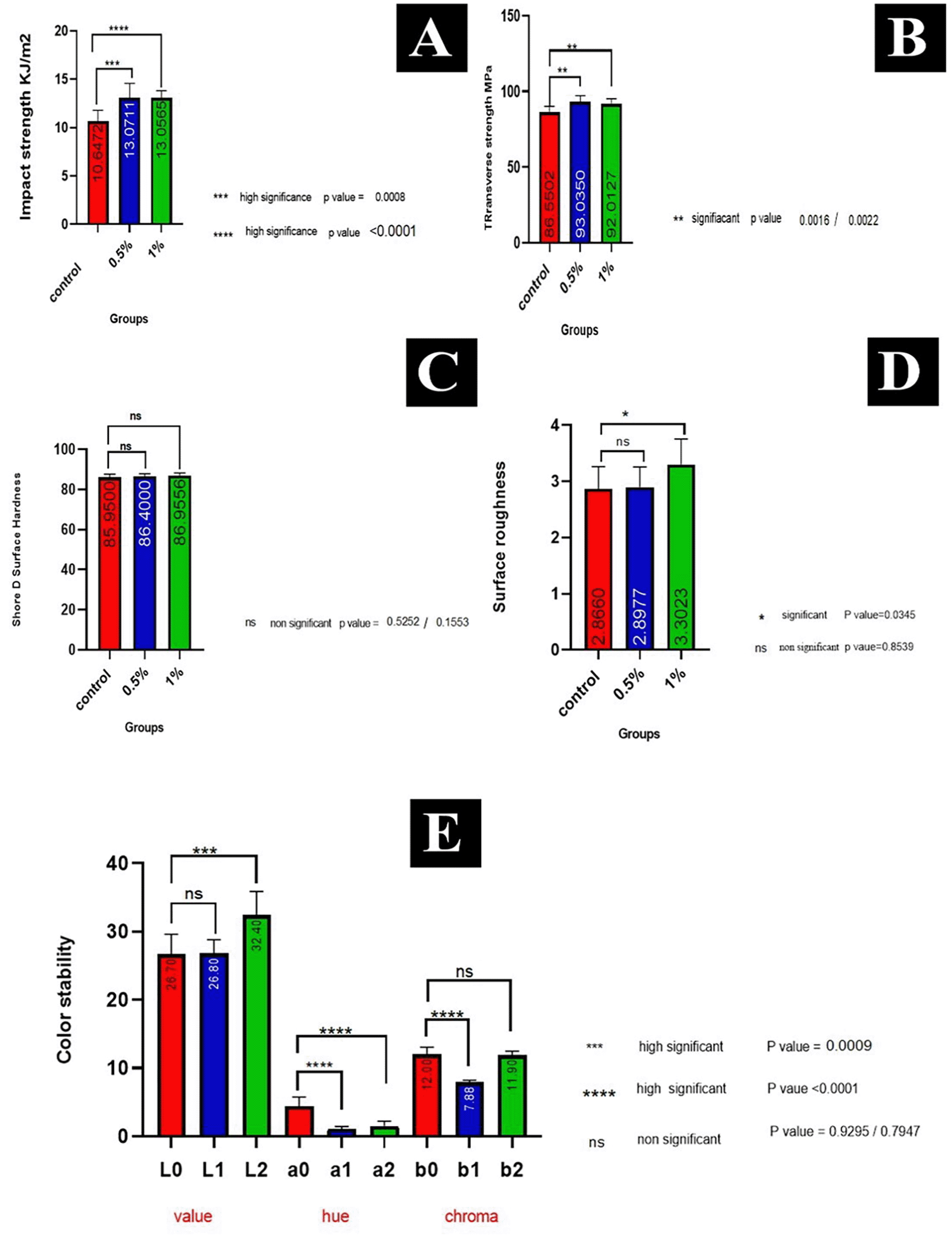
3.1.2 Transverse strength test
The transverse strength of the control group (PMMA without CNF addition), two reinforced groups (0.5% and 1%), and the results of the unpaired t-test are shown in Figure 2B.
Compared to the control group, the experimental groups showed a considerably increased transverse strength at 0.5% and 1%, respectively. As a whole, the 0.5% group scored the highest, then the 1% group, and lastly the control group.
3.1.3 Shore D surface hardness test
Figure 2(C) displays the results of an unpaired t-test and descriptive statistics applied to the Shore D surface hardness test; these reveal that the control group’s surface hardness did not significantly increase with the addition of 0.5% and 1% by weight CNF, respectively.
3.1.4 Surface roughness test
After 48 hours of incubation in distilled water, the surface roughness test revealed that the control group had a value of 2.8660 μm for surface texture roughness, while the 0.5% and 1% groups had values of 2.8977 and 3.3023 μm, respectively. The control group had lower mean values than the experimental groups (0.5 and 1%).
Figure 2(D) displays the findings of the descriptive statistics and statistical test of surface roughness, which compared the control and experimental groups’ means using an unpaired t-test. With a p-value of just 0.0345, the 1% difference between the two groups was statistically significant. The difference between the control group and the group that received specimens with 0.5% reinforced acrylic was not statistically significant (P value = 0.8539).
3.1.5 Color stability test
For color data studies of color stability, the means and standard deviations of the dependent variables ΔL*, Δa*, and Δb* were determined. The results of the color stability test, including the average and value (L), are shown in Figure 2(E). In terms of mean value, the L2 experimental group came out on top with 32.40, followed by the L1 group with 26.80, and finally the L0 control group with 26.70. While the L0 and L1 groups did not vary statistically, the L0 and L2 groups did, thus the material become lighter in color.
The a0 group had an average color value of 4,400, the a2 group of 1,500, and the a1 group of 1,100. A statistically significant difference in hue was found between the a0 control group and the experimental groups (a1, a2). This was established using an unpaired t-test. The b chroma was detected. The b0 control group had the highest mean chroma value of 12.00, followed by the b2 group with a value of 11.90 and the b1 group with a value of 7.88.
Based on an unpaired t-test, the chroma levels experienced a significant drop in the b1 group, whereas there was no significant change seen in the b2 or control groups.25
3.1.6 Fourier transforms infrared (FTIR) spectroscopy
The CNF’s structure includes the functional group OH. As can be seen in Figure 3, the OH stretching vibration can be seen at 3342 cm−1 in the FTIR spectrum of CNF powder, while the OH bending vibration can be seen at 1606 cm−1.
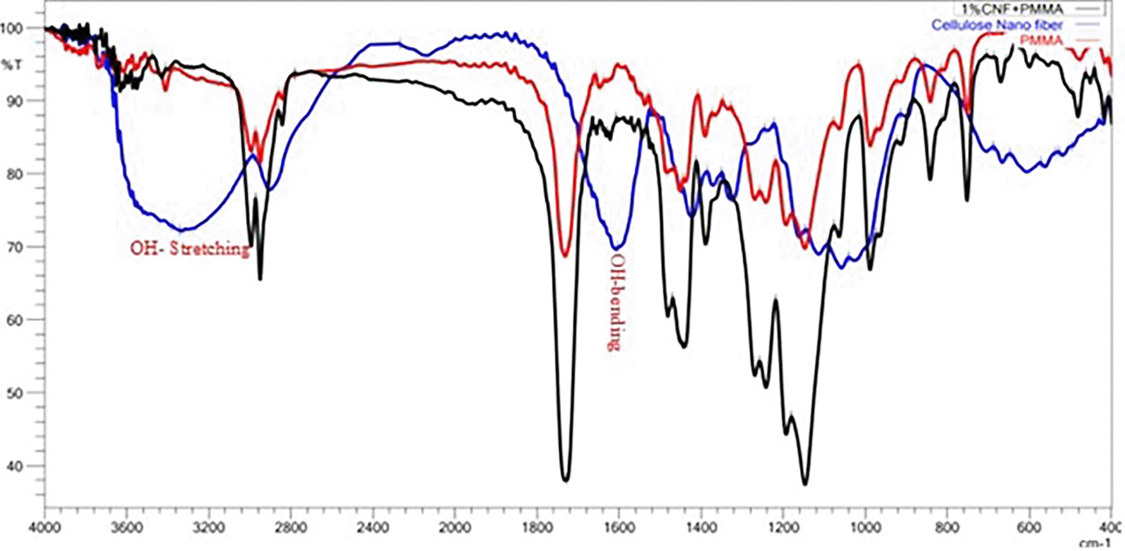
The fingerprint vibration bands of PMMA are seen at 1732 cm−1 C=O stretching mode. Band at 2949 cm−1 are associated with methylene C-H stretching. The spectra of the nanocomposite (PMMA with 1% CNF) are remarkably similar to that of pure PMMA.
3.1.7 Field-Emission scanning electron microscopy (FE-SEM)
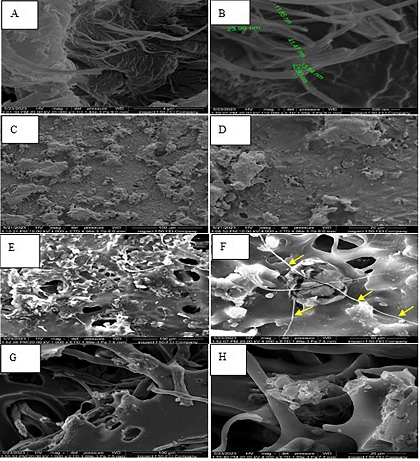
The field emission scanning electron microscopy (FE-SEM) pictures clearly demonstrated that the fibers had a diameter in the nanometer range, as shown in Figure 4(A&B). The C and D in Figure 4 displayed acrylic in its original state, prior to any modifications. Furthermore, Field Emission Scanning Electron Microscopy (FESEM) demonstrated the successful integration of nanofibers into the PMMA resin by the use of a probe sonicator mixer.
Denture bases are affected by the moist conditions of the oral environment as a result of the constant presence of saliva in the oral cavity, as well as other adverse conditions, such as load from mastication. Improvements in the features of denture acrylic can be achieved through the utilization of nanomaterials. The inclusion of nanofibers or nanotubes has been found to yield more substantial improvements in the properties of polymethyl methacrylate (PMMA) compared to the use of nanoparticle fillers. This is mostly due to the fact that nanotubes and nanofibers possess a greater ratio of surface area to volume than nanoparticle fillers.
This study was designed to evaluate the effects of various loadings of CNF fibers (0.5% and 1%) on the impact strength, transverse strength, shore D surface hardness, surface roughness, and color stability of PMMA heat-cured denture base material. Additionally, the morphological and structural characteristics were examined using field-emission scanning electron microscopy (FE-SEM) and Fourier transform infrared spectroscopy (FTIR).
Considerable improvements in the mechanical characteristics were reported in all experimental tests with 0.5% and 1% CNF addition. This was due to the homogenous dispersion of CNFs in the polymeric matrix, which was critical to the composite’s performance. The addition of nanofibers or nanotubes dramatically improved the impact strength of PMMA compared to nanoparticle fillers. This was primarily due to the fact that nanotubes and nanofibers have a higher surface area to volume ratio than nanoparticles (the ratio of nanofibers can be up to 103 times that of microfibers). Superior mechanical performance (such as stiffness and strength) tends to be achieved compared to any other form of the material.31 The nano metric scale can generate massive and extended specific surface areas of up to 1000 m2/g.32 Thus, nanocomposites have a larger interfacial matrix material surface (interphase), which has been reported to play an important role in the interface quality of nanocomposite properties, thereby achieving efficient load transfer from the matrix to the CNF.33 In one study, addition of cellulose nanofibers to epoxy composite increased the impact strength of the epoxy.34 Another study discovered that adding a modest amount (0.3 wt.%) of unmodified cellulose nanofibers from pineapple leaves to PMMA nanocomposite enhanced the impact strength significantly. The researchers attributed this to the hollow structure of the fiber giving an anti-vibration effect.35
The addition of 0.5% and 1% by wt. CNFs to PMMA significantly increased the transverse strength when compared to the control group. This effect was due to the high tensile strength and elastic modulus of CNFs. These results were consistent with previous findings for PMMA denture base materials combined with microcrystalline cellulose fiber derived from natural oil palm empty fruit bunches.36 They were also consistent with the findings of a study that added cellulose nanofibers to epoxy composites, which resulted in an increase in the transverse strength and epoxy modulus.34 Similar findings were reported for the addition of cellulose nanofibers to thermoplastic and injection molded PMMA heat polymerized acrylic resin denture base material.37 Another study found a significant increase in transverse strength after the addition of 0.5% and 1% by wt. sisal nanofibers to PMMA denture base material.38
The addition of 0.5% and 1% CNFs caused a small increase in the shore D surface hardness of specimens, which may be attributed to the homogenous distribution of CNFs in the acrylic matrix. The nanofibers were dispersed uniformly throughout the polymer, as shown in Figure 4 (E&F), and developed networks within the polymer over time, thereby reducing the inter-aggregate space and increasing the material’s toughness, stiffness, and hardness.
Interlaminar toughness enhancement in fiber-reinforced composites has been the subject of research for some time, as it is directly related to the composite’s dynamic and damage tolerance performance. A number of strategies have been explored, including stitching,39 Z-pinning,40,41 and interleaving,42 resulting in a substantial increase in toughness and enhancements to mechanical qualities like fatigue life. To achieve the desired interlaminar fracture toughness, other approaches have focused on adjusting the matrix or interface properties. Importantly, toughening the matrix can be achieved either through chemical modification or, more recently, through the addition of chemicals to the matrix material. Grafting can also be utilized to make the fibers and matrix more chemically compatible.43 One study added 0.5% and 1% cellulose nanofibers to maxillofacial silicon material and found a statically significant increase in the shore A surface hardness of the silicon matrix.44 Hussein reported that an increasing nano-filler concentration of zirconia nanoparticles significantly increased the surface hardness.45 The addition of 0.25 wt.% and 0.2 wt.% TiO2 nano-filler, respectively, to the VST50F and Cosmesil M511 elastomers led to a statistically significant increase in their mean values.46 In contrast to these results, PMMA surface hardness was decreased insignificantly with the addition of 1% sisal nanofibers.38
In this investigation, a profilometer device was utilized, which has been acknowledged as a highly effective method for examining the roughness of the surface of restorative materials. The device provides readings that can be evaluated through statistical analysis and comparison.47 The mean value of surface roughness in the 1% group was the highest, followed by the 0.5% group and the control group. When compared to the control group, adding 0.5% by weight of cellulose nanofibers (CNF) did not make the surface rougher in a way that was statistically significant. However, a significant increase in surface roughness was seen in the 1% group when compared to the control group. The increase in surface roughness of the PMMA denture base material can be attributed to the agglomeration of fibers on the surfaces of the samples, which occurs as the concentration of fibers increases. Additionally, it should be noted that CNFs have small whiskers that extend outward from their surface. The arrangement of CNF fibers during sample preparation is presumed to be random. The presence of various orientations and scattered projecting whiskers on the surface of the PMMA may contribute to the observed increase in the average surface roughness following the addition of cellulose nanofibers. The null hypothesis for surface roughness in this study was rejected because there was a statistically significant difference between the group that did not have nanofibers (PMMA) and the group that had 1% CNFs added to it. Selective dissolving is employed in certain instances to increase the surface roughness of PMMA nanofibers. In this process, polyethylene oxide (PEO) is taken out of a mix of PMMA and PEO. This leaves holes and other irregularities on the surface of the nanofibers.48 Adding nanofibers to PMMA (polymethyl methacrylate) denture base material could have different effects on the surface roughness, depending on the fibers and processing methods used. Adding nanofibers to polymethyl methacrylate (PMMA) has been shown in several studies to make the surface rougher.49 It was found in one study that adding plasma-treated polypropylene fibers to PMMA heat-cured denture bases made the surfaces of the samples rougher.50 In another study,51 it was shown that adding 7% by wt. of ZrO2 nanoparticles (NPs) to PMMA resin made the surface a little rougher. On the other hand, it has been observed that the addition of a specific amount of silicon carbide filler decreases surface roughness.52 The addition of synthesized inorganic, organic, and hybrid nanofibers to PMMA denture base material did not result in a significant effect on the surface roughness of reinforced specimens.53 Addition of 0.5% and 1% nano sisal fiber to PMMA denture base resulted in a significant decrease of surface roughness,38 and it was also found that the addition of burnt sienna intrinsic pigment to silicone elastomer for maxillofacial prostheses did not significantly affect the surface roughness of the silicon matrix.54
For aesthetic reasons, denture base material should be transparent and have pigmentation allowing it to blend in with natural teeth and gums.55 Additionally, it needs to have excellent color stability in the dynamic oral environment. Adobe Photoshop is often used for digital color analysis in scientific studies of PMMA (polymethyl methacrylate) color stability. Several researchers have examined the effects of various solutions on the optical behavior of 3D-printed resins,56 using Adobe Photoshop as a color assessment tool to evaluate the color stability and wear resistance of provisional restoration.56 In another study, Ali and Safi conducted an assessment of the color stability of maxillofacial silicone following the incorporation of cellulose nanofibers. The evaluation was also performed using Adobe Photoshop software v22.5.8.998 Adobe Systems, USA), https://www.adobe.com/products/photoshop/free-trial-download.html. As indicated by Ref. 44.
Adobe Photoshop can be used for the comparative analysis of color alterations before and after exposure to varying environmental conditions. The software facilitates accurate measurement and analysis of color fluctuations, enhancing the general understanding of the color stability of polymers.
Digital cameras generate images by capturing photographs on a light-sensing medium, resulting in images composed of blue, green, and red RGB values for each individual pixel.57 Based on the findings derived from this investigation, it was evident that color instability occurs in the PMMA, irrespective of whether it is pigmented or not, as shown by ΔE > 1.5. It is worth noting that both intrinsic and external factors have the potential to induce modifications in color value and chromatic alterations.26
Within the polymer matrix, a denser network forms as the fiber load increases. The fibers tend to occupy any voids or gaps within the polymer. Due to the interaction between light and the polymer, light transmission may be limited. This can cause some light to be partially absorbed and some to be partially reflected. It is the scattering effect of CNFs that reduces light transmission. Because the nanofiber and acrylic polymer have distinct indices of refraction, they cause light to scatter. The scattering effect diminishes the transparency and lightness of the material. Each of the reinforced groups had reduced Chroma and lighter color values compared to the control group.
The decrease in color saturation (Chroma) was because nanofibers absorb or scatter light.26 This can be caused by the rough surface scattering light in a way that leads to lower color intensity. Additionally, the uneven distribution of nanofibers in the polymer may scatter light more, resulting in lower color intensity.
Highly significant decreases in color hue can have several causes. The size and distribution of nanofillers within the PMMA matrix can impact the way light interacts with the material. Nanofillers of certain sizes may preferentially scatter or absorb specific wavelengths, leading to changes in color. The uniform dispersion of nanofillers is crucial. Aggregation can lead to uneven coloration and affect the overall color hue of the material. In addition, the conditions under which the nanofillers are incorporated into the PMMA matrix can influence their dispersion and, consequently, the optical properties of the resulting material.
The null hypothesis of this study was rejected, because significant differences was detected among the studied groups. In agreement with this study, significant color change were found to occur after the addition of 1% cellulose nanofibers to vulcanized maxillofacial silicon.39 Significant color variations were also observed between the control group and specimens reinforced with ZrO2 nanoparticles (NPs) in various immersion solutions.58
In contrast to this study, Safi et al.59 investigated the effect of zirconia nanoparticles on the color properties of polymethyl methacrylate (PMMA), and found no obvious color alterations.
The FTIR revealed that after adding CNF to PMMA, the peak of the OH stretching vibration disappeared and its intensity diminished. The OH bending vibration at 1606 cm-1 in the CNF spectrum appeared as a new peak after the addition of fibers to the polymer owing to the interaction the OH- functional groups of the CNFs with each other to form physical blending, and the interaction with each (O) pair’s electron in PMMA, as shown in Figure 3. Moreover, the presence of more than one active site of the functional-OH group leads to physical interactions (molecular interaction) by Van de Waals forces and hydrogen bonds, which enhance bonding strength. Both of these increase the adhesive forces and shear strength.60
FE-SEM demonstrated that a 0.5% CNF concentration was well-dispersed and agglomeration-free, as shown in Figure 4 (E&F). Agglomeration and poor distribution of nanofibers was noted in 1.5% CNFs, as shown in Figure 4 (G&H).
Incorporating different weight percentages of cellulose nanofibers (0.5–1%) wt. into heat-cured denture base material significantly improved some of its mechanical properties, namely, its impact strength and transverse strength, with the optimum improvement obtained at a concentration of 0.5% CNFs by weight. Cellulose nanofibers increased the hardness and surface roughness of the acrylic resin. The increase was directly proportional to the concentration of the nanofibers, and was within an acceptable clinical range, with no effect on the other material properties. The addition of CNFs at 0.5% wt. had no effect into the translucency of the acrylic material, but a concentration of 1% wt. led to a decrease in translucency. The addition of 0.5% and 1% CNFs led to a significant increase in the color hue (a), while 0.5% CNF increased the color chroma (b) significantly.
The cellulose nanofibers were effectively dispersed inside the acrylic resin according to FE-SEM, and the FTIR analysis demonstrated highly physical blending between the CNFs and the heat-cured acrylic denture base material.
Conceptualization, Ihab Safi; Data curation, Maysem Fadhel; Formal analysis, Maysem Fadhel; Investigation, Maysem Fadhel; Methodology, Maysem Fadhel; Project administration, Ihab Safi; Resources, Maysem Fadhel; Software, Maysem Fadhel; Supervision, Ihab Safi; Validation, Maysem Fadhel; Visualization, Ihab Safi; Writing – original draft, Maysem Fadhel; Writing – review & editing, Ihab Safi.
| Views | Downloads | |
|---|---|---|
| F1000Research | - | - |
|
PubMed Central
Data from PMC are received and updated monthly.
|
- | - |
Provide sufficient details of any financial or non-financial competing interests to enable users to assess whether your comments might lead a reasonable person to question your impartiality. Consider the following examples, but note that this is not an exhaustive list:
Sign up for content alerts and receive a weekly or monthly email with all newly published articles
Already registered? Sign in
The email address should be the one you originally registered with F1000.
You registered with F1000 via Google, so we cannot reset your password.
To sign in, please click here.
If you still need help with your Google account password, please click here.
You registered with F1000 via Facebook, so we cannot reset your password.
To sign in, please click here.
If you still need help with your Facebook account password, please click here.
If your email address is registered with us, we will email you instructions to reset your password.
If you think you should have received this email but it has not arrived, please check your spam filters and/or contact for further assistance.
Comments on this article Comments (0)