Keywords
inner ear, hemorrhage, sudden hearing loss, Magnetic resonance imaging, case report.
Sudden deafness is a common cause of emergency consultation in otology. Usually, despite investigations, no etiology is known. Intracochlear hemorrhage is a rare cause of sudden sensorineural hearing loss (SSNHL) and may be associated with various labyrinthine disorders. In such cases, magnetic resonance imaging (MRI) is the clef of the diagnosis. We report the case of a 70-year-old patient who was referred to our department for sudden hearing loss, tinnitus, and vertigo. Otoscopic and neurological examinations were normal, and pure-tone audiometry revealed left profound sensorineural hearing loss. Videonystagmography (VNG) revealed left vestibular deficit. MRI demonstrated a high signal intensity inside the cochlea on unenhanced T1-weighted images, and no other abnormalities were found; in particular, no enhancement after intravenous administration of gadolinium. No etiology was identified. Vertigo disappeared rapidly with corticosteroid treatment and hyperbaric oxygen therapy, but hearing did not improve. Intra-labyrinthine hemorrhage causing SSNHL is rare, and the hearing prognosis is poor.
inner ear, hemorrhage, sudden hearing loss, Magnetic resonance imaging, case report.
We have submitted the revised version of the manuscript by affecting the corrections suggested by the reviewer.
These are essentially modifications affecting the form and the quality of the medical English used without any substantive problem.
See the authors' detailed response to the review by Khadija Bahrini
Sudden sensorineural hearing loss (SSNHL) is defined as a hearing loss of more than 30 dB in at least three contiguous frequencies occurring in a period of less than 72 hours.1 The investigation of SSNHL requires audiological examination and MRI.2
It is often classified as idiopathic,3 although several causes have been suggested, including viral infections, immune-mediated, logical factors, toxic, neurological, and traumatic microcirculatory problems.1–3 Cochlear or inner ear hemorrhage (IEH) has been reported is a rare cause of sudden deafness, and isolated cases are often described.3,4
A 70-year-old man with a past history of diabetes mellitus and hypertension presented to our department with sudden onset hearing loss in his left ear over the thirty seven days. This was preceded by vertigo of the rotatory type a week earlier, which evolved into short, frequent attacks, and permanent left-sided tinnitus. The patient denied a history of acoustic or physical trauma, medication use, or recent ENT infection. He claimed to avoid both alcohol and tobacco.
Otoscopy was normal. Videonystagmoscopy showed spontaneous right horizontal nystagmus with no neurological deficits.
Laboratory test revealed no abnormalities. We found no infectious or inflammatory syndrome (white blood cell count 9800E/mm3 and CRP, 8 mg/l). No positive results were detected in either the sample or the COVID-19 antibody test. Tonal audiometry revealed left unilateral subcochlear hearing loss with a hearing threshold of 100 dB, confirmed by auditory brainstem evoked potentials (BER) (Figure 1).
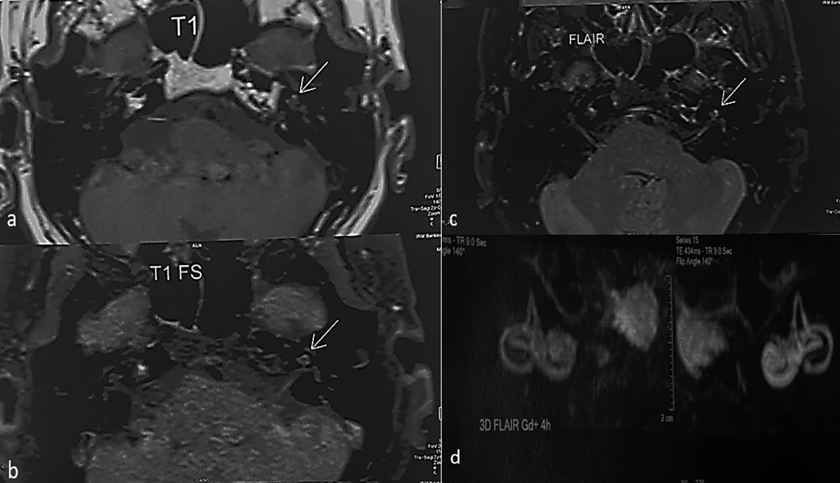
In view of the unilateral sensorineural hearing loss, an additional MRI of the inner ear was performed, including a 3D FLAIR sequence, which showed a high signal intensity in the left cochlea (Figure 2). The patient was diagnosed with a labyrinthine hemorrhage.

Based on the imaging data, we completed the etiological investigation, in particular with a hemostasis laboratory test, a nuclear antibody, and a tumor marker assay.
Given the normal test results and the lack of risk factors, idiopathic cochlear hemorrhage was considered the most likely diagnosis, particularly cervicofacial and cerebral radiotherapy, the use of antiplatelet or anticoagulant agents, or a history of meningitis.
The patient received intravenous corticosteroid therapy at a dose of 1 mg/kg/day for 10 days, followed by gradual tapering of the oral doses. Additionally, the patient was administered vasodilators and underwent 15 sessions of hyperbaric oxygen therapy (2ATA per session, five sessions per week).
On the seventh day of intravenous treatment and third oxygen therapy, the patient noted that the vertigo and tinnitus had disappeared. The improvement in hearing was partial, with a hearing threshold of 75 dB at the end of treatment. A hearing aid was also provided.
The patient reported a marked improvement in their condition and expressed hope to resume normal activities after fitting.
The diagnosis of hemorrhage requires a combination of clinical and imaging data. Clinical data should include severe to profound deafness with a described hearing loss exceeding 80 dB and vertigo.4
MRI allows diagnosis in the form of a spontaneous hypersignal T1, which is not enhanced by gadolinium injection due to the presence of methemoglobin appearing 48 h after the hemorrhagic event. The sequence T2 signal varies according to the age of the hemorrhagic event (hyposignal initially, progressing to isosignal, and then to hypersignal).3 The radiological evolution is variable: persistence of the hypersignal, regression of the images, and normalization or evolution towards sclerosing and ossifying labyrinthitis.5
The pathophysiological characteristics are currently unclear, but various etiologies of inner ear hemorrhage (IEH) have been identified. Vascular aetiologias due to anticoagulants or antiaggregants seem to be the most implicated in cases of overdose.6
In our study, our patient did not receive any treatment that could have affected coagulation.
The second most commonly described etiology in the literature is hematological diseases, such as myeloma, Waldenstrom’s disease, and autoimmune diseases, such as rheumatoid arthritis and systemic lupus erythematosus or leukemia.4
Cell blood count, antinuclear antibodies and rheumatoid factor were negative in our case.
Meningitis with bacterial diffusion may also be involved, but the clinical presentation is different, with neurological symptoms.7 Less frequently reported are radiotherapy to the head and neck, with cases reported 20 years after irradiation,8 and chemical attacks on the inner ear or toxic substances such as cocaine causing IEH by vascular effects.9
Nevertheless, the etiology often remains undetermined, with many cases classified as idiopathic. Our clinical case fell into this category.
Management of IEH is not specific, and patients are under corticoids associated with etiological treatment. However, the prognosis remains poor because of severe cochlea-vestibular lesions in comparison to other etiologies of SSNHL. Many authors have noted no significant improvements in early or late control.4,7,10 Wu et al.,10 in a comparative study of 30 patients with IEH vs. 62 patients with non-hemorrhagic inner ear, noted that the second group had a better hearing recovery in the two weeks three and six months follow up (p<0.05).
Cochlear implantation was necessary for the case reported by Meunier et al.4 because of profound bilateral hearing loss with bilateral IEH. In our case, patient have a partial improvement with 25 dB hearing gain with Tinnitus and vertigo disappears.
To our knowledge, no other author has introduced hyperbaric oxygen therapy for the management of idiopathic or secondary IEH.
Inner ear hemorrhage is a rare cause of sudden sensorineural hearing loss (SSNHL). The diagnosis of IEH was based on the clinical and imaging data. Before diagnosing idiopathic IEH, it is important to investigate the cause of hemorrhage. The prognosis of hearing loss in patients with IEH is uncertain.
| Views | Downloads | |
|---|---|---|
| F1000Research | - | - |
|
PubMed Central
Data from PMC are received and updated monthly.
|
- | - |
Is the background of the case’s history and progression described in sufficient detail?
Yes
Are enough details provided of any physical examination and diagnostic tests, treatment given and outcomes?
Yes
Is sufficient discussion included of the importance of the findings and their relevance to future understanding of disease processes, diagnosis or treatment?
No
Is the case presented with sufficient detail to be useful for other practitioners?
Yes
Competing Interests: No competing interests were disclosed.
Reviewer Expertise: Otology
Competing Interests: No competing interests were disclosed.
Reviewer Expertise: clinical research; hyperbare oxygen and immunology
Is the background of the case’s history and progression described in sufficient detail?
Yes
Are enough details provided of any physical examination and diagnostic tests, treatment given and outcomes?
Yes
Is sufficient discussion included of the importance of the findings and their relevance to future understanding of disease processes, diagnosis or treatment?
Yes
Is the case presented with sufficient detail to be useful for other practitioners?
Yes
Competing Interests: No competing interests were disclosed.
Reviewer Expertise: clinical research; hyperbare oxygen and immunology
Alongside their report, reviewers assign a status to the article:
| Invited Reviewers | ||
|---|---|---|
| 1 | 2 | |
|
Version 2 (revision) 02 Sep 24 |
read | read |
|
Version 1 21 Jun 24 |
read | |
Provide sufficient details of any financial or non-financial competing interests to enable users to assess whether your comments might lead a reasonable person to question your impartiality. Consider the following examples, but note that this is not an exhaustive list:
Sign up for content alerts and receive a weekly or monthly email with all newly published articles
Already registered? Sign in
The email address should be the one you originally registered with F1000.
You registered with F1000 via Google, so we cannot reset your password.
To sign in, please click here.
If you still need help with your Google account password, please click here.
You registered with F1000 via Facebook, so we cannot reset your password.
To sign in, please click here.
If you still need help with your Facebook account password, please click here.
If your email address is registered with us, we will email you instructions to reset your password.
If you think you should have received this email but it has not arrived, please check your spam filters and/or contact for further assistance.
Comments on this article Comments (0)