Keywords
Abdominal actinomycosis, actinomyces, acute intestinal obstruction, antibiotic therapy, surgery.
Abdominal actinomycosis, actinomyces, acute intestinal obstruction, antibiotic therapy, surgery.
We added that the patient is not an alcohol or tobacco user.
We also added Serum HbA1c level at admission.
We added that there are no immunosuppressive factors other than diabetes mellitus.
We made a grammatical correction by replacing “we underwent” with “the patient underwent”.
We corrected a mistake in the abstract, replacing “subtotal colectomy” with “total colectomy”.
See the authors' detailed response to the review by Mhasisielie Zumu and Dr Arun RS
See the authors' detailed response to the review by Prajwal Dahal
Actinomycosis is an uncommon inflammatory bacterial disease caused by Actinomyces Israeli, a Gram-positive anaerobic bacterium typically found in the digestive and genital tracts. This condition is often mistaken for a tumor or presents as an inflammatory mass. It can also lead to the formation of abscesses.1 The progression is slow and insidious, with local inflammation extending across different organs without confinement to a single one.2 Actinomyces typically colonizes the oral cavity, bronchi, gastrointestinal and female genital tracts. In the gut, it preferentially involves the stagnated zones, notably the caecum, the appendix, and the sigmoid colon. Clinical manifestations and radiological findings are nonspecific.3 Since acute and complicated forms require early treatment, most forms are diagnosed postoperatively. In this case report, we present a rare occurrence of colonic actinomycosis mimicking neoplasm and causing acute large bowel obstruction.
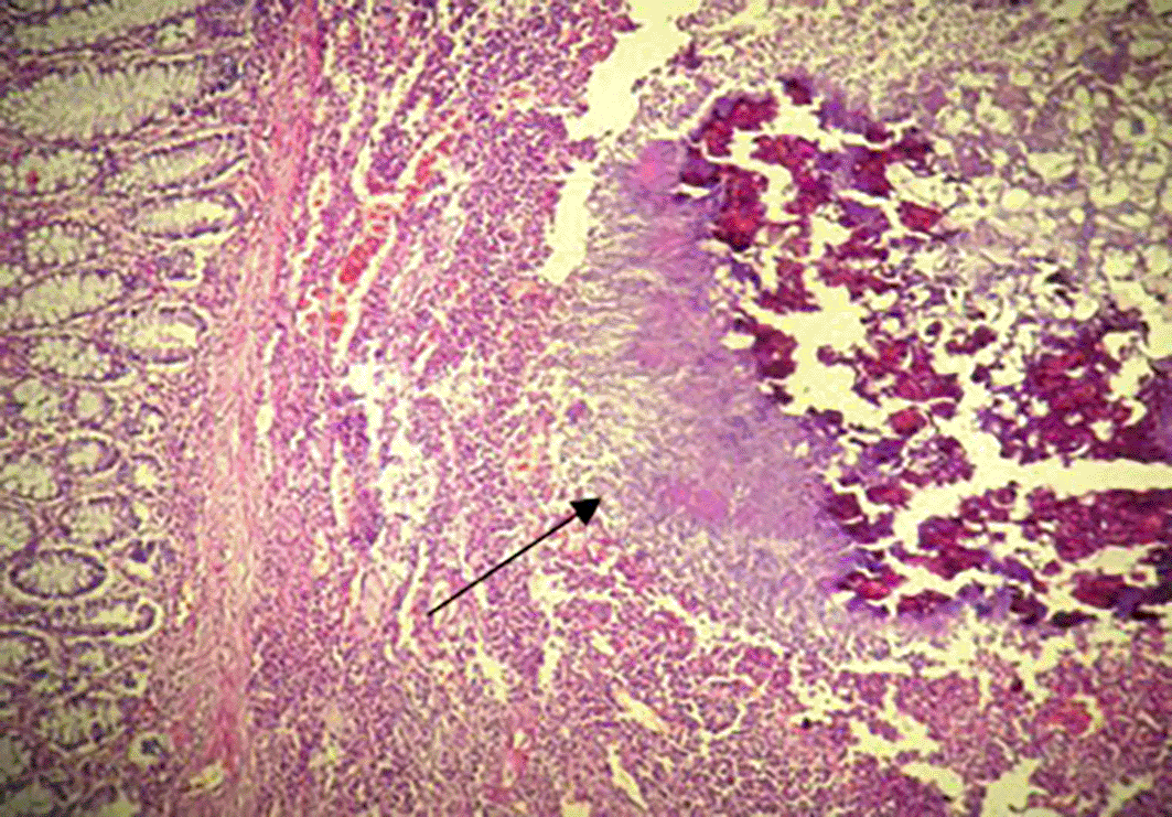
A 68-year-old non-smoking non-alcoholic Tunisian man, with a history of diabetes, hypertension, penicillin allergy, and renal failure, with no prior surgical history, presented to the emergency department with abdominal pain, vomiting, and bowel obstruction. The patient reported a similar symptomatology over the last two months, which resolved spontaneously. He also complained of chronic abdominal discomfort. On examination, he was hemodynamically stable. There was no fever. The abdominal examination revealed a distended and resonant abdomen, tender throughout, with a palpable mass in the left iliac fossa. The laboratory tests were normal, except for a previously known renal failure. Serum HbA1c level was 6%. We followed up with an abdominal CT scan without contrast, which revealed an acute intestinal obstruction upstream of a suspected obstructive tissue process at the sigmoid colon, with associated satellite lymph nodes and a dilated cecum measuring 12 cm (Figure 1). Emergency surgery was decided after a brief resuscitation. Exploration of the abdomen by midline laparotomy revealed a mass in the sigmoid loop, measuring 7 cm along its major axis, adherent to the omentum, the parietal peritoneum, and the posterior wall, with dilation of the entire upstream colonic frame. The cecum was dilated to 13 cm with a weakened, pre-perforative wall. The patient underwent a total colectomy with ileostomy and distal end closure (Figure 2). The postoperative recovery was uneventful. Gross pathologic examination of the surgical specimen revealed a stenosing lesion of the sigmoid colon with ulcerated surface mucosa. Histological examination showed acute inflammatory reaction and abscess formations surrounding clumps of short branching basophilic filaments stained with PAS (Periodic Acid Schiff). Dense fibrosis was associated. There was no granulomatous inflammation (Figures 3, 4). Actinomyces infection of the sigmoid colon was confirmed. The patient was then placed on long-term doxycycline and Bactrim. Upon follow-up, he was seen regularly for 9 months. No recurrence has been diagnosed. The restoration of bowel continuity was postponed until completing a full year of antibiotic therapy.


Actinomycosis is a rare granulomatous inflammation caused by Actinomyces species, especially Actinomyces Israeli, gram-positive anaerobic bacteria that are part of the normal human flora, colonizing the oral, digestive, and urogenital tracts.4,5
Actinomyces species have low virulence potential and require mucosal barrier disruption. This can occur after surgery, trauma, or in the presence of a foreign body, or in immunosuppression situations.6
All tissues may be infected, and we can distinguish four types of pre-ponderant infections, cervicofacial 50 to 60%, thoracic 15%, abdominopelvic 20%, and rarely disseminated disease.7
Actinomycosis commonly occurs between the ages of 20 and 60 years old and affects men three times more than women.8 Nevertheless, its incidence in women is increasing, associated with the rising use of intrauterine devices, reaching 75% of patients with pelvic actinomycosis in some studies.8
Abdominopelvic forms can mimic malignant tumors due to their chronic evolution. They have no specific clinical presentation and patients can consult for various symptoms such as chronic abdominal pain, abdominal mass, nausea, vomiting, anorexia, weight loss, and bleeding.9
This explains that it can evolve insidiously and manifest as a voluminous mass at the time of diagnosis.
Our case described an unusual presentation of abdominopelvic actinomycosis characterized by large bowel obstruction occurring in a 75-year-old patient with no risk factors cited above, except diabetes mellitus. It was due to a pseudotumoral sigmoid mass with a pre-perforative cecum.
Differential diagnoses in patients presenting with abdominal forms include appendicitis, diverticulitis, inflammatory bowel disease, tuberculosis, and bowel malignancies.10
Being a chronic suppuration, abdominal actinomycosis leads to the formation of multiple adjacent abscesses and to a large inflammatory reaction that can potentially invade neighboring tissues, appearing as a locally advanced tumor.6,10
In addition, being able to spread through hematogenous ways, actinomycosis may cause distant infections, mimicking distant metastasis.10
The management of abdominopelvic actinomycosis depends on its presentation.
The diagnosis can be suspected on CT scan findings, and confirmed after undergoing CT-guided puncture where Actinomyces species can be identified.
In such cases, the patient will undergo long-term antibiotherapy, such as parenteral penicillin G, followed by oral penicillin V or amoxicillin for up to 12 months. Alternative antibiotics like Tetracycline, Erythromycin or Clindamycin can be given in patients with penicillin allergy.
Generally, the prognosis is favorable and treatment efficacy is verified through ultrasonography or computer tomography.8,11,12
However, in most cases, actinomycosis is only diagnosed postoperatively. Indeed, confusion with a malignant mass, or manifestation in complicated forms, as in our observation, often leads to primary surgery.
According to the literature, actinomycosis involving the colon and presenting as acute abdomen or acute large bowel obstruction is rarely reported.
A review of the literature was conducted using the PubMed Database. We used “actinomycosis”, “colon”, “intestinal obstruction”, and “acute abdomen” as keywords. We excluded articles that reported extrinsic invasion of the colon, non-complicated colonic actinomycosis treated with antibiotics, and manuscripts not written in English.
A total of 15 articles were found between 1980 and 2024.
The most commonly affected colonic segments were the ascending colon and the transverse colon. In almost all reported cases, emergency surgery was performed and the diagnosis was made postoperatively. All patients received prolonged antibiotic therapy after surgery, with no reported recurrence.
In only one case, as detailed by Lin et al.,13 the diagnosis was made through endoscopic biopsy conducted during an episode of acute infectious colitis, suspected to be caused by actinomycosis infection. The patient was successfully treated with antibiotics and did not require surgery.
The details are summarized in Table 1.
| Year | Author | Age | M/F | Size | Localization | Presentation/Complication | Diagnosis | Treatment |
|---|---|---|---|---|---|---|---|---|
| 2024 | Our case | 68 | M | 7 cm | Sigmoid colon | Acute large bowel obstruction | Postoperative anatomopathological examination | Surgery + antibiotic therapy |
| 2023 | Lyew et al.14 | 48 | F | 7.7 × 4.8 × 4.5 cm | Transverse colon, small bowel, abdominal wall | Abdominal pain, epigastric mass | Postoperative anatomopathological examination | Surgery + antibiotic therapy |
| 2021 | Pamathy et al.6 | 40 | F | 9.7 × 4.5 cm | Transverse colon + descending colon | Acute large bowel obstruction | Postoperative anatomopathological examination | Surgery + antibiotic therapy |
| 2020 | Morais-Kansaon et al.15 | 46 | F | 3.2 × 3.6 × 2.8 cm | Transverse colon | Acute abdominal pain | Postoperative anatomopathological examination | Surgery + antibiotic therapy |
| 2020 | Jabi et al.16 | 48 | M | _ | Sigmoid colon | Acute abdominal pain, fever, general health state deterioration | Postoperative anatomopathological examination | Surgery + antibiotic therapy |
| 2019 | Hui et al.17 | 35 | F | 8 cm | Caecum | Abdominal discomfort, fever, right iliac fossa mass | Postoperative anatomopathological examination | Surgery + antibiotic therapy |
| 2018 | Yang et al.10 | 55 | F | _ | Sigmoid colon | Colon perforation | Postoperative anatomopathological examination | Surgery + antibiotic therapy |
| 2016 | García-Zúñiga et al.18 | 41 | M | _ | Distal ileum + ascending colon | Acute abdominal pain, fever, diarrhea, weight loss | Postoperative anatomopathological examination | Surgery + antibiotic therapy |
| 2008 | Valko et al.19 | 38 | F | 10 cm | Sigmoid colon | Acute large bowel obstruction | Postoperative anatomopathological examination | Surgery + antibiotic therapy |
| 2007 | Saha et al.20 | _ | _ | _ | Transverse colon | Acute abdominal pain | Postoperative anatomopathological examination | Surgery + antibiotic therapy |
| 2006 | Jung21 | 27 | F | 6 × 7 cm | Sigmoid colon + small bowel + mesentery | Colon perforation | Postoperative anatomopathological examination | Surgery + antibiotic therapy |
| 2005 | Filippou et al.22 | 72 | F | 5 × 5 cm | Caecum | Pericolic abscess | Postoperative anatomopathological examination | Surgery + antibiotic therapy |
| 2005 | Işık et al.23 | 28 | M | 8 × 6 cm | Ascending colon | Acute abdominal pain + vomiting | Postoperative anatomopathological examination | Surgery + antibiotic therapy |
| 2004 | Bittencourt et al.24 | 58 | M | _ | Caecum + distal ileum | Acute large bowel obstruction | Postoperative anatomopathological examination | Surgery + antibiotic therapy |
| 2003 | Lin et al.13 | 45 | F | _ | Caecum (mass) Entire colon (colitis) | Acute abdominal pain, weight loss, diffuse colitis | Endoscopic biopsy | Antibiotic therapy |
| 2000 | T.C.A Ferrari et al.25 | 56 | F | 3 cm 15 cm | Transverse colon Descending colon | Colon fistulization | Postoperative anatomopathological examination | Surgery + antibiotic therapy |
In our particular case, emergency surgery was inevitable given the obstructive character of the sigmoid colon lesion. We underwent a total colectomy due to the pre-perforative lesions appearing on the cecum. Due to anatomopathological findings, he was prescribed long-term antibiotherapy based on doxycycline and Bactrim regarding his penicillin allergy.
Despite appropriate treatment, abdominal actinomycosis may recur and patients should be followed up. Currently, there is no standardized protocol for this monitoring. However, patients should at least undergo ultrasonography or computed tomography after treatment.12
Abdominal actinomycosis, though rare, presents diagnostic challenges. It can be mistaken for malignancy, leading to unnecessary surgery in non-complicated cases. The diagnosis should be considered when there is an abdominal mass with local invasion signs, whether or not an infectious syndrome is present. As a result, all efforts should be made to confirm the diagnosis. Once the diagnosis is certain through microbiological or pathological examinations, antibiotic treatment with penicillin should be started, lasting for six to 12 months, depending on the extent of the infection. This extended treatment duration helps reduce the risk of recurrence and often completely resolves the lesions. In complicated cases, a combined approach involving surgery and antibiotic therapy is necessary until the infection is completely eradicated.
| Views | Downloads | |
|---|---|---|
| F1000Research | - | - |
|
PubMed Central
Data from PMC are received and updated monthly.
|
- | - |
Provide sufficient details of any financial or non-financial competing interests to enable users to assess whether your comments might lead a reasonable person to question your impartiality. Consider the following examples, but note that this is not an exhaustive list:
Sign up for content alerts and receive a weekly or monthly email with all newly published articles
Already registered? Sign in
The email address should be the one you originally registered with F1000.
You registered with F1000 via Google, so we cannot reset your password.
To sign in, please click here.
If you still need help with your Google account password, please click here.
You registered with F1000 via Facebook, so we cannot reset your password.
To sign in, please click here.
If you still need help with your Facebook account password, please click here.
If your email address is registered with us, we will email you instructions to reset your password.
If you think you should have received this email but it has not arrived, please check your spam filters and/or contact for further assistance.
Comments on this article Comments (0)