Keywords
fungal keratitis; rare fungus; natamycin; tissue adhesive; bandage contact lens
This article is included in the Eye Health gateway.
fungal keratitis; rare fungus; natamycin; tissue adhesive; bandage contact lens
Fungal keratitis typically results from the external acquisition of fungal agents, usually resulting from the traumatic implantation of fungal fragments or spores into the cornea. Notably, plant debris-related injuries account for a substantial 60% of fungal keratitis cases, making them the predominant causative mechanism. Fungal keratitis can be attributed to a spectrum of cornea-pathogenic fungi. These include species within the genera Fusarium, Aspergillus, Candida, Curvularia, and Penicillium. The prevalence of these fungi may vary based on geographical, occupational, and host-specific factors. Nevertheless, several fungal species are consistently detected as a cause of keratitis.1 An emergent pathogen Medicopsis romeroi is allocated to phylum Ascomycota according to National Center for Biotechnology Information. These organisms are widely distributed in the soil and plants, and they are able to infect people by direct inoculation. It is an emerging fungus causing subcutaneous phaeohyphomycosis. It has mostly been reported in immunocompromised patients receiving immunosuppressive drugs for hematologic malignancy, renal transplantation, rheumatoid arthritis, leprosy, and asthma; however, there has been one report of a healthy, immunocompetent host.2 Comprehensive discussions about Medicopsis romeroi are lacking in existing medical literature. Therefore, this case report aims to shed light on the clinical features and management strategies employed in a case of corneal ulcer caused by Medicopsis romeroi.
A male in his late teens presented to cornea clinic with complaints of redness, pain and decreased vision in his left eye. He had undergone corneal tear repair elsewhere after trauma with vegetative matter while swimming. At the time of presentation, his visual acuity was 20/20 in right eye and counting finger at 1 metre in the left eye. Upon detailed examination using slit lamp biomicroscopy, we observed a sutured corneal tear which was surrounded by a 2.5*3.3mm full thickness corneal infiltrate. [Figure. 1a] In order to assess the status of the posterior segment, a B-scan ultrasound was conducted, which showed no evidence of posterior segment involvement.
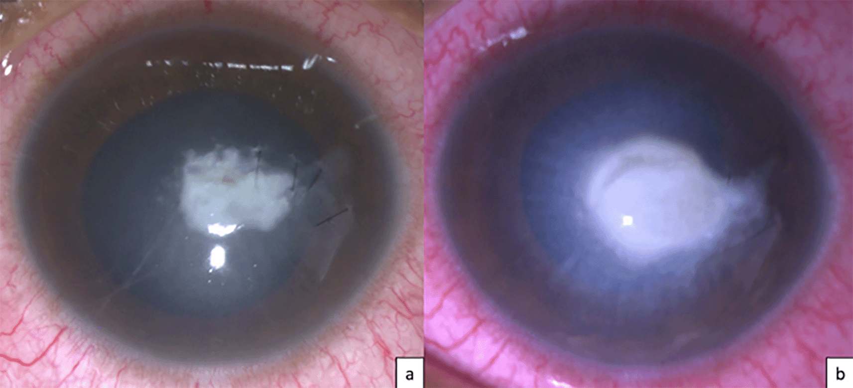
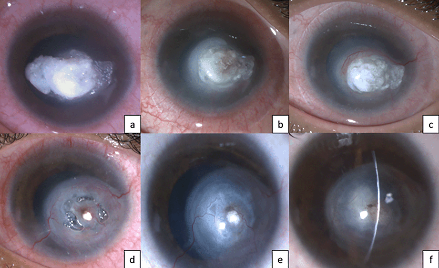
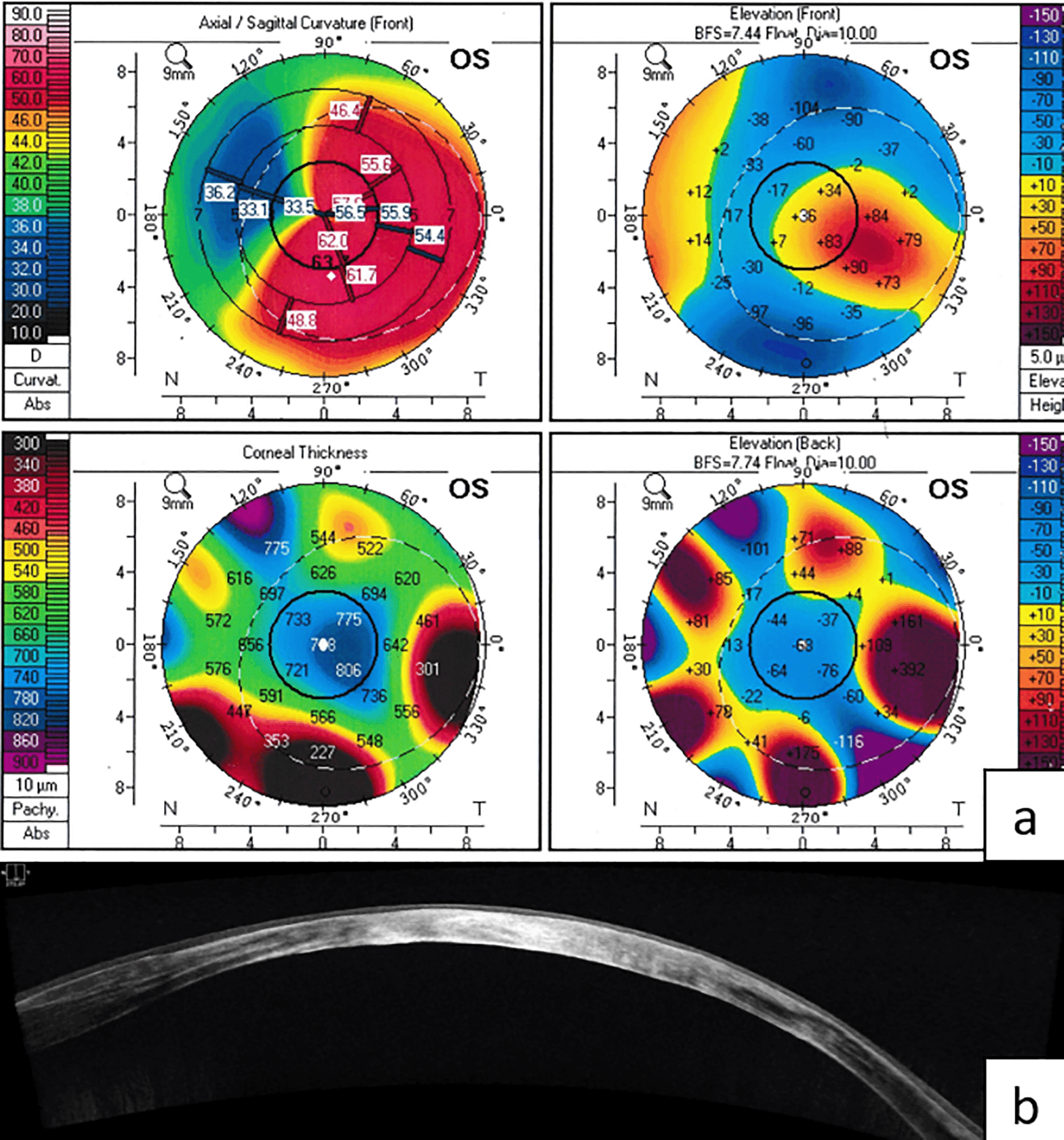
Corneal scraping was done, and the obtained material was then prepared with potassium hydroxide (KOH) for microscopic examination, which unveiled septate fungal filaments. The patient was treated with natamycin 5% eyedrops, administered hourly, coupled with oral ketoconazole 200mg twice a day and cycloplegics. Further the material was cultured but did not grow and organisms. Worsening was noted with the infiltrate increasing in size and severity and anterior chamber (AC) leak was noted. [Figure 1b] Repeat corneal scraping with AC reformation, TA and BCL was done. Topical Voriconazole 1% drops was added to the treatment. This time the culture revealed significant growth of dematiaceous fungi which was later identified as medicopsis romeroi. Over four months, there was significant improvement with scar formation with vascularization. [Figure. 2] Visual acuity also improved to 20/80. Corneal topography followed by contact lens trial was conducted, and the patient accepted the lens, improving vision to 20/100. [Figure 3a] Anterior segment OCT was done which revealed dense stromal scar tissue along the full thickness and he also developed an anterior capsular opacification in the visual axis. [Figure 3b] Synechiolysis was performed to release the anterior synechiae formed. The visual acuity improved to 20/50 and in view of zonular dialysis, pars plana lensectomy was planned.
In the presented case, our patient was diagnosed with fungal keratitis caused by Medicopsis romeroi, highlighting the persistent challenge of fungal keratitis as a leading cause of visual impairment in developing nations. An understanding of the regional epidemiological features, risk factors, and etiological agents is important in the prevention and appropriate management of this disease entity.3 Fungal keratitis manifests in two primary presentations: filamentous fungal keratitis and yeast-like fungal keratitis. Filamentous fungal keratitis encompasses a spectrum of potential pathogens, including Fusarium, Aspergillus, Curvularia, Scedosporium apiospermum, Phaeohyphomycetes, and Paecilomyces. Identified risk factors for fungal keratitis encompass corneal trauma, contamination with plant material, immunosuppression, contact lens-related trauma, corneal surgery, and chronic keratitis. Diagnosis of fungal keratitis is traditionally established through culture techniques involving blood agar, Sabouraud agar, or brain–heart infusion broth, with corneal biopsy yielding a positive organism isolation rate of up to 82%.4
Dematiaceous fungi, also referred to as melanized fungi, derive their dark pigmentation from the presence of melanin within the hyphal cell wall. More than 150 species and 70 genera of dematiaceous fungi have been implicated in human or animal diseases, and the list is expanding due to advances in the techniques applied to modern taxonomy. Dematiaceous fungi are primarily saprophytic, commonly inhabiting soil and plant environments. While they infrequently cause clinical disease, their significance as pathogens becomes pronounced, particularly in immunocompromised individuals.5 M. romeroi (formerly Pyrenochaeta romeroi), belongs to the saprophytic dematiaceous fungi and is classified within the order Pleosporales and the class Coelomycetes. These filamentous fungi are distinguished by their production of conidia within specialized fruiting structures called pycnidia. Coelomycetes are known to cause various superficial or subcutaneous infections, including onychomycosis, subcutaneous phaeohyphomycosis (PHM), and eumycetoma. They exhibit a wide geographic distribution, commonly thriving in soil and plants, particularly in tropical and subtropical regions, with the capacity to infect humans through direct inoculation into the skin. Human cases of M. romeroi infections are rare and primarily manifest as subcutaneous PHM or black grain eumycetoma.6
Regardless of the clinical manifestation, the most common approach to management of M. romeroi infection was treatment with azole antifungals along with surgical excision. The challenge lies in the fact that most azole antifungals have shown a relatively high failure rate in the face of M. romeroi. It is unclear which antifungal is the drug of choice, whereas the low reported minimum inhibitor concentrations and favourable side effect profile of voriconazole support its use against M. romeroi7 In the secondary analysis of the mycotic ulcer treatment trial II, the presence of hypopyon at baseline was associated with a 2.3-fold increase in the odds ratio of needing TPK. However, our patient did not present with hypopyon, neither at diagnosis nor at follow-up, and, no surgical procedure except TA+BCL with AC reformation was necessary for the resolution of the fungal infection, demonstrating inactivity after four months of follow-up.1,8
| Views | Downloads | |
|---|---|---|
| F1000Research | - | - |
|
PubMed Central
Data from PMC are received and updated monthly.
|
- | - |
Provide sufficient details of any financial or non-financial competing interests to enable users to assess whether your comments might lead a reasonable person to question your impartiality. Consider the following examples, but note that this is not an exhaustive list:
Sign up for content alerts and receive a weekly or monthly email with all newly published articles
Already registered? Sign in
The email address should be the one you originally registered with F1000.
You registered with F1000 via Google, so we cannot reset your password.
To sign in, please click here.
If you still need help with your Google account password, please click here.
You registered with F1000 via Facebook, so we cannot reset your password.
To sign in, please click here.
If you still need help with your Facebook account password, please click here.
If your email address is registered with us, we will email you instructions to reset your password.
If you think you should have received this email but it has not arrived, please check your spam filters and/or contact for further assistance.
Comments on this article Comments (0)