Keywords
Gallstone ileus, Cholecysto-duodenal fistula, Small bowel obstruction, Case report
Gallstone ileus, Cholecysto-duodenal fistula, Small bowel obstruction, Case report
Small bowel obstruction triggered by gallstones is a rare complication of cholelithiasis.1,2 It is defined as a mechanical obstruction of the intestine caused by the entrapment of a sizable gallstone that perforates the gallbladder wall, leading to the formation of a biliary digestive fistula.1 This condition arises in approximately 0.3 to 0.5% of individuals with cholelithiasis.3,4 It is associated with comparatively elevated rates of morbidity and mortality.1,2 There is no consensus regarding the optimal management of gallstone ileus.5,6 The present case involved a subacute small bowel obstruction caused by a gallstone, which was addressed through open enterolithotomy. Through this case report and literature review, our aim was to underscore the conditions leading to the development of this pathology, as well as the challenges associated with clinical and paraclinical diagnosis and therapy.
We report the case of an 88-year-old female patient with a history of type 2 diabetes on oral antidiabetic medication and no surgical history. The patient presented to the emergency department with diffuse abdominal pain accompanied by early postprandial vomiting and cessation of bowel movements and gas, which persisted for the last two days. She denied having fever, jaundice, melena, discolored urine, or abdominal trauma preceding the onset of symptoms.
Physical examination revealed that the patient was hemodynamically stable without jaundice or fever. Abdominal examination revealed a distended tympanic abdomen with tenderness on palpation in the suprapubic and periumbilical regions. The hernial orifices were free, and rectal examination indicated normal-colored stools.
Laboratory results indicated a biological inflammatory syndrome with a white blood cell count of 15,000 cells/mm3 and C-reactive protein (CRP) level of 100 mg/L. The remaining laboratory findings, including liver function test results, were within the normal range.
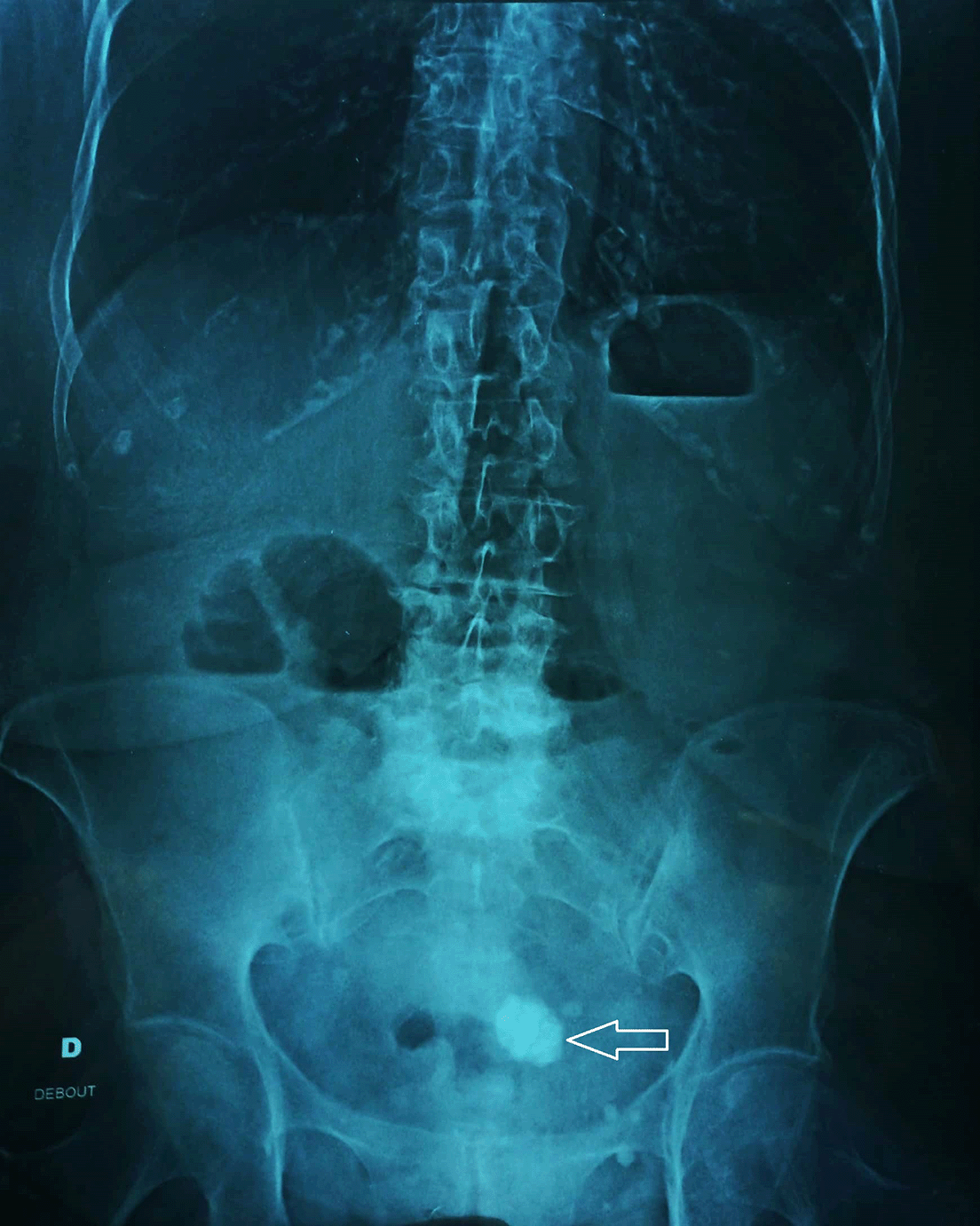
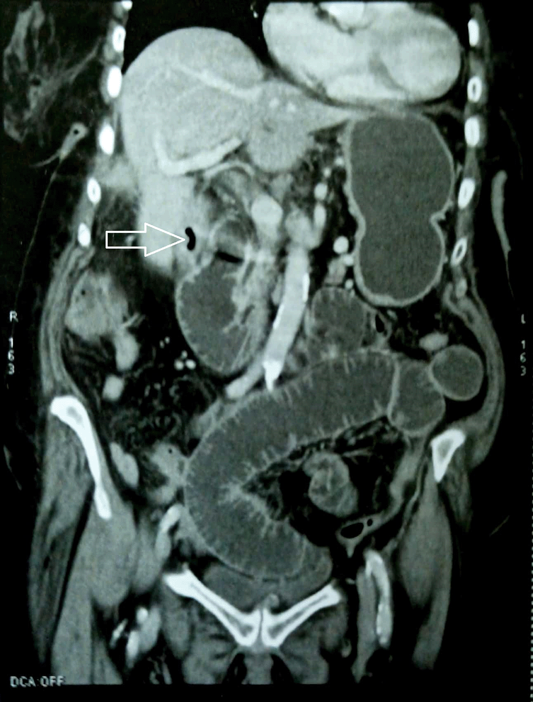
An unprepared abdomen showed small bowel air-fluid levels with an approximately 4 cm round pelvic opacity (Figure 1). Further exploration using an abdominal CT scan revealed a dilated proximal small bowel with a maximum measured diameter of 40 mm upstream of a transitional level consisting of a distended loop with a flattened appearance. At this level, an intraluminal stone measuring 3 cm in diameter was observed. In addition, there were no radiological signs of intestinal distress. The scan also indicated a multilithiatic gallbladder with a cholecystoduodenal fistula confirmed by an intra-vesicular air bubble (Figure 2). Furthermore, no intra-abdominal fluid was found. A diagnosis of biliary ileus was made.
After admission, the patient underwent a hydration and analgesia protocol. A urinary catheter and nasogastric tube for gentle aspiration were inserted, yielding a fecaloid-looking fluid. Subsequently, the patient underwent emergency surgery via a midline approach.
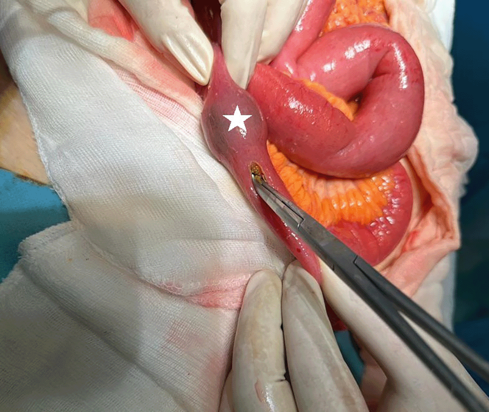
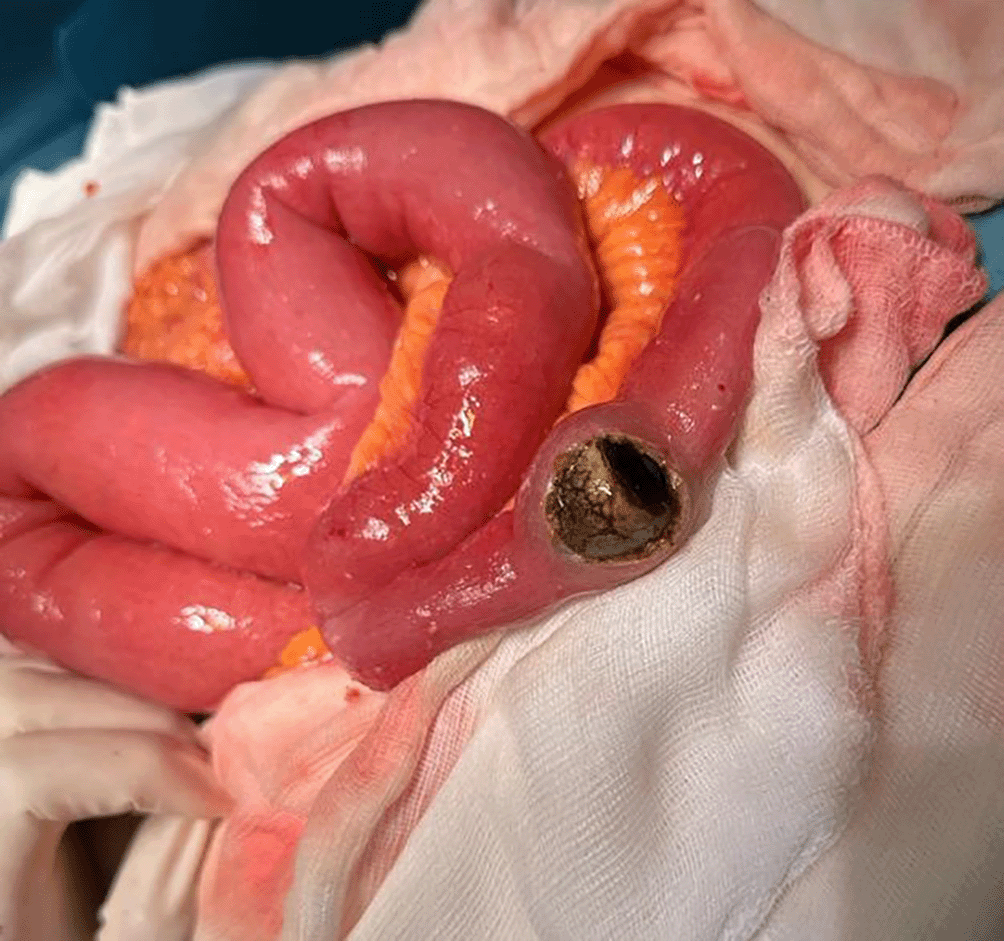
Intraoperative exploration revealed a dilated and viable proximal small bowel upstream of an intraluminal stone located 1 m 30 cm from the ligament of Treitz and 1 m 50 cm from the ileocecal valve (Figure 3). The distal small bowel and colon were flat and normal, respectively. An inflammatory mass involving the gallbladder, duodenum, right colic angle, and omentum was identified. An enterotomy was performed in a non-inflammatory healthy area, through which the stone was extracted (Figure 4). The opening was closed in two layers using Vicryl 3/0. We decided to postpone any additional procedures, such as cholecystectomy or closure of the cholecystoduodenal fistula, given the patient’s age and intraoperative findings. The intervention included peritoneal lavage using warm saline and drainage of the Douglas pouch. Postoperative recovery was uneventful, with the patient being allowed a regular diet from postoperative day 1 and discharged on postoperative day 4. The patient remained asymptomatic during one year of follow-up at the outpatient clinic, and radiological control showed spontaneous closure of the biliary digestive fistula.
Gallstone ileus is a rare complication of cholelithiasis, occurring in less than 0.5% of patients who present with mechanical small-bowel obstruction.2,3,7 It has a higher incidence in females and older individuals, as in our case.1,8
Gallstone ileus (GI) is typically preceded by an initial acute cholecystitis.1,2,9 Inflammation affecting the gallbladder and its adjacent structures, compounded by the pressure exerted by the gallstone, can result in erosion of the gallbladder wall, ultimately leading to fistula formation.1,10 Fistulas connecting the gallbladder and gastrointestinal tract commonly manifest in the duodenum owing to their proximity.4 Other regions of the gastrointestinal tract may also be implicated, including the stomach, small intestine, and transverse colon.1,11 Because of their narrow lumens, the terminal ileum and ileocecal valve are frequently the primary locations of gallstone ileus.1 Gallstones causing small bowel obstruction (SBO) typically exceeding 2.5 cm.1,4,6 In our case, gallstone ileus manifested in the jejunum due to the considerable size of the gallstone (4 cm).
Diagnosis is often challenging because of nonspecific or incomplete symptoms, such as intense abdominal pain, nausea, vomiting, inability to pass gas or stool, and hematochezia.6 Physical examinations may reveal abdominal distension and tenderness, decreased or absent bowel sounds, jaundice, and signs of dehydration.6
Biochemical markers may appear normal or lack specificity, potentially featuring findings such as leukocytosis and abnormalities in electrolyte levels, as observed in our patient.9
CT scans exhibit high sensitivity and specificity in diagnosing gallstone ileus, with rates of 93% and 100%, respectively.1,10 Therefore, a heightened level of suspicion is essential in individuals with a history of gallstones who present with bowel obstruction.9 Rigler’s triad delineates classical imaging features indicative of gallstone ileus: (1) intestinal obstruction, (2) pneumobilia, (3) gallstone within the intestinal lumen, and more recently, a change in the position of the gallstone observed in serial imaging.3,9
There is no consensus regarding the surgical management of gallstone ileus.1,5,12 Laparotomy is commonly considered to be the preferred method.1 The treatment objective is to alleviate obstruction by focusing on gallstone extraction.9 Surgical options encompass a one-stage approach involving enterolithotomy, cholecystectomy, and fistula closure or a two-stage procedure with enterolithotomy, cholecystectomy, and subsequent fistula closure.1 In our case, we performed only an enterotomy with stone extraction, considering the patient’s condition and the highly inflammatory mass formed by the gallbladder, thereby reducing postoperative morbidity. The one-stage procedure helps prevent the recurrence of gallstone ileus, with rates ranging from 8 to 33%.1 Nevertheless, this approach is technically challenging and associated with increased morbidity and mortality, particularly in older patients with comorbidities.1,6,8 In contrast, the two-stage procedure is more straightforward and requires a shorter operative time.1 As a result, it has emerged as a safer option for patients with compromised general conditions, dehydration, sepsis, and shock.1 Recently, laparoscopic management has been reported cases of laparoscopic management.1 This approach has proven to be both effective and safe, particularly in the context of a two-stage procedure.
In conclusion, we present a novel case of gallstone ileus that was successfully treated using a two-stage procedure. The choice of surgical approach should be tailored to factors such as the patient’s overall health, hemodynamic status, local conditions, and surgeon’s expertise.1
Gallstone ileus (GI) is a rare complication of cholelithiasis, particularly in females and older individuals.5,9 Our case, successfully treated with an enterotomy, highlights the challenges in diagnosis and emphasizes the importance of individualized surgical approaches based on patient characteristics. The choice between one- and two-stage procedures depends on the overall health of the patient.1 Recent reports have suggested that laparoscopic management is a safe and effective option. This case contributes to our understanding of gallstone ileus and underscores the need for tailored treatment.
| Views | Downloads | |
|---|---|---|
| F1000Research | - | - |
|
PubMed Central
Data from PMC are received and updated monthly.
|
- | - |
Provide sufficient details of any financial or non-financial competing interests to enable users to assess whether your comments might lead a reasonable person to question your impartiality. Consider the following examples, but note that this is not an exhaustive list:
Sign up for content alerts and receive a weekly or monthly email with all newly published articles
Already registered? Sign in
The email address should be the one you originally registered with F1000.
You registered with F1000 via Google, so we cannot reset your password.
To sign in, please click here.
If you still need help with your Google account password, please click here.
You registered with F1000 via Facebook, so we cannot reset your password.
To sign in, please click here.
If you still need help with your Facebook account password, please click here.
If your email address is registered with us, we will email you instructions to reset your password.
If you think you should have received this email but it has not arrived, please check your spam filters and/or contact for further assistance.
Comments on this article Comments (0)