Keywords
Endovascular Aortic Repair, Abdominal Aortic Aneurysm, Stent Graft, B-EVAR, Contralateral Gate Cannulation
Endovascular Aortic Repair, Abdominal Aortic Aneurysm, Stent Graft, B-EVAR, Contralateral Gate Cannulation
Abdominal Aortic Aneurysm (AAA) is defined as a dilation equal to or greater than 3 cm in size or an increase of ≥1.5 times the normal diameter at the renal artery level.1,2 Its prevalence increases with age. The current global prevalence of AAA in patients aged 60 years or older is estimated to be around 1.2-3.3%.2 While in the Asian population, the overall prevalence was around 2.56% in patients with cardiovascular risk factors.3 In 2017, it was reported that AAA caused 167,000 deaths and 3 million disability-adjusted life years worldwide.4
Intervention is recommended for AAA patients with rapid aneurysm growth (>5 mm/6 months) or a fusiform aneurysm with a maximum diameter of 5.5 cm or more. Studies have shown that endovascular aortic repair (EVAR) provides better results than open repair in terms of perioperative mortality rate, 30-day mortality rate, procedure time, blood loss, and length of stay (LOS).5 EVAR procedures require experienced operators in well-equipped centers with proper devices and operating rooms. They are expected to have sufficient training to perform catheter-based interventions with the recommended minimum number of endovascular procedures, including 80 endovascular therapeutic procedures, 100 endovascular diagnostic procedures, and 20 EVARs.6,7 Furthermore, EVAR has been shown to use considerable contrast when performed.8 Thus, various graft configurations have been developed over time to subjugate the limitations that come with EVAR, including one of the most challenging and time-consuming parts of EVAR, which is the contralateral gate cannulation (CGC).9 We propose a new device and technique intended to simplify endovascular AAA repair alongside reports of its application on six patients.
The procedures were performed at the National Cardiovascular Center in Harapan Kita, Jakarta, Indonesia, which saw approximately 55 EVAR and thoracic endovascular aortic repair (TEVAR) cases per year. A vascular intervention consultant cardiologist with more than ten years of experience performed the procedures, assisted by a fellow vascular intervention student.
Prior to conducting this study, we sought ethical approval from the National Cardiovascular Center in Harapan Kita. The stages involved in obtaining ethical approval included submitting the research proposal, developing the initial study protocol, creating a prototype, conducting stent graft trials, and ultimately presenting at a full board meeting. We received ethical clearance from the National Cardiovascular Center in Harapan Kita under the reference number DP.04.03/KEP161/EC086/2023 on September 12, 2023. The ethics committee authorized the study, ensuring that the research subjects’ rights are protected and confirming that the research adheres to the ethical, legal, social, and non-clinical standards outlined in the Nuremberg Code and the Helsinki Declaration.
Taofan & Kang bidirectional endovascular aortic repair (T&K B-EVAR) is a newly developed universal device intended to convert the bifurcated EVAR technique to a tubular/trunk-type EVAR with limb extensions deployed according to Taofan-Kang Modified Mitsudo’s Kissing Balloon Formula. The device consists of a main body stent graft with a bare stent and encapsulated two-limb extensions (Figure 1A-B).
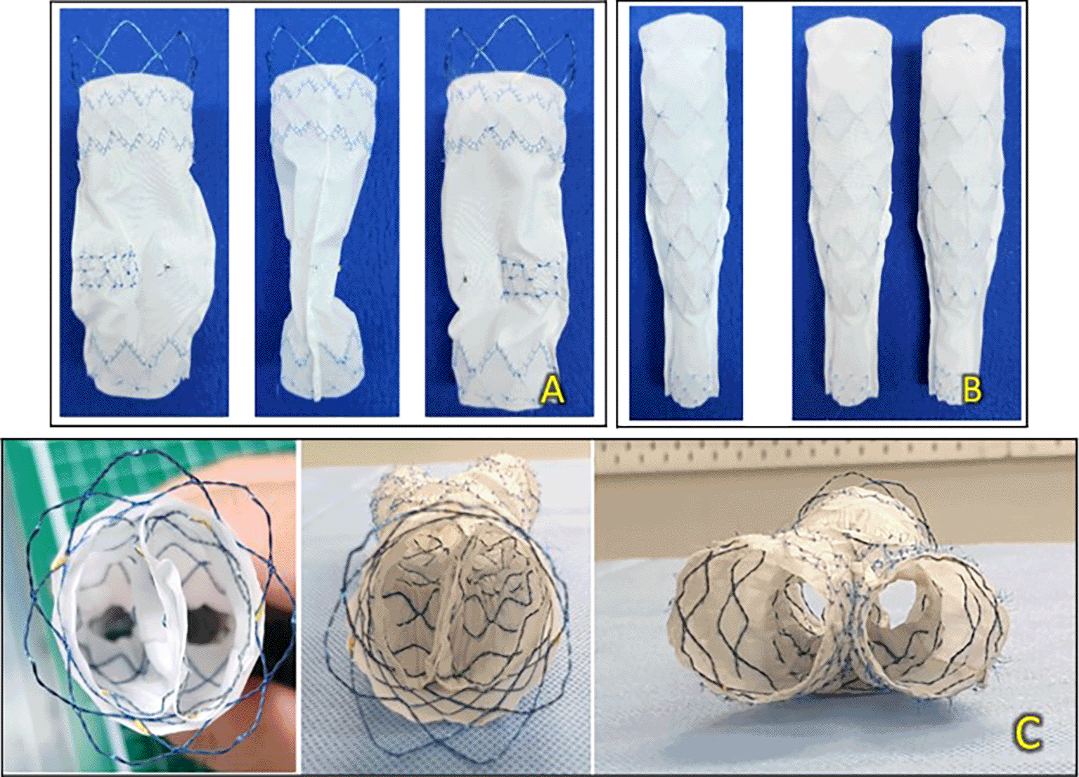
(A) Primary body stent graft from the front, side, and back. (B) Limb extensions from the front and side. (C) Axial view of the device.
The standard procedure requires two access sites from the bilateral common femoral arteries, which are achieved by surgical cutdown. Standard aortography was performed using a 5 Fr marker pigtail catheter through the contralateral femoral artery. The guidewires were placed in the ascending aorta. Both wires were replaced with an extra stiff wire. Subsequently, both contralateral and ipsilateral femoral artery sheaths are exchanged with larger sheath sizes ranging from 16 F to 18 F SEAL NOVUS Body Stent Graft™ (S&G Biotech, Yongin, Korea) deployment is performed through the more patent, more anatomically ideal access site. A balloon catheter is placed on the ipsilateral side. At the same time, the contralateral gate was cannulated using a 0.035 hydrophilic coated wire with the assistance of a multipurpose catheter, and another balloon catheter was placed. Both balloons were inflated simultaneously to ensure that they were in different lumens. If both balloons are successfully inflated, it can be inferred that both wires successfully cannulated the separate lumens. The contralateral and ipsilateral limb extensions were deployed using a SEAL NOVUS Limb stent graft™ (S&G Biotech, Yongin, Korea). The standard aortography evaluation procedure was performed to evaluate the graft stent position, apposition, endoleaks, and patency of the renal arteries. Many follow the same procedure in the six cases presented. However, certain cases have highlighted the applicability of T&K B-EVAR, even with challenging anatomical morphology.
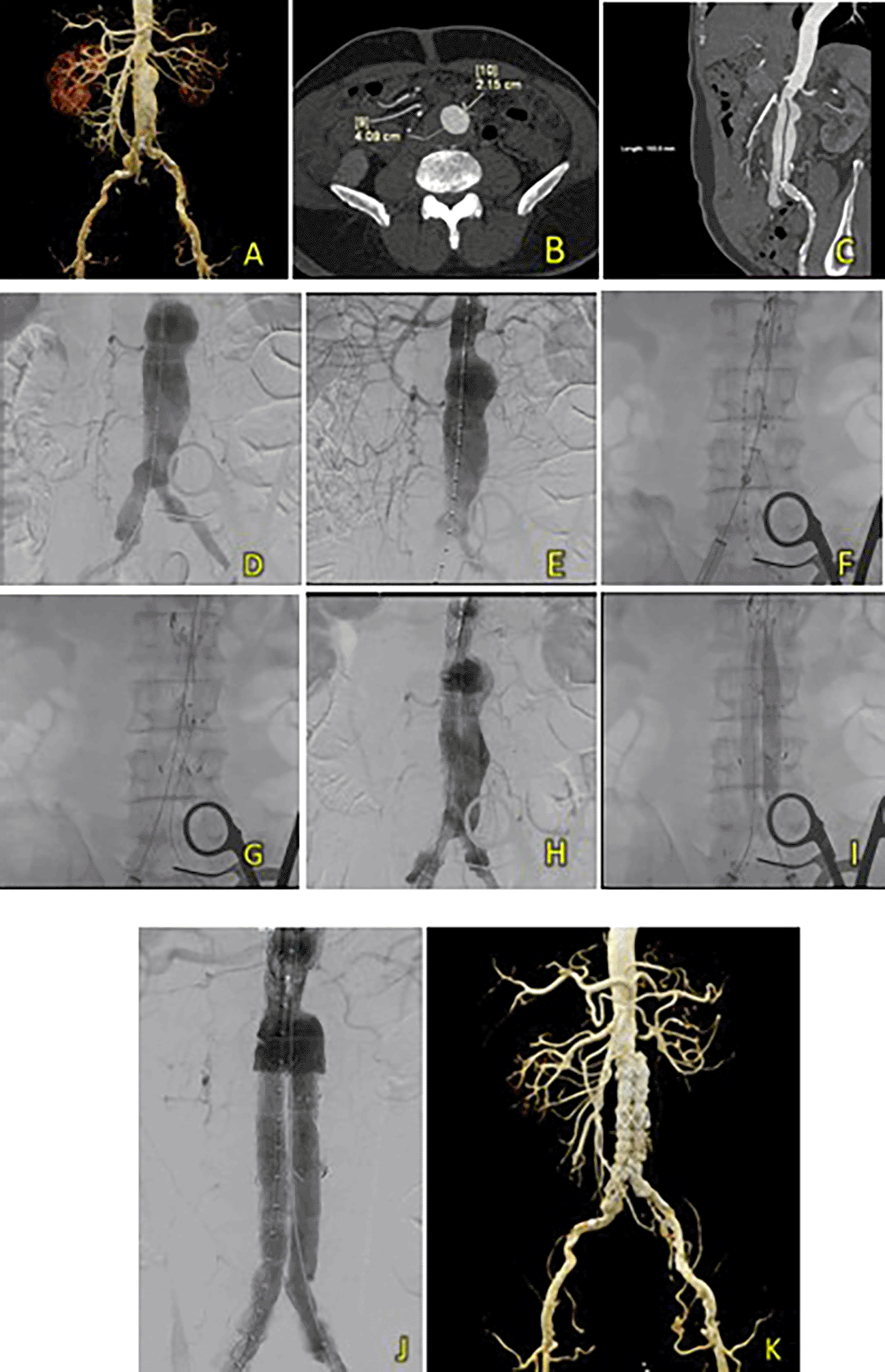
An 83-year-old man was admitted with sharp, pulsing abdominal pain 1 year prior to presentation. The patient also had a history of hypertension. Laboratory results showed normal hemoglobin counts (Hb 12.2 mg/dL) and mildly reduced eGFR (Ureum 35.6 mg/dL, Creatinine 1.33 mg/dL, and eGFR 53 ml/min/1.73 m2). A CT-Scan angiography (CTA) was performed. It revealed an AAA from the infrarenal region to the aortic bifurcation 79 mm in length and a maximum sac diameter of 58.2 mm, and dilatation of the right common iliac artery with a diameter of 21.1 mm (Figure 2A). A T&K B-EVAR procedure was then performed for the patient.
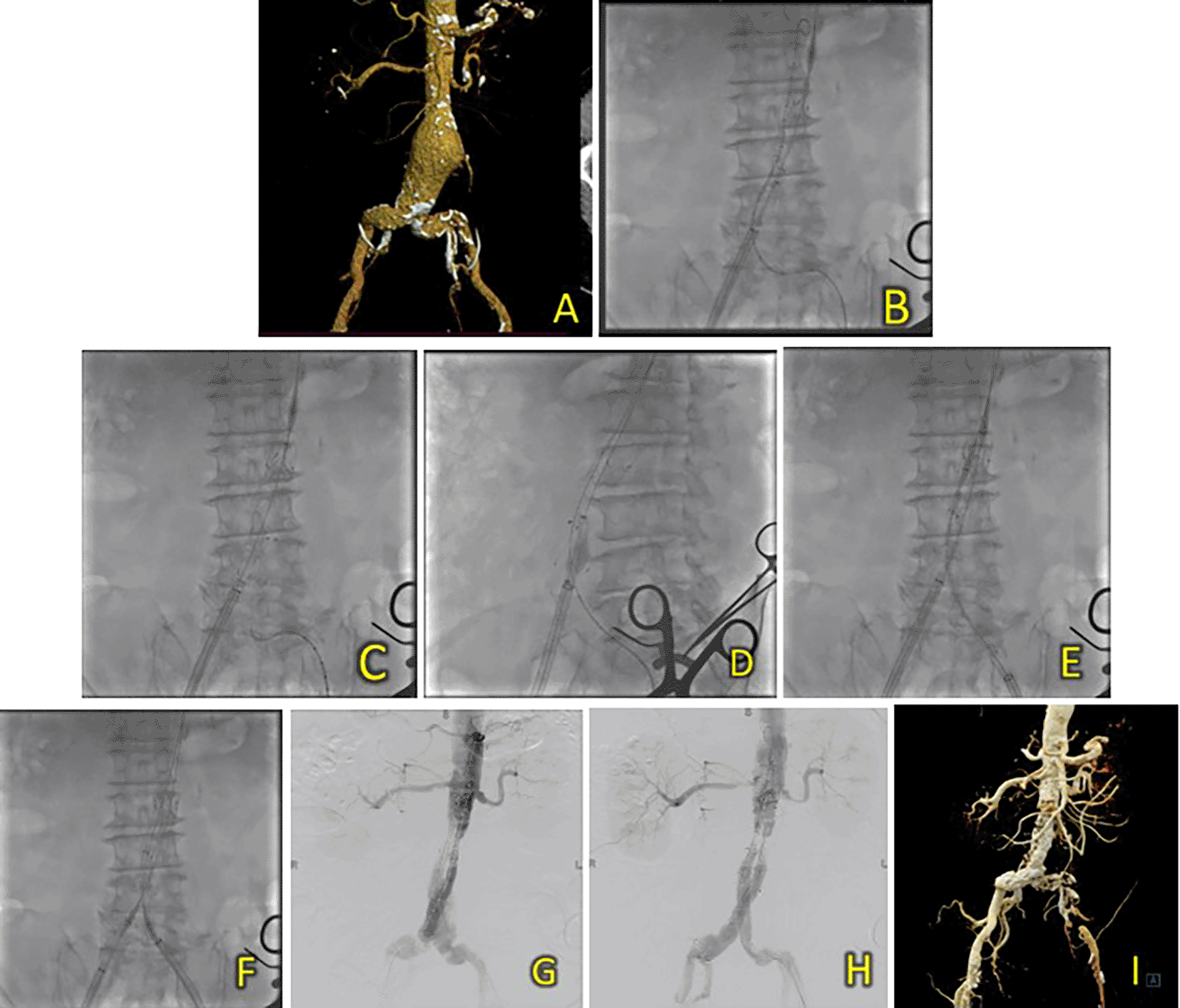
(A) CT of the 1st patient’s aortic pathology. (B) Diagnostic angiography. (C-H) T&K B-EVAR step-by-step with limb extension deployment using a ballerina technique. (I) Post-procedure follow-up CT.
Surgical cutdown was performed to gain access to the right and left femoral arteries. Initial aortography revealed an infrarenal fusiform abdominal aorta aneurysm (Figure 1B). A SEAL NOVUS Body Stent Graft 26(20) × 90 mm × 15 × 550 (S&G Biotech, Yongin, Korea) was deployed through the right femoral artery. Subsequent wiring was performed using a 0.035 hydrophilic coated wire, guided by an MP2/5F catheter. Deployment of the SEAL NOVUS Body Stent Graft 2210 × 120 mm 18 × 550 mm (S&G Biotech, Yongin, Korea) was performed through ipsilateral access (Figure 2C-G). Aortography showed the stent in a good position, a type 1 B endoleak, and stenosis in the intrastent abdominal aorta. Contralateral and ipsilateral limb extensions using the SEAL NOVUS Limb Stent Graft 12 (24) × 80 mm 15 × 550 mm (S&G Biotech, Yongin, Korea) were deployed (Figure 2H). When withdrawing the olive tip through ipsilateral femoral access, it was difficult to retract the apparatus. Hence, we continued to snare from the brachial access site, which allowed us to free the apparatus. Aortography revealed a minimal endoleak from the right iliac artery and intrastent stenosis at the abdominal aorta. Post-dilatation of the contralateral iliac stent was performed using a balloon catheter 10-46 mm inflated to 9 cc. Contralateral and ipsilateral intrastent post-dilatation were performed simultaneously using peripheral 10 × 60 mm and 10 × 60 mm balloons, respectively. Final angiography showed good stent position and no residual endoleaks or intrastent stenosis (Figure 2H). The total contrast was 130 mL hexiol, and the dose area product (DAP) was 205.48 Gy.cm2. The total procedure time was 1 h and 50 min, and the fluoroscopy times were 41 min and 56 s, respectively.
The patient was discharged without any complaints after four days. Follow-up CTA performed 3 days and 2 months after the procedure showed a deployed stent in the infrarenal aorta until the left and right common iliac arteries without endoleaks (Figure 2I).
A surgical cutdown was performed to gain access to the right and left femoral arteries, a puncture of both arteries was subsequently performed, and 8F sheaths were inserted. Wiring via left femoral artery access was performed using an exchange wire 0.035” 260 mm, placed in the abdominal aorta, and then exchanged with a marker 5F Pigtail catheter. Wiring through the contralateral access site using an extra stiff wire was performed and placed in the abdominal aorta. Initial aortography revealed an infrarenal fusiform AAA. The contralateral femoral access sheath was exchanged between 8F and 16F. SEAL NOVUS Body Stent Graft 32(26) × 90 mm 18 × 550 mm (S&G Biotech, Yongin, Korea) was deployed through contralateral femoral access (Figure 3B-C). Balloon dilation was performed using a peripheral balloon catheter (10 mm × 80 mm) through the right access, placed inside the right side of the proximal main body, and inflated to 4 atm. Simultaneous cannulation of the main body was performed using a 0.035 hydrophilic coated wire, guided by an MP2/5F catheter (Figure 3D-E). Once subsequent wiring of the ipsilateral and contralateral lumens was ensured, limb extensions were deployed using the SEAL NOVUS Limb Stent Graft 12 (24) × 100 mm 15 × 550 mm (S&G Biotech, Yongin, Korea) and SEAL NOVUS Limb Stent Graft 12 × 120 mm 15 × 550 mm (S&G Biotech, Yongin, Korea), respectively. Stenosis of the left iliac artery was found during angiography (Figure 3F), and balloon dilatation was performed to resolve this issue (Figure 3G-I). Evaluation aortography showed that the stent was in a good position, with no visible endoleak or patent renal arteries (Figure 3J). Iopamiro 370 contrast medium was used, totaling 70 mL, and the dose area product (DAP) was 201.02 Gy.cm2. The total procedure time was 1 h and 10 min, and the fluoroscopy times were 33 min and 37 s, respectively.
(A) Pre-procedure CT of the 2nd patient’s aortic pathology. (B-E) Deployment of T&K B-EVAR. (F-J) Stenosis of the left iliac artery and subsequent ballooning with good results. (K) Post-procedure CT scan evaluation.
After five days, the patient was discharged without any complaints. Follow-up CTA within the same month showed good graft position and no endoleak (Figure 3K).
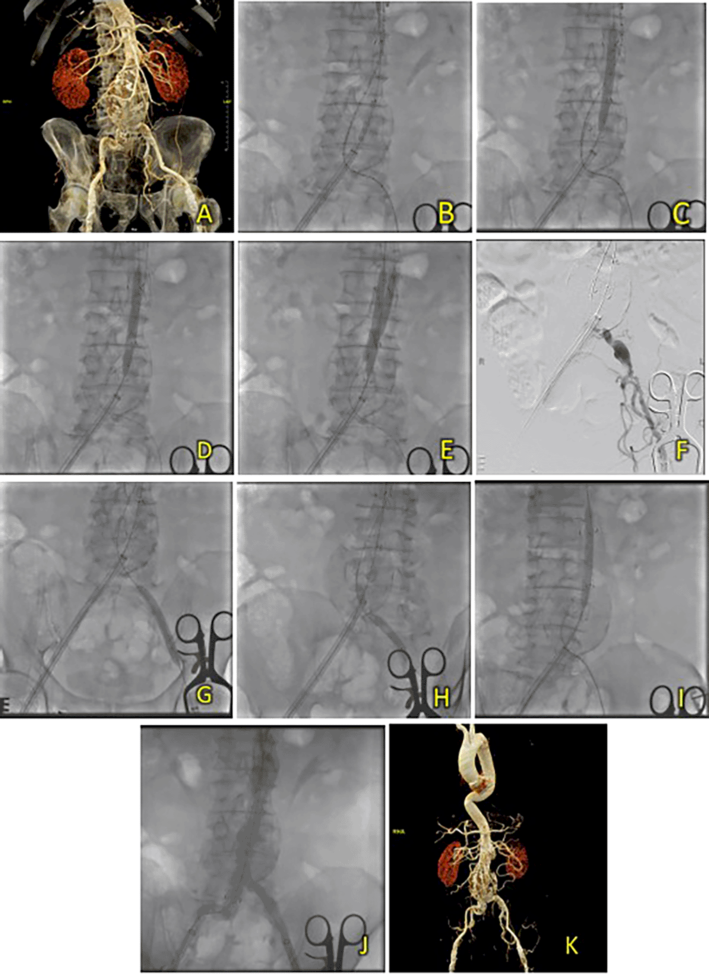
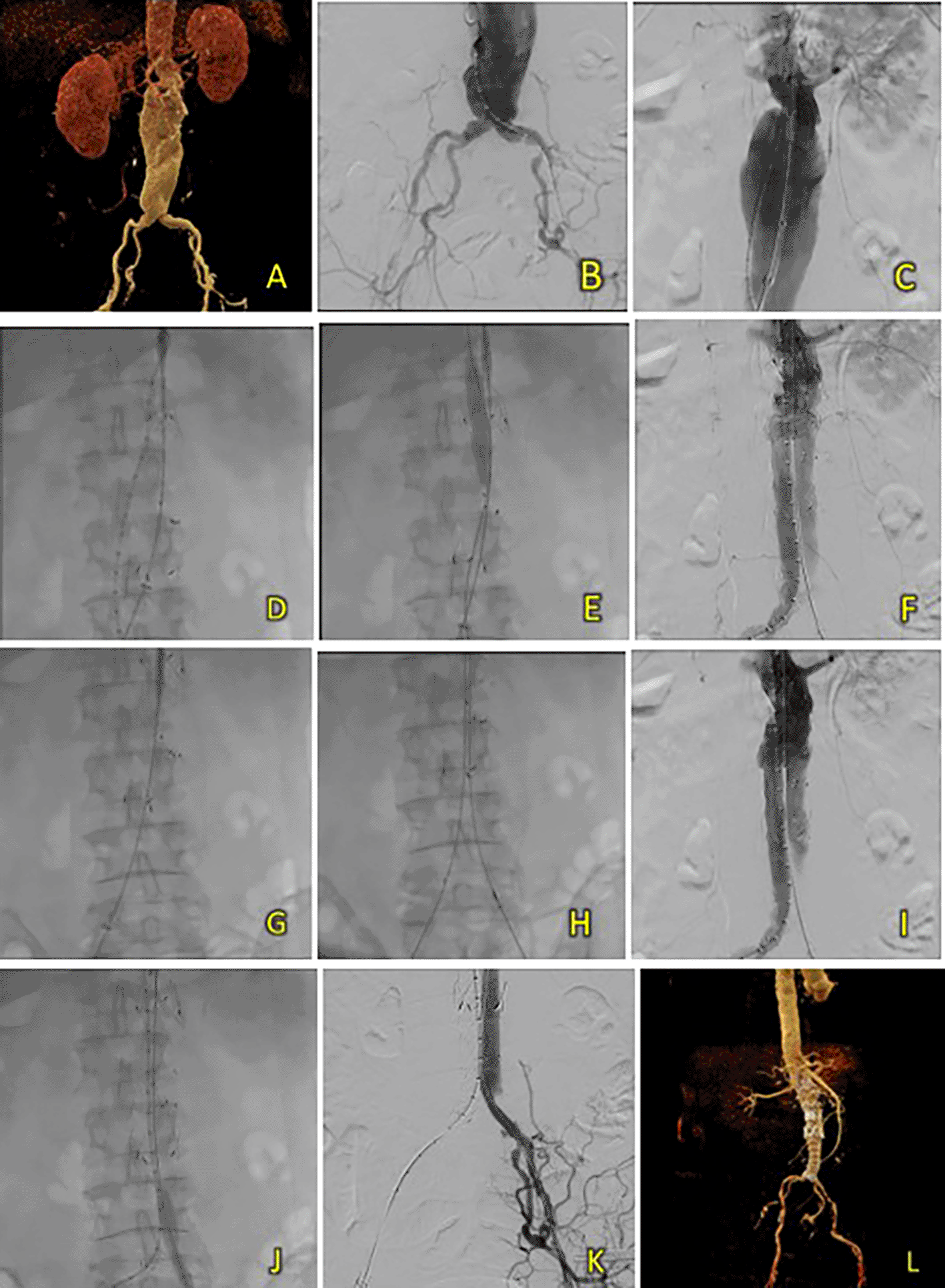
A 67-year-old male was initially suspected of having an abdominal aortic aneurysm due to recurrent abdominal pain. Ultrasonography revealed an abdominal mass. The patient had a history of hypertension, diabetes, and dyslipidemia. Laboratory results showed normal hemoglobin (14.3 Â g/dL) and mildly reduced eGFR (Ureum 37.3 mg/dL, Creatinine 1.19 mg/dL, eGFR 67 ml/min/1.73 m2). CT-scan Angiography (CTA) revealed a challenging infrarenal AAA anatomy with a very short neck (9 mm), a maximum aneurysm diameter of 70.6 mm, and a length of 127 mm (Figure 4A).
(A) Pre-procedure CT scan of the 3rd patient’s aortic pathology. (B) Diagnostic angiography. (C-D) Standard deployment of T&K B-EVAR with angiography evaluation. (E-F) Coiling of right internal iliac artery and coronary angiography evaluation. (G-H) The deployment of limb extensions and evaluation showed slow flow through the graph stent. (I-J) Balloon dilatation intrastent. (K) Evaluation angiography without endoleaks. (L) Post-procedure CT scan evaluation.
The Standard T&K B-EVAR procedure was performed in this patient, and surgical cutdown was performed for access to the right and left femoral arteries. Aortography confirmed several challenging anatomical features on the CT scan: a very short neck, neck angulation, and right common iliac aneurysm extending to the right internal iliac artery (Figure 4B-D). After the main body was deployed, a JR 3.5/6F catheter was introduced via ipsilateral femoral access and crossed over into the right internal iliac artery. To occlude the vessel, several coils were used: 6 × 6.5 mm (2 coils), 5 × 5.5 mm (2 coils), and 4 × 3.7 (1 coil) (Figure 4E-F). Standard cannulation of both lumens was successful and both balloons were successfully inflated. However, the balloon in the contralateral limb could not be inflated maximally because of wire twisting. An additional bail-out strategy was performed using antegrade wiring and guidewire via contralateral brachial access. The peripheral balloon was inflated to 4 atm in the ipsilateral limb. At the same time, the wire was advanced into the contralateral limb and externalized towards the ipsilateral femoral artery access (Figure 4G-H). Simultaneous ballooning of both ipsilateral and contralateral limbs was successful, and twisting of the wire was not observed, as confirmed by the LAO, AP, and RAO views (Figure 4I-J). Finally, limb graft extension using SEAL Novus Flared™ 12 (14) × 100 mm, 15 × 550 mm (S&G Biotech, Yongin, Korea) was deployed from the abdominal aorta with the distal portion right above the left internal iliac artery. The final aortography evaluation showed a good stent position and minimal type 1 B endoleak. After additional ballooning, final aortography showed no endoleaks (Figure 4K). The total contrast used was 230 cc, DAP 95350.47 mikroGy.m2, and fluoroscopy time was 1 h and 23 min. CT scan tomography revealed promising results, good graft stent position, and no endoleaks.
The patient was discharged without any complaints after five days. Follow-up CTA 3 months after the procedure showed an excellent stent position at the infrarenal aorta until the left and right common iliac arteries without endoleaks (Figure 4L).
A 71-year-old man was admitted for EVAR due to a pulsing sensation in the stomach. The patient had a history of undergoing a CABG procedure approximately 11 years ago and had a history of hypertension. Laboratory results revealed a normal hemoglobin level (13.6 g/dL) and slightly decreased eGFR (Ureum 38.7 mg/dL, Creatinine 1.41 mg/dL, and eGFR 53 ml/min/1.73 m2). CT-Scan Angiography (CTA) was performed. It showed an AAA with thrombus from the infrarenal region after the bilateral renal arteries to the aortic bifurcation around 103.55 mm in length with a maximum sac diameter of 40.9 mm, and right common iliac artery aneurysm with thrombus, as well as intermittent subtotal thrombus with irregularity in the bilateral internal iliac artery (Figure 5A-C). The T&K B-EVAR procedure was performed.
(A) Pre-procedural CT scan. (B) Axial CTA. (C) Sagittal CTA. (D-I) T&K B-EVAR step-by-step. (J) Post-procedure aortography with good stent apposition and no endoleaks. (K) Post-procedural CT scan evaluation had good results with no signs of endoleaks.
Surgical cutdown was performed to gain access to the right and left femoral arteries. Initial aortography revealed an infrarenal fusiform abdominal aortic aneurysm (Figure 5D-E). A SEAL NOVUS Body stent graft (24 × 90 mm 18 × 550 mm (S&G Biotech, Yongin, Korea) was deployed through contralateral femoral access (Figure 5F). After successful wiring, the SEAL NOVUS Limb Stent Graft 12 (16) × 80 mm 15 × 550 mm (S&G Biotech, Yongin, Korea) was deployed through contralateral femoral access. The SEAL NOVUS Limb Stent Graft 12 × 100 mm 15 × 550 mm (S&G Biotech, Yongin, Korea) was deployed through ipsilateral femoral access (Figure 5G-I). Aortography evaluation showed the stent in a good position and no endoleaks (Figure 5J). The total contrast was 90 mL Ultravist370, and the dose area product (DAP) was 51.722 Gy.cm2. The fluoroscopy times were 17 min and 9 s, respectively.
After four days, the patient was discharged without any complaints. A follow-up CTA 1 month after the procedure revealed promising results: a well-expanded and apposed graft stent without an endoleak (Figure 5K).
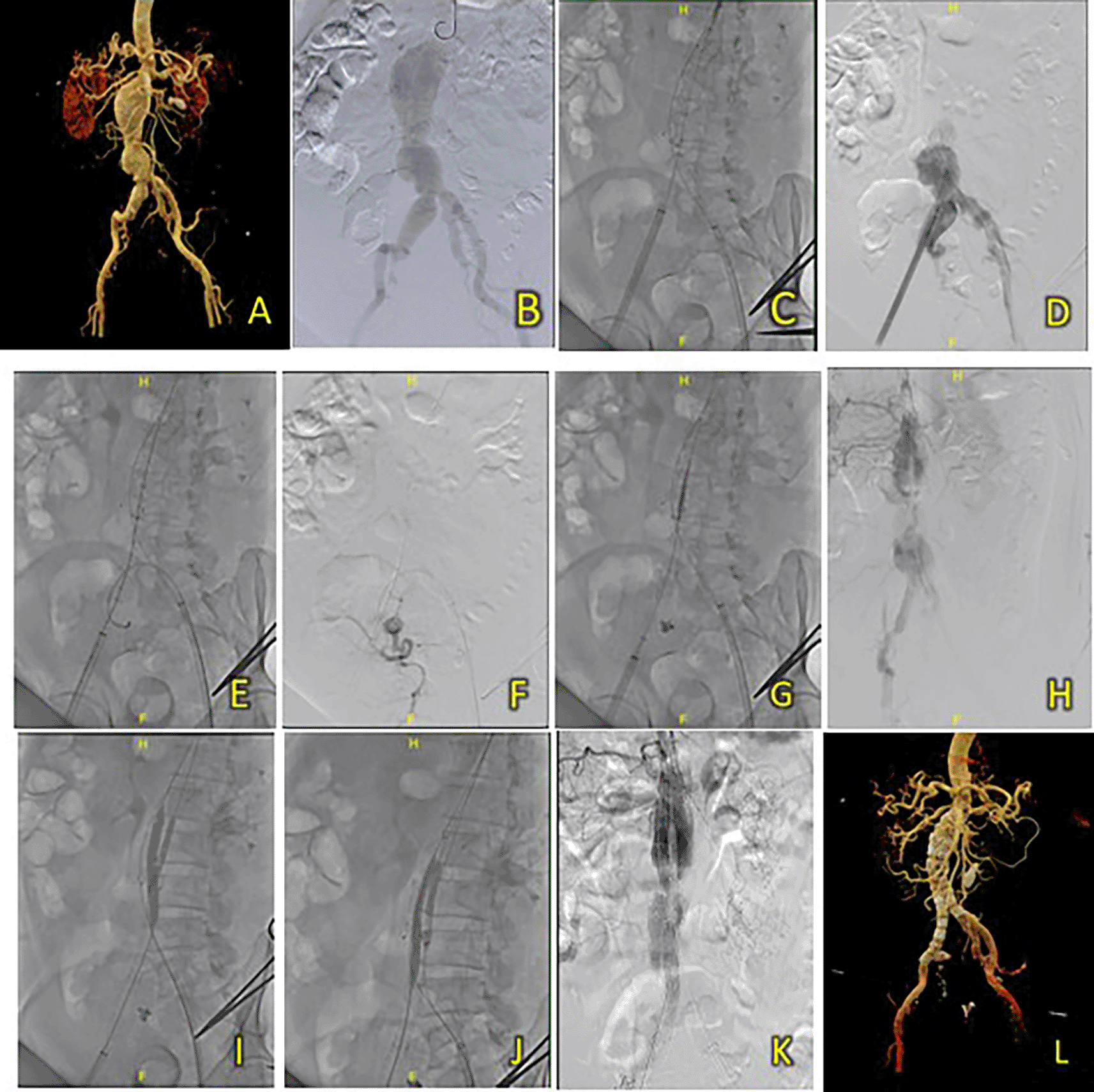
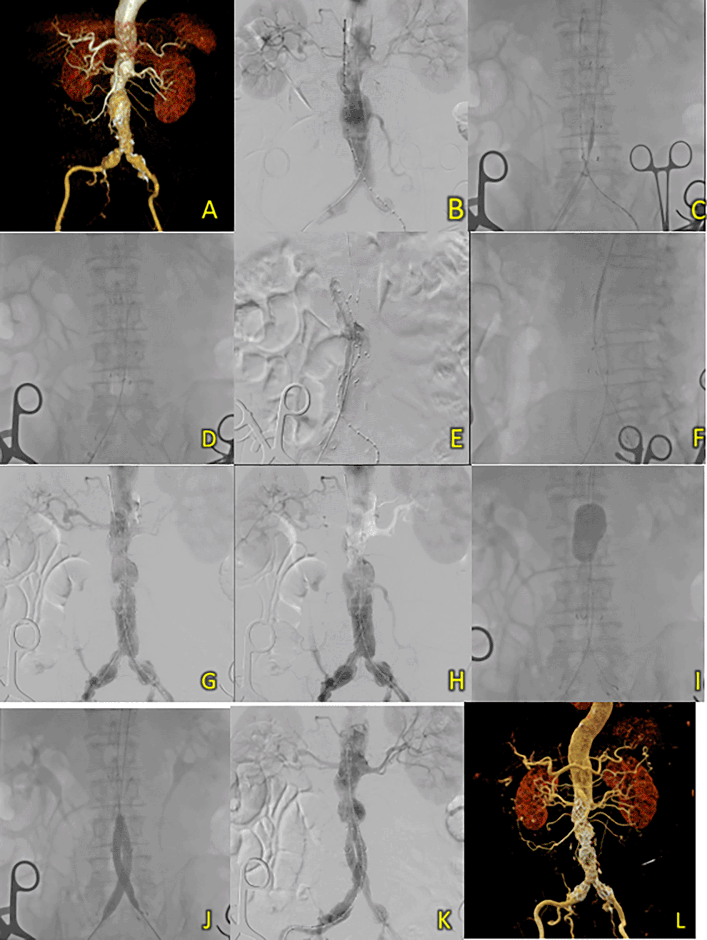
A 67-year-old man was admitted to our hospital with AAA and a history of hypertension and dyslipidemia. Laboratory results showed normal hemoglobin level (15.3 g/dL) and signs of renal insufficiency (Ureum 56.2 mg/dL, Creatinine 1.85 mg/dL, and eGFR 40 ml/min/1.73 m2). A CT-Scan angiography (CTA) revealed a challenging AAA morphology with a conical-shaped neck, thrombus throughout the whole aneurysm from the infrarenal region to the aortic bifurcation around 113.7 mm in length, a maximum sac diameter of 58 mm, a very short common iliac artery (15.4 mm), and a small access diameter (Figure 6A).
(A) Pre-procedure CT scan. (B-C) Procedural diagnostic angiography. (D-E) Deployment of main body stent. (F) Angiographic evaluation revealing the expansion of graft stent. (G-H) deployment and ballooning of extensions. (I) Angiographic evaluation with reduced flow through the left extension limb. (J) Ballooning of limb extension. (K) After the angiography evaluation after additional limb extension deployment, the results showed good flow and no endoleak. (L) Post-procedure CTA was showing promising results.
Surgical cutdown was performed to gain access to the right and left femoral arteries. A double puncture was performed in the right and left femoral arteries. Initial aortography showed an infrarenal fusiform abdominal aortic aneurysm (Figure 6B-C). For this patient, a sheathless approach was used because of the relatively small size of the right iliac artery. A SEAL NOVUS Body Stent Graft 24 × 90 mm 18 × 550 mm (S&G Biotech, Yongin, Korea) was deployed, and subsequently, both limb extension grafts were deployed (Figure 6D-E). Aortography evaluation showed under-expansion of the contralateral limb extension, particularly near the left common iliac artery, suspected to be due to arterial stenosis (Figure 6F). Balloon inflation was performed using a 10 mm × 40 mm peripheral balloon catheter, inflated several times up to a maximum pressure of 6 atm, at the main graft stent up to the left common iliac artery (Figure 6G-J). Aortography revealed that the stent coverage was insufficient; hence, another 12 × 40 mm 8 × 900 mm (S&G Biotech, Yongin, Korea) was deployed through the left femoral artery. Final aortography revealed good stent position, no endoleak, and good flow in the left iliac artery (Figure 6K). Ultravist 370 contrast was used at approximately 90 mL, and the dose area product (DAP) was approximately 92.4 Gy.cm2. The total procedure time was 1 h and 32 min, and the fluoroscopy times were 34 min and 9 s, respectively.
The patient was discharged without any complaints after six days. A follow-up CTA 4 months after the procedure revealed promising results, a well-expanded and apposed graft stent without any endoleak (Figure 6L).
A 76-year-old man was admitted with a burning sensation in the chest and epigastric areas. He was suspected to have AAA and had a history of hypertension. Laboratory results showed a normal hemoglobin level (13.4 g/dL) and signs of chronic kidney disease (Ureum 26.1 mg/dL, Creatinine 1.83 mg/dL, and eGFR 38 ml/min/1.73 m2). CT-Scan Angiography (CTA) was then performed, and it showed an AAA from the infrarenal region to the aortic bifurcation around 114.4 mm in length and a maximum sac diameter of 45.1 mm, and dilatation of the right common iliac artery with a diameter of 21.1 mm (Figure 7A).
(A) Pre-procedure CTA. (B) Angiography reveals an infrarenal fusiform aneurysm. (C-F) T&K B-EVAR step-by-step. (G-H) Underexpansion of graft stent. (I-J) Balloon dilatation of main body graft and limb extensions. (K) Evaluation angiography reveals good graft placement and no endoleak. (L) Post-procedure CTA shows good results.
Surgical cutdown was performed on the right and left femoral arteries. Initial aortography revealed an infrarenal fusiform abdominal aortic aneurysm (Figure 7B). A SEAL NOVUS Body Stent Graft 30 (24) × 90 mm 18 × 550 mm (S&G Biotech, Yongin, Korea) was deployed from the ipsilateral femoral access, and subsequently, a SEAL NOVUS limb Stent Graft 12 (20) × 100 mm 15 × 550 mm (S&G Biotech, Yongin, Korea) was deployed in the right common iliac artery. A SEAL NOVUS limb Stent Graft 12 (24) × 100 mm 15 × 550 mm (S&G Biotech, Yongin, Korea) was placed in the left common iliac artery. Aortography evaluation revealed under-expansion of the graft stent, particularly in the main body and areas covering the right common iliac artery (Figure 7C-H). Balloon dilation was performed using a 10 × 46 mm peripheral balloon inflated several times up to 10 cc in the right common iliac artery. The proximal and main body graft stents were expanded using a 10 × 80 mm peripheral balloon catheter and inflated up to 10 cc several times. Double-inflated balloon dilatation was performed using a 10 × 80 mm peripheral balloon catheter, inflated several times at the right and left common iliac arteries (Figure 7I-J). Evaluation aortography showed that the stent was in a good position, with no endoleak and better flow in the left and right iliac arteries (Figure 7K). The total contrast was 180 mL Metacosfar 320, and the dose area product (DAP) was 17.140 Gy.cm2. The fluoroscopy times were 34 minutes and 38 seconds.
After six days, the patient was discharged without any complaints. Follow-up CTA approximately 2 weeks after the procedure revealed minimal contrast leakage at the 4th and 5th lumbar vertebrae and a penetrating atherosclerotic ulcer (PAU) in the aortic isthmus (Figure 7L).
EVAR has evolved rapidly over the past decade, leading to many advances in configuration and design. Cannulation of the contralateral gate remains one of the most challenging and time-consuming procedures in EVAR deployment, increasing procedural time, radiation exposure, and material costs resulting from any potential bailout strategy.11,12 Even with experienced operators, this rate-limiting step can still be challenging as it requires the most wiring and catheter skills. This is especially relevant as the number of patients with complex aortoiliac anatomy who undergo EVAR continues to increase. Several anatomic features have been reported to prolong the time spent for CGC, namely maximal aneurysm diameter, active thrombus-free lumen, iliac tortuosity, and aortic bifurcation angulation.10 Several experiments have shown a correlation between aortic neck angulation and prolonged CGC. Despite device improvements, it still depends on the operator’s skills and technical expertise.
The various cases presented highlight the potential universal applicability of the T&K B-EVAR device and method. Of the six reported cases, all had successful outcomes despite the difficult anatomy in some cases. Only one patient in our cohort required an additional adjunctive procedure using a snaring technique from the right brachial access to untwist the contralateral femoral wire. None of the patients experienced serious complications or were discharged with an acceptable LOS. In our case series, difficult cases were reported to have a longer fluoroscopy time and an overall longer procedure time. This finding was consistent with the current literature, which states that a more complex aortoiliac anatomy is associated with an increased procedure time.
Compared to normal bifurcated EVAR, traditional bifurcated EVAR is influenced by many factors, all of which increase contralateral gate cannulation time. Free-hanging bifurcation is susceptible to severe aneurysm neck angulation or highly splayed iliac arteries, which may cause narrowing or bending of gates and require additional adjunctive approaches. T&K B-EVAR offers a sophisticated and simplified approach in which the main body graft fabric extends to encapsulate the bifurcation. In theory, this special design enables easier cannulation, as cannulation is performed on the main body with a larger diameter. This design also means that the positioning strategies normally needed in traditional bifurcated EVAR are no longer required. Furthermore, the graft design offers extra structural support and ballooning of the ipsilateral gate enables easier wiring into the contralateral gate. The extra advantage offered by the device design can enable faster procedure time, reduce fluoroscopy time, and potentially reduce contrast usage and its associated complications, such as contrast-induced nephropathy. The potential reduction in cannulation difficulty also reduces the need for bail-out procedures, which adds to the procedure time and cost. Finally, this device was designed to be universally applicable and can be used in patients with various anatomical features.
T&K B-EVAR aims to simplify the endovascular AAA repair. The device design enables easier cannulation of the contralateral limb, thereby reducing the procedure time, radiation exposure, and the risk of endoleaks. This technique has been proven to be safe in six patients, reproducible, and potentially universally applicable. However, further research with a larger sample size is needed to validate these results.
| Views | Downloads | |
|---|---|---|
| F1000Research | - | - |
|
PubMed Central
Data from PMC are received and updated monthly.
|
- | - |
Provide sufficient details of any financial or non-financial competing interests to enable users to assess whether your comments might lead a reasonable person to question your impartiality. Consider the following examples, but note that this is not an exhaustive list:
Sign up for content alerts and receive a weekly or monthly email with all newly published articles
Already registered? Sign in
The email address should be the one you originally registered with F1000.
You registered with F1000 via Google, so we cannot reset your password.
To sign in, please click here.
If you still need help with your Google account password, please click here.
You registered with F1000 via Facebook, so we cannot reset your password.
To sign in, please click here.
If you still need help with your Facebook account password, please click here.
If your email address is registered with us, we will email you instructions to reset your password.
If you think you should have received this email but it has not arrived, please check your spam filters and/or contact for further assistance.
Comments on this article Comments (0)