Keywords
COVID-19, Viral pneumonia, Critical illness, Adult respiratory distress syndrome, Biomarkers, Prognosis
COVID-19, Viral pneumonia, Critical illness, Adult respiratory distress syndrome, Biomarkers, Prognosis
In December 2019, a novel coronavirus caused viral pneumonia cases in Wuhan, China, spreading globally. Termed SARS-CoV-2, it was declared a “public health emergency of international concern” in January 2020, impacting various levels. Tunisia reported imported COVID-19 cases in March 2020, with a milder initial wave due to strict measures. The second wave strained care facilities in July 2020 due to eased preventive measures.1 Tunisia’s limited healthcare resources posed challenges, necessitating hospital reorganization and resource optimization. Intensive care units expanded, and departments like pulmonology were dedicated to COVID-19 care. Identifying high-risk patients and predictors of severe outcomes has become of paramount importance. Severe SARS-CoV-2 cases exhibited vascular processes caused by inflammation, leading to complications such as acute respiratory distress syndrome (ARDS) and cardiovascular issues.2,3 Urgent identification of predictors guided risk assessment and intervention studies. This contributed to the effective allocation of resources and informed clinical decision-making. Moreover, distinguishing severe cases using parameters like haematological and inflammatory markers improved clinical management.4,5 This is a study performed at University Hospital Center Mongi Slim, focused on COVID-19 patients hospitalized between October 2020 and June 2021 in the COVID-19 unit comprising Pulmonology and Rheumatology departments. The objectives of this study was to describe the clinical and evolutionary characteristics of hospitalized patients with COVID-19 and to determine predictive biological markers of a critical condition on the 7th day of admission.
This was a retrospective study, conducted at University Hospital Center Mongi Slim, including patients hospitalized for COVID-19 pneumonia between October 2020 and June 2021 in the COVID-19 unit: Pulmonology Department and Rheumatology Department, which were jointly managed in collaboration between the two teams.
Inclusion and exclusion criteria:
Eligible patients were those admitted to the COVID-19 unit between October 2020 and June 2021 with moderate to severe COVID-19 pneumonia at admission, confirmed for SARS-CoV-2 via Reverse Transcription-Polymerase Chain Reaction (RT-PCR), Rapid Antigen Test (RAT), or positive SARS-CoV-2 serology (IgM or IgG) for vaccine-naive patients.
Patients were not included if they had a mild COVID-19 infection throughout their hospitalization and another reason for hospitalization, if they were patients admitted to the COVID-19 unit after an initial stay in the intensive care unit, or if they remained in the emergency department for more than 7 days before being admitted to the COVID-19 unit.
We excluded patients who were admitted to the COVID-19 unit with hypoxemic pneumonia and suggestive findings of COVID-19 pneumonia on thoracic CT scan but with negative virological results (RT-PCR, RAT, and SARS-CoV-2 serologies), and an alternative cause was identified during the course of the disease. Additionally, patients were excluded if virological documentation data could not be located.
Outcome measure:
We defined the development of a critical state before or on the 7th day of hospitalization (D7) as the outcome measure. Patients were categorized into two groups:
Definition of a Critical State: We defined a critical state as:
• The occurrence of Acute Respiratory Distress Syndrome (ARDS) requiring invasive or non-invasive respiratory support. For this criterion, we defined the oxygen requirement level of 10 liters with a high-concentration mask as the threshold for evolving into ARDS.
• Vital distress or shock, sepsis, and/or organ failure. In all cases, intensive care unit admission is indicated. We relied on the definitions from the World Health Organization (WHO) version of November 20216 and the National Evaluation and Accreditation Agency for Health (INEAS) version of April 20217 to establish the definition of a critical state.
Definition of ARDS: We defined the oxygen requirement level of 10 liters with a high-concentration mask as the threshold for evolving into ARDS. This threshold was considered throughout the COVID unit’s activity based on the medical team’s experience as an indicator of a critical state and warranting alerting the intensive care units.
Data were retrospectively and anonymously collected between June 2023 and January 2024. All demographic, clinical, biological, radiological, therapeutic, and progression-related data were extracted from the medical records. The medical records followed a standardized format with predefined items consistently used in both departments of COVID-19 unit. The collected information included demographic data, clinical details recorded at admission and D7, and biological data obtained at admission. For the latter, all patients underwent a routine standard biological assessment at admission complete blood count (CBC) including leukocytes, lymphocytes, neutrophils, platelets, hemostasis profile (Prothrombin Time, Activated Partial Thromboplastin Time, D-Dimers), renal profile (Urea, Creatinine, Blood Ionogram), liver profile (Aspartate Aminotransferase (AST), Alanine Aminotransferase (ALT)), markers of muscle injury (Creatine Kinase (CK), Lactate Dehydrogenase (LDH)), and C-Reactive Protein (CRP). Some parameters were unavailable due to reagent shortages, and others were optional based on clinical presentation, such as Troponin and N-terminal prohormone of brain natriuretic peptide (NT-ProBNP). Chest computed tomography (CT) data were recorded at admission and upon aggravation during evolution. Therapeutic data included oxygen therapy, antibiotic treatment, corticosteroids, anticoagulation, and other measures, adhering to a uniform protocol guided by INEAS recommendations. Evolvement data covered hospital discharge, transfers to intensive care, COVID-19 unit deaths, and complication occurrences.
For the analysis of the association between two qualitative variables, we used Pearson’s Chi-squared test to compare two frequencies when the conditions for its application were met, and the Fisher’s test otherwise. For the analysis of the association between a qualitative variable and a quantitative variable, we employed the Student’s t-test to compare two means in the case of a normal distribution, and the non-parametric Mann Whitney test otherwise. Correlations between two quantitative variables were calculated using the Pearson correlation coefficient, with significance tested bilaterally. In the multivariate study, a binary logistic regression model was followed, and the risk was computed as the Odds Ratio (OR) with a retained 95% confidence interval (CI 95%). To assess performance, we utilized parameters related to the Receiver Operating Characteristic (ROC) Curve, as well as sensitivity and specificity. Pre- and post-test probabilities were determined through likelihood ratios represented on the Fagan nomogram. We set the significance threshold at p ≤ 5%.
This retrospective study received approval from the Ethics Committee of University Hospital Center Mongi Slim, under approval number 57/2023 on Friday, 22 December 2023.
We followed strict ethical committee guidelines that allowed for exemption of consent, due to the non-intrusive nature of the study and the use of non-identified data to ensure confidentiality and anonymity of participants. All personally identifiable data were anonymized prior to analysis to protect individuals’ privacy. We also carefully assessed the risks and benefits of our research, ensuring to minimize the former and maximize the latter for participants and the scientific community. No financial or other conflicts of interest were identified by the authors of this study. Policies regarding the future use of data and their potential sharing with other researchers were strictly established in accordance with ethical guidelines.
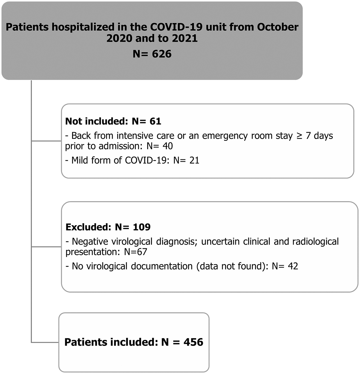
Among 626 patients hospitalized in the COVID-19 unit of University Hospital Center Mongi Slim for a period of 9 months (October 2020 - June 2021), we selected 456 patients after applying the inclusion and exclusion criteria. The number of included, not included, and excluded patients is summarized in the flowchart below (Figure 1).
1) Patient characteristics
On the 7th day of hospitalization, 115 (25.2%) evolved to a critical condition. Among the patients in the study, 260 (57%) were males; 196 (43%) were females. The gender of the patients was not statistically significant between critical and non-critical groups (p=0.106). As shown in Table 1, Chi-square outcome indicated that the age differed significantly between the critical and non-critical patients (p=0.004). In fact, the mean age for the non-critically ill patients was 61.5 ± 13.2 years while the critically ill patients had a mean age of 65.7 ± 14. Chronic concomitant diseases were found among 322 patients (70.6%), with no significant difference between both groups. The mean time from onset of symptoms to admission into the COVID-19 unit was 8.5 ± 4.12 days. This lapse was significantly shorter for the critically ill patients: 7.9 vs 8.8 days (p=0.022). Among the total cases, 31.4% had an extent of lesions greater than 50% on chest CT, the critical group had a higher proportion of cases with an extent exceeding 50% (37% versus 25.6%, p=0.021).
2) Laboratory test outcomes
The CBC findings shown in Table 2 indicate that concentration of the lymphocytes were statistically lower among critically ill patients (0.85 ± 0.24) compared to non-critically ill patients (1.10 ± 0.33, p<0.001). Lymphopenia also occurred among 89.5% of patients in critical group (p=0.002) (Table 3). Similarly, the platelet count was lower among the critical group (203 vs 235, p<0.001). The level of WBC and neutrophil did not statistically differ between both groups.
The mean Neutrophil-to-Lymphocyte ratio (NLR) was 7.52. The NLR at admission was higher in the critical group with a marginally significant difference (p=0.073). There was also a significant correlation between the NLR and the extent of lesions on the chest CT (r=0.213, p<10−3).
The CRP concentration outcome is the other biochemistry test that was statistically significantly different between critically ill (122 ± 78) and non-critically ill patients (106 ± 72, p=0.047). Upon admission, data regarding levels of D-dimer (full data available for 136 patients), hypersensitive troponin (full data available for 117 patients), B-type natriuretic peptide (full data available for 67 patients) were obtained. As shown in Table 2, in comparison to the non-critically ill patients, patients with critical illness had significantly higher levels of D-Dimer, hypersensitive and NT-pro-BNP. Besides, patients with severe pneumonia had higher rates of rhabdomyolysis markers than patients exhibiting less critical forms of the disease; (CK: 127 vs 83, p=0.039), (LDH: 831 vs 701, p=0.04). However, the findings presented in Table 2 indicate that the concentration of AST and ALT did not vary between both groups.
The univariate logistic regression indicated that laboratory findings between non-critical and critical groups showed numerous differences including C-reactive protein (p=0.047), D-Dimer (p=0.011) and creatinine (0.026) as well as, creatine kinase (p=0.039), lactate dehydrogenase (p=0.04), troponin (p=0.001) and NT-pro-BNP level (p=0.011) which were all higher among patients in critical condition. On the other hand, lymphocyte (p<0.001) and platelet (p<0.001) counts, natremia (p=0.011) and kalemia (p=0.029) were significantly lower among the critical group, whereas liver enzymes showed no significant differences. Table 3 summarizes the biological abnormalities on admission.
Based on the multivariate logistic regression model, four variables were demonstrated as independent risk factors. As shown in Table 4, the results indicated that lymphopenia (OR=2.771, 95%CI=1.482-5.181, p=0.001), NLR (OR=2.286, 95%CI=1.461-3.578, p<0.001), thrombocytopenia (OR=1.944, 95%CI=1.092-3.459, p=0.024), and CRP>71.5 (OR=1.598, 95% CI=1.042-2.45, p=0.032) were associated with critical outcome.
| Parameters | OR | 95%CI | p-value |
|---|---|---|---|
| Lymphopenia | 2.771 | 1.482-5.181 | 0.001 |
| NLR > 5 | 2.286 | 1.461-3.578 | <0.001 |
| Thrombocytopenia | 1.944 | 1.092-3.459 | 0.024 |
| CRP > 71.5 mg/L | 1.598 | 1.042-2.45 | 0.032 |
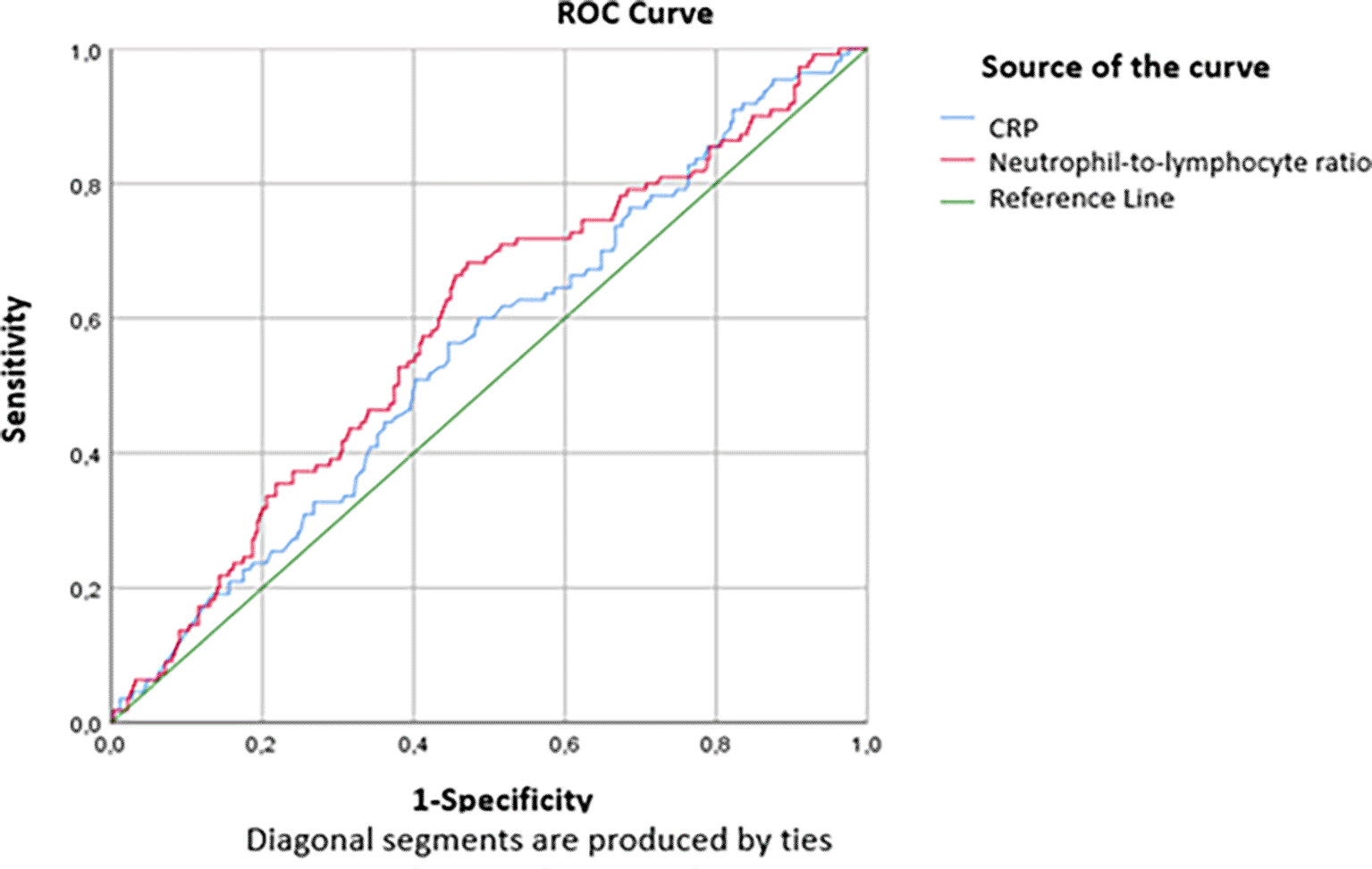
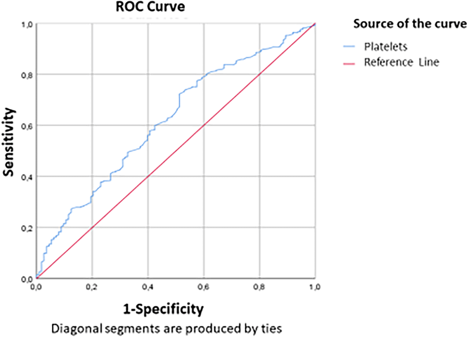
The threshold values of 71.5mg/L for the CRP level and 5 for the RNL were defined according to the study of the ROC curve according to the critical and non-critical groups. The findings in Table 4 show that statistically significant AUCs were obtained for CRP (AUC=0.533, p=0.032) and NLR (AUC=0.589, P<0.001). Thus, the performance of the NLR was slightly superior to that of the CRP in predicting a critical condition on day 7 (Figure 2). For a threshold equal to 71.5 mg/L, the sensitivity and specificity of CRP to predict critical illness were respectively equal to 66.1% and 38.1%, while for a threshold equal to 5, the sensitivity and specificity of the NLR to predict the critical illness were respectively equal to 68.7% and 51% (Table 5). Figure 2 and Figure 3 show the area under the ROC curve of CRP and NLR (Figure 2) and platelet count (Figure 3). The platelet count was negatively correlated with the occurrence of a critical condition and it was quite efficient in predicting non-critical cases; (AUC=0.62; CI95%=0.56-0.679) (Figure 2). The best threshold was estimated at 187.5*103 elements/mm3 corresponds to a sensitivity and specificity equal to 72.4% and 48.7% respectively with a quite good accuracy (Table 6).
| Cut-off | AUC (95%CI) | Sensitivity | Specificity | |
|---|---|---|---|---|
| CRP (mg/L) | 71.5 | 0.533 (0.492-0.614) | 66.1% | 38.1% |
| NLR | 5 | 0.589 (0.528-651) | 68.7% | 51% |
Based on the univariate analysis of our study, some biomarkers were predictive of the progression to a critical state on the 7th day of hospitalization for COVID-19 pneumonia.
A significant difference in biochemical and CBC test parameters was found between critically and non-critically ill patients, with elevated CRP, D-dimer, creatinine, CK, LDH, troponin and NT-Pro-BNP among the critical group compared to the non-critical group. Added to that, lymphocytes and platelet count were higher among the non-critical group. Our findings showed that the parameters with good specificity and sensitivity in predicting a critical outcome among COVID-19 patients are: CRP>71.5 mg/L and NLR>5.
Analysis of demographic characteristics revealed that critically ill patients were older than non-critically-ill ones which aligns with previous studies’ results.8 However, this age difference was smaller compared to that reported previously, noted as 20 years.9
We underline the fact that the study sample of COVID-19 patients cared during the study involved more males than females, as well among both groups (critical and non-critical), which supports previous observations that indicate gender as a potential determinant of likelihood to develop serious complications and unfavorable outcome. In fact, a multicenter study conducted in China, including 1099 patients hospitalized for COVID-19 pneumonia in 552 hospitals,10 had shown a clear male predominance (58%). This male susceptibility has been clearly elucidated in an Italian study,11 in which, two significant factors incriminated in the initiation of viral infection were emphasized, namely angiotensin-converting enzyme 2 (ACE2) and transmembrane serine protease 2 (TMPRSS2), both of which are influenced by gender.
Moreover, this observed difference could also be attributed to behavioral differences between the sexes, notably that smoking and alcohol consumption are more often seen among men12 heightening the risk of chronic comorbidities such as cardiovascular and chronic pulmonary diseases, which have been associated with a greater likelihood of developing severe illness with COVID-19.13
Our univariate analysis findings regarding the CBC parameters were different to previous findings.14,15 Previous studies concluded that increased neutrophils count is characteristic of patients with severe cases.16 Evidence suggests that severe COVID-19 is associated with elevated neutrophil levels, which increase inflammation via cytokine storm, and hemorrhages especially in the lungs, which occur as a result of neutrophil-induced tissue damage.
According to our data, lymphopenia occurred in 78% of cases. The lymphocyte count ranged between 500 and 1000/mm3 among 36.6% of our patients while 8.8% had deep lymphopenia (< 500/mm3). Furthermore, in the critical group of patients, lymphopenia was more common (89%). Our results also showed, using the multivariate logistic regression model, that lymphopenia was an independent risk factor for a severe illness. These findings are in line with the literature. Indeed, several studies have assessed the impact of lymphopenia on disease severity. For example, Zhang and al.17 reported an average blood lymphocyte count of 1200/mm3, and Jiang and al.9 showed that 41% of patients had a lymphocyte count of less than 1000/mm3. This count was significantly lower in patients with an unfavorable outcome. Additionally, Bellan and al.18 demonstrated that a low lymphocyte count at admission was correlated with the risk of mortality due to SARS-CoV-2 infection. Two Chinese meta-analyses19,20 have concluded that patients with severe COVID-19 pneumonia had lower blood lymphocyte counts compared to non-severe forms, and lymphopenia was associated with a three-fold higher risk of severe infection, particularly ARDS. They, therefore, suggest that lymphocyte count and lymphopenia could be an effective indicator for quickly identifying patients at risk of severe pneumonia. Indeed, lymphocytes play a fundamental role in maintaining adaptive immune responses against viral infections. When viral particles resulting from the replication of SARS-CoV-2 spread through the respiratory mucosa, it leads to a cytokine storm, generating a cascade of immune responses primarily involving CD4 and CD8 T lymphocytes.21 Four main mechanisms have been recognized as responsible for lymphopenia22: Sequestration of lymphocytes in target organs and their direct infection by the virus, as these cells express the ACE2 receptor; Damage of lymphatic organs such as the thymus and spleen by the virus; Apoptosis of lymphocytes due to the increase of pro-inflammatory cytokines (TNF-α, IL-6, etc.) and Inhibition of lymphocytes by substances produced during certain metabolic disorders.
In our study, thrombocytopenia was present among 13% of cases and was correlated with the progression to a critical condition (p=0.023). We demonstrated that thrombocytopenia was associated with an odds ratio (OR) of 1.94 of developing a critical condition. Our findings are consistent with those of the literature. Indeed, a meta-analysis of 21 studies including 3,377 SARS-CoV-2 positive patients4 found that platelet count was negatively correlated with the severity of the infection as well as mortality. Another meta-analysis involving 1,779 COVID-19 patients23 revealed that thrombocytopenia was associated with a threefold higher risk of severe disease, and mortality. Moreover, the platelet count was also correlated with disease severity scores and the risk of mortality in intensive care units.24,25 Platelet count was considered as an independent risk factor for COVID-19 mortality. This count was significantly lower in COVID-19 patients who died compared to survivors.18,26 Platelet kinetics could also help to identify patients at risk of an unfavourable outcome.27 In COVID-19 patients, thrombocytopenia appears to be multifactorial. In the case of ARDS, there is a significant platelet consumption resulting from the combination of viral infection and mechanical ventilation, leading to endothelial damage and platelet activation, aggregation, and thrombus formation in the lungs.23,27 Moreover, the lung can be a site for platelet release from mature megakaryocytes, and alteration of the pulmonary capillary bed can affect platelet degranulation and pulmonary megakaryocytopoiesis.23,27
In our study, the mean NLR was 7.52. This ratio was higher in the critical group patients (p=0.073). As we previously mentioned, lymphopenia was more common among patients in a critical condition. It appears logical to observe a trend of increasing NLR in severe forms. This finding was consistent with studies of both Yang and al.28 and Yan and al.,29 where a higher NLR was correlated with severe forms and mortality, respectively (p<10−3). NLR is a biomarker that integrates two subtypes of leukocytes representing two inversely related immune pathways. It provides information about systemic inflammation and is a useful biomarker for predicting bacterial infection, including pneumonia, better than absolute leukocyte, lymphocyte, or neutrophil counts.29 A high NLR reflects a discrepancy in the inflammatory response.29 Numerous studies and meta-analyses30–32 have proved that an increased NLR is associated with a poor prognosis in various diseases, such as cardiovascular diseases, solid cancers, and infections.
Increased NLR during COVID-19 could be a result of the expression of inflammatory cytokines, an unusual increase in pathological low-density neutrophils, and the upregulation of genes involved in the lymphocyte cell death pathway caused by SARS-CoV-2 infection.33 Furthermore, two studies34,35 have proposed integrating the NLR into nomograms to prove the prognostic value of this biomarker. In these studies, the authors demonstrated that NLR values at admission were higher in severe or critical cases34 and that patients with an NLR greater than 4.87 were 8 times more likely to develop a severe or critical form of the disease.35 The NLR threshold varies according to studies. An NLR threshold >3.7 on Day 7 was a major predictor of progression to severe illness, while an NLR>11.75 was significantly correlated with in-hospital mortality from all causes.29
Our study showed that for a threshold ≥ 5, the sensitivity and specificity of NLR in predicting critical status on Day 7 were 68.7% and 51%, respectively. This sensitivity was 56.52% in the study by Liu and al.35 Moreover, we further demonstrated that patients with an increased NLR (>5) were as much as 2.286 times (95% CI: 1.461-3.578) more likely to develop the severe/critical type than those with a low NLR (≤5). Therefore, NLR is an objective, simple, and inexpensive parameter that could be used in routine practice as a prognostic biomarker.
Thrombotic complications and coagulopathy represent a major event during SARS-CoV-2 infection. An increase in D-dimer and fibrinogen levels and a decrease in prothrombin time indicate a state of hypercoagulability. Many studies have shown that high levels of D-dimer are associated with severe36,37 and critical8 forms, the need for intensive care management,2,15 and in-hospital mortality due to COVID-19 pneumonia.26,38 This hypercoagulability in COVID-19 patients could result from several mechanisms39: in viral infections, there is often an unbalance between pro-inflammatory response and anti-inflammatory response; this can lead to endothelial cell dysfunction and excessive thrombin production; hypoxia contributes to thrombosis by increasing blood viscosity. In addition to that, hospitalized patients, typically have risk factors for hypercoagulability due to age, comorbidities, extended hospital stay, and invasive treatments.
Increased D-dimer levels indicate that the fibrinolytic system is activated in COVID-19 patients. Furthermore, the increased release of cytokines during viral infections stimulates coagulation cascade.40 Moreover, D-dimer >2.0 mg/L at admission were an independent predictor of death, and dynamic changes in serum D-dimers were closely associated with disease severity. A reduction in D-dimer levels was observed in recovered patients, independently of anticoagulant treatment, while a continuous increase in D-dimer levels was predictive of a higher risk of thrombosis and unfavorable outcomes.39,41 Thus, D-dimers are an early and reliable marker for predicting a poor prognosis in COVID-19 hospitalized patients.39
In our study, thromboembolic events occurred among 11 patients. The incidence of pulmonary embolism (PE) in COVID-19 patients is still unknown and likely underestimated.42 Bilaloglu and al. reported a prevalence of PE of 3.2% in a study involving 3,334 COVID-19 patients in New York,43 while according to a French study,44 the incidence of PE was 23.7%. This difference could be explained by differences in disease severity, patient characteristics, and the limited use of CT angiography among patients.
Biochemical tests findings, showed that the critical group of patients had elevated level of CRP, D-Dimer, creatinine, lactate dehydrogenase, creatine kinase, troponin and NT-Pro-BNP, which corroborates the conclusion made by Feng and al.37
The CRP was elevated (>8 mg/L) in 93.5% of patients with an average of 110 ± 73 mg/L. We noted that the CRP on admission was significantly higher in the critical group (p=0.047). In multivariate analysis, an elevated CRP level was correlated with an increased risk of progressing to a critical condition on the 7th day of admission (OR: 1.598; 95% CI: 1.042-2.45; p=0.032), and for a threshold of 71.5 mg/L, the sensitivity and specificity of CRP in predicting a critical condition were 66.1% and 38.1% respectively. Many studies have shown that elevated CRP levels are correlated with critical forms,8,36 disease progression,45 and mortality from COVID-19 pneumonia.18 As known, CRP is a non-specific marker of inflammation induced by interleukin-6 secretion. In clinical practice, it is used as a biomarker for various inflammatory and infectious conditions. High CRP levels have been directly correlated with the inflammation’s degree and disease severity.46 Moreover, CRP levels among dead COVID-19 patients were decuple higher than survivors.47
A meta-analysis of 20 studies including 4,843 COVID-19 patients and focusing on the clinical utility of CRP,48 emphasized that high CRP level was associated with a fourfold higher risk of an unfavorable outcome (p<10−3). In fact, at the early stages of COVID-19 infection, an increase in CRP was directly associated with the development of lung lesions, reflecting the severity of the disease.48,49 Furthermore, Ali and al.50 demonstrated that CRP level could predict disease worsening among non-severe cases, indicating 5% risk of progressing to a severe form for each unit increase in the CRP rate. Added to that, the CRP level has also been reported as a reliable biomarker for treatment responses in COVID-19 patients16; in fact, this marker could be used to select patients who would benefit from treatment with tocilizumab, another IL-6 receptor inhibitor similar to sarilumab.51,52
Our findings indicated that NLR and CRP are good predictors of unfavorable outcome. The reported excellent accuracy of these parameters in the prediction of COVID-19 patients’ outcome corroborates findings of the studies of Yang and al.28 and. Liu and al.35
We found that blood creatinine (p=0.026) and urea levels (p=0.061) were higher in the critical group. Furthermore, urea levels were significantly higher in elderly patients (> 70 years old) (p<10−3) and in patients with high blood pressure (p=0.012). Elevated blood urea (≥ 7 mmol/L) was noted in 46.6%. These findings align with the literature, where it has been demonstrated that elevated blood creatinine and urea values are associated with severe disease, unfavorable prognosis, and significant mortality.38,53,54 The mechanism of renal involvement in COVID-19 is likely to be multifactorial.53 It involves direct cytopathic effects on kidney tissue by the virus leading to renal cell necrosis as well as indirect damage by cytokines and metabolites induced by hypoxia, shock, or rhabdomyolysis.
Increased liver enzymes level was found in 39.5%, especially in male patients (p<10−3), while AST and ALT rates did not vary between both groups. Numerous studies have demonstrated the association between high transaminase levels and the severity of the disease,36,37,54,55 transfer to the intensive care unit2,15,56,57 and death26,38 due to COVID-19. Moreover, the good accuracy of AST and ALT as a predictor of ICU admission have been clearly shown with AUC>0.7.58 Added to that, Malik and al. demonstrated in their meta-analysis48 that high levels of AST and ALT (> 40 IU/L) were associated with a threefold higher risk of a poor prognosis. Some studies have shown that COVID-19 only transiently increases transaminases. Cytolysis is rather due to liver damage secondary to systemic inflammatory processes or to the use of hepatotoxic drugs, especially antivirals such as Lopinavir and Ritonavir during patient management.48,59 However, viral RNA has been detected in the liver at high titers, exceeding viremia, during autopsies, suggesting that SARS-CoV-2 hepatic infection can contribute to elevation of transaminase levels in patients with severe forms of COVID-19.41,60 In addition, hypoxia observed in COVID-19 patients induces hepatocellular necrosis through the production of free radicals that increase the release of hepatotoxic pro-inflammatory factors.48
Regarding rhabdomyolysis markers, we found an elevation of LDH and CK levels among 98% and 24% of our patients, respectively. We also demonstrated that muscle lysis enzymes were significantly higher in critical group patients. Our data were consistent with the literature, where it has been shown that high levels of LDH and CK were associated with the severity5,61 and progression62 of the disease, transfer to intensive care units, and mortality54 from COVID-19. In a meta-analysis assessing the prognostic value of LDH levels,38 it was found that elevated LDH levels were associated with a risk of mortality (OR: 16) and severe disease (OR: 6). Furthermore, a study conducted on COVID-19 patients63 showed that increased LDH levels at the early stage of the disease can predict lung damage and severe cases of COVID-19. These high LDH levels may result from decreased tissue oxygenation leading to stimulation of the glycolytic pathway or from damage of multiple organs in case of multi-organ failure.48 Additionally, severe infections can induce cytokine-mediated tissue damage and LDH release. Since LDH is present in alveoli, it is common to observe elevated LDH levels in patients with severe forms of COVID-19.64 Moreover, CK is a marker of muscle injury. Thus, an acute elevation of CK indicates rhabdomyolysis. The mechanism of viral myositis is unclear, but some authors suggest that myocyte damage is immune-mediated through the deposition of immune complexes in muscles.48 Therefore, LDH and CK levels are considered as important biomarkers in the prognosis of patients with severe and critical COVID-19.41
Concerning cardiac injury markers, we found that elevated levels of cardiac troponins were correlated with critical conditions (p=0.001). Indeed, troponins have a prognostic value in sepsis65 and have been proposed as severity markers15 of the disease and predictors of COVID-19 mortality.26,38 These disturbances in cardiac enzymes can result from viral myocarditis, which is more frequent during the cytokine storm, myocardial damage caused by cytokines or microangiopathies and coronary spasms secondary to hypoxia.66
Furthermore, according to our data, we observed an increase in NT-Pro-BNP levels among 25 patients (5.5%), indicating a left heart failure. Additionally, the mean NT-Pro-BNP value was higher in the critical group of patients (p=0.011). NT-Pro-BNP is a hormone secreted by the left ventricular cardiomyocytes in response to increased stretching of myocardial fibers. This biomarker helps diagnose and estimate the severity of heart failure. The mechanisms of heart failure can be attributed to an imbalance between increased cardiac output and reduced oxygen supply, with the possibility for type 2 myocardial infarction.67 According to Li and al.,36 NT-Pro-BNP>500pg/L were significantly associated with severe forms of COVID-19, and Hong and al.,8 demonstrated that critically ill patients had higher NT-Pro-BNP levels (p=0.002). Therefore, cardiac biomarkers, including troponins and NT-Pro-BNP, can reflect cardiovascular involvement in COVID-19, since they are independent risk factors for poor prognosis and mortality.67,68
Our study has shown that some biochemical and CBC tests are important in predicting COVID-19 patients’ need for ICU care. Specifically, laboratory tests that should be prioritized to determine patient risk of developing severe COVID-19 pneumonia, include lymphopenia, NLR, thrombocytopenia, CRP, D-Dimer, creatinine, LDH, CK, troponin and NT-Pro-BNP. NLR is most preferred as it was noted to be a very good test.
However, limitations should be considered in the interpretation of our findings. First, our study is retrospective and monocentric. Second, some biological parameters were missing in the medical records and other specialized tests are not commonly performed in our hospital such as Interleukine-6 and Procalcitonin given their cost. Also, the lack of systematic data on blood gas analysis on Day 7, did not allow us to evaluate PaO2/FiO2 ratio, to define ARDS. Besides, the study did not consider the influence of pre-existing health conditions. Despite its limitations, this study has provided insights into laboratory parameters that can be used to predict the severity of COVID-19 cases allowing prediction of severe illness at the time of admission.
It is therefore recommended that healthcare providers consider these parameters in making evidence-based decisions regarding patient management especially where there are limited ICU facilities.
In conclusion, we have identified certain biological markers that can be used to assess the risk of COVID-19 pneumonia progressing to a critical state. In patients hospitalized with moderate to severe forms, we recommend close monitoring of leukocyte count, lymphocyte count, platelet count and CRP.
Since the beginning of the pandemic, it has been scientifically important to analyze the discriminatory capacity of hematological, biochemical, inflammatory and immunological biomarkers in patients with COVID-19, with or without a severe or critical form. Determining risk categories after the diagnosis of COVID-19 is essential for better resource allocation, improved clinical management and prevention of serious complications.
This retrospective study received approval from the Ethics Committee of University Hospital Center Mongi Slim, under approval number 57/2023 on Friday, 22 December 2023.
We followed strict ethical committee guidelines that allowed for exemption of consent (institute policy), due to the non-intrusive nature of the study and the use of non-identified data to ensure confidentiality and anonymity of participants. All personally identifiable data were anonymized prior to analysis to protect individuals’ privacy. We also carefully assessed the risks and benefits of our research, ensuring to minimize the former and maximize the latter for participants and the scientific community Policies regarding the future use of data and their potential sharing with other researchers were strictly established in accordance with ethical guidelines.
| Views | Downloads | |
|---|---|---|
| F1000Research | - | - |
|
PubMed Central
Data from PMC are received and updated monthly.
|
- | - |
Provide sufficient details of any financial or non-financial competing interests to enable users to assess whether your comments might lead a reasonable person to question your impartiality. Consider the following examples, but note that this is not an exhaustive list:
Sign up for content alerts and receive a weekly or monthly email with all newly published articles
Already registered? Sign in
The email address should be the one you originally registered with F1000.
You registered with F1000 via Google, so we cannot reset your password.
To sign in, please click here.
If you still need help with your Google account password, please click here.
You registered with F1000 via Facebook, so we cannot reset your password.
To sign in, please click here.
If you still need help with your Facebook account password, please click here.
If your email address is registered with us, we will email you instructions to reset your password.
If you think you should have received this email but it has not arrived, please check your spam filters and/or contact for further assistance.
Comments on this article Comments (0)