Keywords
Key words : Empyema, Pleural Tissue, Epigallocatechin-3-gallate, Transforming Growth Factor-β, SCUBE3
Key words : Empyema, Pleural Tissue, Epigallocatechin-3-gallate, Transforming Growth Factor-β, SCUBE3
In the new version of this article, we added some explanations to the methods and discussion sections.
In the method section, we added an explanation regarding the basis for calculating the number of samples.
While in the discussion section, there are three important points that we added including, the first we explained more about why we used two different sizes of preparates. Secondly, we also explained why there was a significant difference in SCUBE3 expression between the 0-hour control and the 24-hour incubated control sample, but there was no significant relationship with the 72-hour incubated control. The third point, we also explained why in the 1x1 cm preparation sample incubated for 24 hours found a significant difference in TGF-β1 expression while that incubated for 72 hours was not significant. We also added an explanation why the opposite result was found in the 2x2 cm preparation sample.
In this version, at the end of the discussion section we also added the limitations of this study. we also changed some words or phrases or inefficient and disorganized narratives in the discussion section.
See the authors' detailed response to the review by Amal Ali El-Koa
Empyema is a medical condition characterized by accumulation of pus in the body cavity, particularly within the pleural space surrounding the lungs. The incidence of empyema in the United States is approximately 32,000 cases/year (Bostock et al., 2018). The incidence of empyema continues to grow. According to a retrospective study conducted by Antonio Bobbio and colleagues in 2020, the number of patients with pleural empyema who were treated at the French National Hospital from 2013 to 2017 tended to increase. Bobbio found that the average incidence of empyema in 2013 was approximately 7.15 cases in 100,000 population and increased to 7.75 cases in 100,000 population in 2017 (Bobbio et al., 2021).
There is no clear data on the prevalence of empyema in Indonesia. However, based on the theory that empyema is a complication that accompanies pneumonia, it can be said that the development of pneumonia in Indonesia can be used as a benchmark to estimate the possible development of empyema. The prevalence of pneumonia based on the diagnosis of health workers in Indonesia according to Riskesdas 2018 increased from the previous year, which was 2.0%. Based on 2018 Riskesdas data, the prevalence of pneumonia in West Sumatra Province in 2018 was 1.7%, which increased from 1.4% in 2007. The percentage was calculated based on the number of cases per total population in the same age range (Kemenkes RI, 2018).
Inflammation caused by infection in the pleural space triggers a cascade of immune responses including the release of inflammatory cytokines and growth factors. These immune responses can stimulate the activation and proliferation of fibroblasts, which are responsible for producing collagen and other components of the extracellular matrix (Feller-Kopman and Light, 2018).
Studies have shown that Transforming Growth Factor-β1 (TGF-β1) is a key role in the development of fibrosis in various organs, including the pleura. This factor can bind to type II beta receptors (TβR-II) to activate the smad2 and smad3 pathways that play a role in the Mesothelial Mesenchymal Transition (MMT) process. This process converts pleural mesothelial cells into mesenchymal cells that further develop into fibroblast cells. If TGF-B continues to increase, the fibroblasts will turn into myofibroblasts, which are highly contractile cells that play a major role in the production of collagen and other extracellular matrix components, leading to excessive production of collagen and subsequent pleural fibrosis in empyema (Cui and Liu, 2022).
Activation of smad2 and smad3 pathways influenced by Signal peptide-CUB-EGF domain-containing protein 3 (SCUBE3). SCUBE3 is an endogenous ligand for the TGF-β1 receptor and is also involved in the regulation of MMT. Additionally, SCUBE3 modulates the activity of BMP-3, further contributing to the development of pleural fibrosis (Tu et al., 2014).
Epigallocatechin-3-gallate (EGCG) is a potent antioxidant and anti-inflammatory compound found in green tea, especially in the gambier leaves (Uncaria gambir Roxb). The plant is commonly found and cultivated in West Sumatera. (Sampurno et al., 2007). EGGC has anti-inflammatory properties that can inhibit the formation of pro-inflammatory cytokines and profibrotic factors in inflammatory tissues (Panji et al., 2021).
However, the direct effect of EGCG on the expression of SCUBE3 and TGF-β1 in the context of empyema-induced pleural fibrosis has not been previously reported. This study aimed to examine the effect of EGCG on the expression of SCUBE3 and TGF-β1 in patients with fibrotic pleural empyema.
This in vitro experimental study with a posttest-only controlled group design, primary tissue culture from patients. Because resource is human, we calculate sample size using human sample size, three patients. This study was conducted at Dr. M. Djamil Hospital, Padang, Indonesia between March 1st and April 30, 2024. The inclusion criteria in this study were patients who underwent pleural fibrosis decortication surgery due to empyema and were willing to participate in the study by filling out an informed consent sheet. Exclusion criteria in this study were pleural fibrosis patients accompanied by pleural effusion and patients with a history of pleural fibrosis other than due to empyema. Case samples were obtained by consecutive sampling and pleural tissue in each patient was divided into two large groups based on the incubation time, namely, 24 h and 72 h. Each group consisted of samples with 1 × 1 cm and 2 × 2 cm preparations that were administered three treatments, namely the control group, the group with 50 μg EGCG administration, and the group with 100 μg EGCG administration.
Tissue culture, EGCG treatment, and examination of SCUBE3 and TGF-β genes expression were carried out at the Biomedical Laboratory of the Faculty of Medicine, University of Andalas (UNAND). Data analysis was performed to determine whether there was a difference in the mean SCUBE3 and TGF-β1 gene expression between groups.
SCUBE3 and TGF-β1 cDNA were synthesized using a synthesis kit (Thermo Fisher Scientific, Vilnius, Lithuania). After cDNA synthesis was completed, RT-PCR was performed using gene primers in accordance with the design and temperature optimization using an Applied Biosystems Veriti 96 PCR Thermalcycler machine (PCR gradient) and Thermalcycler PCR PRD Flex PCR System. The primary outcomes were changes in SCUBE3 and TGF-β1 gene expression. The secondary outcome was the prevention of the development of fibrosis in the pleural tissue.
The data normality test uses the Shapiro Wilk test to determine the normality of the data. If the data were normally distributed, then proceed with the Levene test to test the homogeneity of the data. If the data were normally distributed and homogeneous, it was continued with the ANOVA test. This test was conducted to assess whether there was a difference in the mean expression of SCUBE3 and TGF-β1 between all groups. A multiple comparison test or Least Significant Difference (LSD) was conducted after the ANOVA test to compare the expression of SCUBE3 and TGF-β1 in one group and another. If the data were not normally distributed and not homogeneous, Kruskal Wallis and Mann Whitney tests were performed.
After monitoring all patients who underwent decortication surgery for two months, four patients met the inclusion criteria. Of the four samples, three samples were found to have viable pleural tissue for study.
The results of the Saphiro-Wilk–Wilk normality test in each group indicated that the data were normally distributed. The Levane homogeneity test was then performed, and the results demonstrated that the data were homogenous. Analysis of variance (ANOVA) and least significant difference (LSD) tests were run as the data were normally distributed and homogenous.
The results of the ANOVA test showed that there were no significant differences in all groups of 1×1 cm and 2×2 cm preparations (p=0.113) (p=0.678), respectively (Figures 1, 2). As for differences between groups, LSD analysis showed that there was a significant difference between the control with 0 hours incubation and the control with 24 hours incubation in the 1×1 cm preparations (p=0.025) (Figure 1).
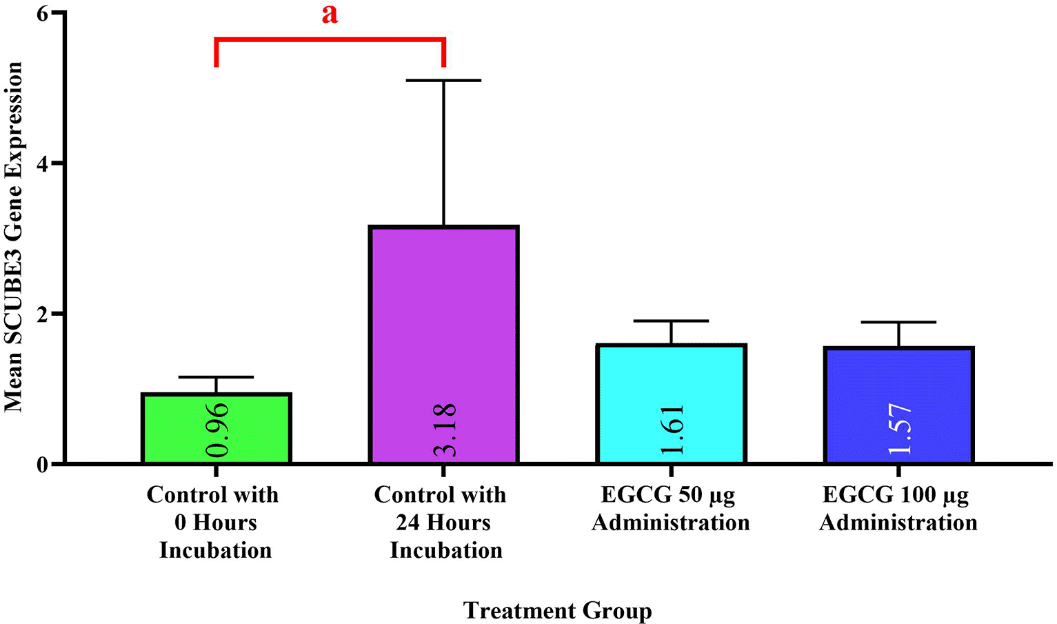
Notes: Significance values in all groups based on ANOVA test (p=0.113); “a” sign indicate significant differences between groups based on LSD comparison test (p<0.05), a=0.025.
Notes: Significance values in all groups based on ANOVA test (p=0.678).
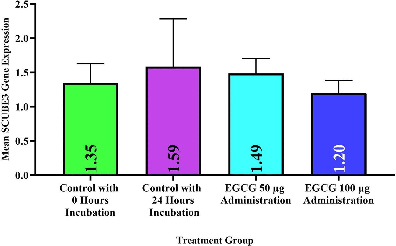
In samples incubated for 72 hours, the ANOVA test showed that there was no significant difference in the 1×1 cm preparation group (p=0.443) (Figure 3). A significant relationship was observed in the 2×2 cm preparation group (p=0.002) (Figure 4). LSD test showed a significant difference between the control with 0 hour incubation and the groups administered EGCG 50 μg and 100 μg in the 2×2 cm preparation (p=0.002) (p=0.001) respectively (Figure 4). Significant differences were also found between the control with 72 hours incubation and the groups administered EGCG 50 μg and 100 μg in 2×2 preparations (p=0.041) (p=0.008) respectively (Figure 4).

Notes: Significance values in all groups based on ANOVA test (p=0.443).
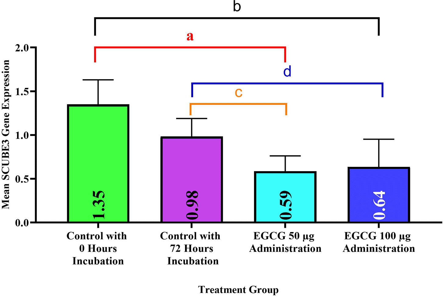
Notes: Significance in all groups based on ANOVA test (p=0.002); different “a,b,c”,…. indicate significant differences between groups based on LSD comparison test (p<0.05), SD: Standard Deviation, a=0.002, b=0.001, c=0.041, d=0.008.
The ANOVA test showed a significant relationship in the 1×1 cm preparation group (p=0.019) (Figure 5) and there was no significant relationship in the 2×2 preparation group (p=0.375) (Figure 6) LSD test showed significant differences between the control with 0 hours incubation and the control with 24 hours incubation (p=0.003) (Figure 5). Significant differences were also observed between the control group with 24 hours incubation and the group administered EGCG 50 μg and 100 μg in 1×1 cm preparation (p=0.016), (p=0.035) respectively (Figure 5).
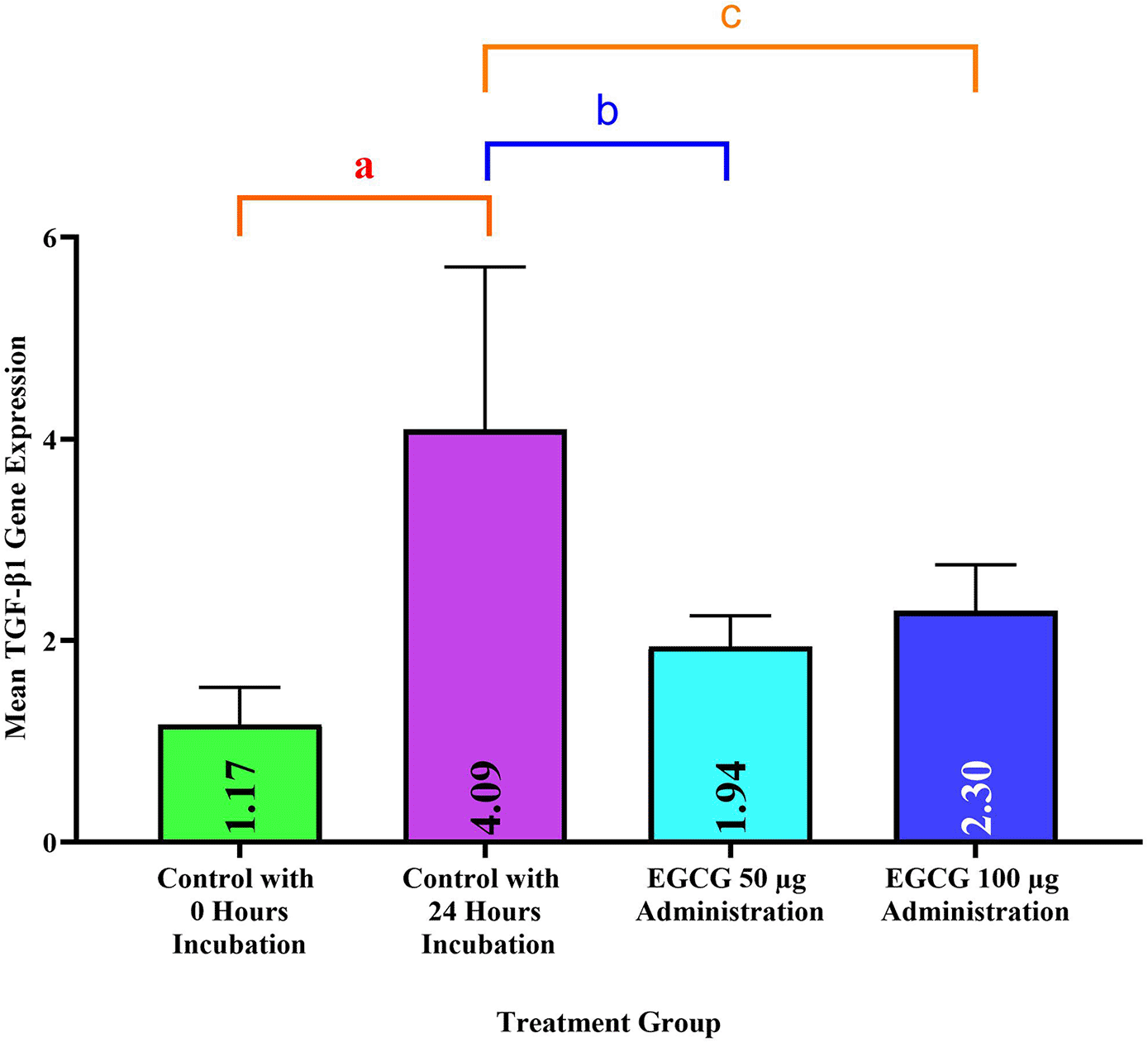
Notes: Significant values in all groups based on ANOVA test (p=0.019); different a,b,c,…. indicate significant differences between groups based on LSD comparison test (p < 0.05), SD: Standard Deviation, a=0.003, b=0.016, c=0.035.

Notes: Significance values in all groups based on ANOVA test (p=0.375).
The ANOVA test showed there was no significant relationship in the group 1×1 cm preparation (p=0.65) (Figure 7). A significant relationship was observed in the 2×2 cm preparation group (p=0.014) (Figure 8). LSD test showed significant differences between the control with 0 hours incubation and the groups administered EGCG 50 μg and 100 μg in 2×2 preparations (p=0.005) respectively (Figure 8).

Notes: Significance values in all groups based on ANOVA test (p=0.65).

Notes: Significant values in all groups based on ANOVA test (p=0.014); different a,b,c,…. indicate significant differences between groups based on LSD comparison test (p<0.05), SD: Standard Deviation, a,b = 0.005.
This study found that pleural tissue from empyema patients were having SCUBE3 gene expression. This was determined by the presence of SCUBE3 gene expression in the control samples. To the knowledge of the researcher, there has not been any study discussing the expression of SCUBE3 in pleural tissue affected by empyema, although theoretically this gene can be expressed due to the inflammatory process. Several studies have shown that SCUBE3 is associated with the development of several human cancers like early stage lung cancer (Chou et al., 2013), osteosarcoma (Liang et al., 2015), liver cancer (Xu et al., 2022), breast cancer (Shen et al., 2024), and the tooth development phase (Huo et al., 2021). Based on these studies, it is possible that SCUBE3 expression can also increase in other types of diseases, such as inflammatory diseases caused by empyema infection.
To the best of our knowledge, research on SCUBE3 expression in patients with empyema has not been conducted. Several studies have shown that empyema indirectly affects SCUBE3 expression. This began with a study conducted by Fiorelli in 2016, who found that pleural fluid levels of MMP-1, MMP-8, and MMP-9 were higher in patients with empyema parapneumonic effusions. MMP-2 levels were higher in patients with effusion but without empyema (Fiorelli et al., 2016).
Regarding its relationship with SCUBE3, research conducted by Qin in 2021 showed that MMP-2 and MMP-9 are proteins known to show the highest correlation with SCUBE3. This research supports the finding that empyema patients with increased levels of MMP-2 and MMP-9 also show an increase in the expression of SCUBE3 (Huo et al., 2021). It is also possible that MMP-1 and MMP-8 which are also widely secreted in empyema patients can also be related to SCUBE3, this can be material for future research.
In this study, we found a significant difference between SCUBE3 expression in 1×1 cm 0-hour incubation control and the 24-hour incubation control. However, there was no difference between the 0-hour incubation control and the 72-hour incubation. This is possible because SCUBE3 expression is greater in the control with 24 hours incubation than 72 hour incubation. We have not found literature that specifically discusses the changes of SCUBE3 expression at certain times in pleural tissue culture. But, some study found that gene expression in tissue culture can change over time.
Research has demonstrated that gene expression is dynamic and can vary significantly depending on various factors, including the duration of culture, cellular environment, and specific treatments. Studies have shown that gene expression profiles can change at different time points during cell culture. For instance, one study observed significant transcriptional changes in cells treated with a carcinogen over time, with specific genes showing increased or decreased expression at various intervals from one hour to eight days post-treatment (Ohmori et al., 2022). Another study highlighted that the transcriptional profiles of human adipose-derived stem cells differ significantly between freshly isolated cells and those expanded in culture (passage 0 vs. passage 1). This indicates that the process of culturing cells can lead to substantial alterations in gene expression profiles (Januszyk et al., 2015). The results obtained in this study indicate that the expression of the SCUBE3 gene in the control samples decreases as the incubation time increases.
Regarding the effect of EGCG on SCUBE3, no studies have directly analyzed the relationship between the two. However, several studies have discussed the effect of EGCG on MMPs, a protein that is correlated with SCUBE3 (Huo et al., 2021). Research conducted by Garbisa et al. showed that EGCG can suppress the gelatinolytic activity of MMP2 and MMP9 (Garbisa et al., 2001). An explanation of this process was explained in Maeda-Yamamoto’s study which found that EGCG inhibited phosphorylation of extracellular signal-regulated kinase 1/2 (ERK) and suppression of p38 activity which is required for the upregulation of MMP9 (Maeda-Yamamoto et al., 1999).
These findings suggest that the suppression of ERK phosphorylation by EGCG is involved in the decrease in MMP2 and MMP9 mRNA expression. Similar results were obtained by Cheng-hung and colleagues, who found that EGCG also reduced ERK phosphorylation and the levels of AP-1 and Sp1, leading to the downregulation of MMP2 and MMP9 (Chou et al., 2013).
The decreased expression of MMP2 and MMP9, which are proteins that correlate with SCUBE3, suggests that the decreased expression of MMP2 and MMP9 can also reduce the expression of SCUBE3. This supports the results obtained in this study, where researchers found that samples given EGCG at a concentration of 50 μg or 100 μg had a lower SCUBE3 expression than the control sample. In addition to the influence of EGCG as previously explained, a 72-hour incubation period resulted in lower SCUBE3 expression compared to a 24-hour incubation. This may be due to the fact that the longer EGCG is present in a tissue, the more it decreases the expression of SCUBE3 in that tissue. These results lead to a new assumption that SCUBE3 expression in pleural tissue samples from empyema patients may decrease over time, with a more significant reduction observed with EGCG administration.
This research found that the 2×2 cm samples treated with EGCG showed lower expression of SCUBE3 compared to the 1×1 cm samples. Based on the results obtained, we suspect that the empyema pleura samples have a better absorption capacity for EGCG in the 2×2 cm samples. This indicates that acquiring tissue to achieve the best potential of EGCG in pleural tissue is through culturing 2×2 cm samples.
Ideally, the optimal size for tissue culture involves small tissue fragments, since this facilitates enhanced perfusion and absorption. Larger tissue pieces possess a higher surface area, resulting in less efficient absorption compared to smaller fragments. On the other hand, if the tissue sample taken is too small, there are concerns that the tissue acquisition may not be optimal. Consequently, an optimal metric is essential for attaining better results in organ cultivation (Cheng et al., 2022).
Several studies indicates that the best size for culturing human pleural mesothelial cells and pleural fibroblasts is 8.5 mm (Metelmann et al., 2022). Therefore, for easier measurement, we standardized it to 1×1 cm. Meanwhile, for the 2×2 cm preparation, we were trying to expand the size to see if a larger size can still serve as a reference for making tissue preparations. Numerous studies indicate that bigger specimen sizes are susceptible to necrosis; however, our findings reveal that tissue samples measuring 2×2 cm did not exhibit necrosis. We suggest that utilizing a 2×2 cm preparation could encourage novel approaches in the examination of pleural tissue collection methodologies.
The increase in TGF-β1 expression in pleural control samples from empyema patients in this study is in line with the findings of several previous studies. Research conducted by Scott et al. in 2003 on rabbit-induced empyema found that there was an increase in TGF-β1 concentration in the pleural fluid of mice (Sasse et al., 2003). One year later this research was supported by Craig and friends who found that intrapleural injection of anti-TGF-β1 in the pleura of mice induced by empyema was known to inhibit the development of empyema and significantly reduce the process of rabbit pleural fibrosis (Kunz et al., 2004).
The opposite result was found by Samanta and colleagues in 2018, who observed an increase in TGF-β1 levels in mice with induced pleural empyema. The study found no significant difference in TGF-β1 concentration between the experimental and control groups (da Silva et al., 2018).
Differences in research results may be due to differences in sample types and research objects. Research conducted by Scott and Craig examined the concentration of TGF-β1 in rabbit pleural fluid and Samanta examined the concentration in mouse pleural fluid. Meanwhile, this study used human pleural tissue to observe the presence of TGF-β1 expression. The difference in sample types between pleural tissue in both humans and experimental animals may have affected the results obtained, although there are studies that show that there are highly homologous TGF-β1 amino acid sequences among mammalian species (Terrell et al., 1993).
We speculate that the increased expression of TGF-β1 in pleural tissue in this study occurred because of inflammation caused by empyema infection. This is based on the finding of TGF-β1 in mesothelial cells (Gerwin et al., 1987) alveolar macrophages (Assoian et al., 1987), lymphocytes, fibroblasts (Awad et al., 1998) and inflammatory cells in the stimulated pleural fluid. All these cell types are present in the visceral pleura and in some inflammatory exudates during early fibrotic formation in empyema (Martin et al., 2000). Previous studies that examined the relationship between TGF-β1 and pleural disease also suggested that the elevated TGF-β1 in pleural fluid is produced by inflammatory cells (Lee and Lane, 2001).
We found that the 1×1 cm control sample incubated for 24 hours had a higher expression of the TGF-β1 gene compared to the 1×1 cm control sample incubated for 72 hours. This resulted a significant difference between the 0-hour incubation control and the 24-hour incubation control. Researchers suspect that the reason for the significant increase in TGF-β1 expression after 24 hours of incubation is the same as that occurring in SCUBE3 expression. This leads to a new finding where the control samples of tissue culture incubated for 24 hours exhibit greater gene expression than those incubated for 72 hours.
This study also found that there was a decrease in TGF-β1 expression in 2×2 cm samples given EGCG 50 μg and 100 μg in the first 72 hours of incubation. In addition to the factor of EGCG administration, the TGF-β1 expression in the samples incubated for 72 hours was also lower than that in the samples incubated for 24 hours. The reason for this has been explained previously when discuss SCUBE3 expression.
To the best of our knowledge, no study has discussed the effect of EGCG on TGF-β1 in the pleural tissue. Research on this topic is often focused on lung fibrosis and cancer. For example, research conducted by Sriram and colleagues on lung fibrosis in bleomycin-induced mice showed that EGCG can inhibit fibroblast activation and collagen accumulation by inhibiting TGF-β1 expression. Based on these results, EGCG is considered to be an effective therapy for lung fibrosis. In an in vitro study, Sriram and colleagues also revealed that EGCG was able to reverse TGF-β1 induced proliferation and activation of fibroblasts (Sriram et al., 2015).
The same study was conducted by Tsai et al. in a rat lung fibrosis model. Tsai showed that EGCG decreased TGF-β1 expression, MMP-2, and MMP-9 levels, and also had anti-fibrotic effects. This study also found that EGCG reduced mortality; improved lung histological changes; reduced serum levels of TGF-β1, IL-6, IL-10, and TNFα; and reduced collagen deposition and fibroblast proliferation (Tsai et al., 2019).
Although the two studies were only on lung fibrosis, they support the results of this study, which showed that there was a decrease in TGF-β1 expression in the pleura of patients given EGCG as much as 50 μg and 100 μg EGCG in the first 72 hours of incubation. A possible mechanism to explain the inhibitory effect of EGCG on TGF-β1 activation is the suppression of various TGF-β1 activators such as inflammatory cytokines, MMPs, macrophages, and other inflammatory cells (Sriram et al., 2009). The authors also assume that such a mechanism is also involved in the reduction of TGF-β1 expression in pleural tissue.
Similar to the SCUBE3 expression, this study also found that the 2×2 cm preparation showed less TGF-β1 expression than the 1×1 cm preparation. These results further strengthen our previous hypothesis that the potential of EGCG is more potent in samples with a 2×2 cm preparation.
This study did not explore the presence of other profibrotic factors that could potentially influence the occurrence of pleural fibrosis such as matrixmetalloproteinase-1,2,8, and 9 (MMP-1, MMP-2, MMP-8, and MMP-9). 2. The researchers only studied the effects of EGCG on pleural tissue, it is preferable to perform a comparative trial of the effect of EGCG on the potential fibrosis in pulmonary tissue due to empiema.
In Summary, our research indicated that EGCG has the potential to lower SCUBE3 and TGF-β1 expression in patients with pleural empyema. This discovery might have applications in EGCG treatment to prevent pleural fibrosis in patients with empyema in the future.
The research was approved by the Universitas Andalas Ethical Clearance Committee (Protocol number: DP.04.03/D.XVI.XI/26/2024), and the gatekeeper’s consent letters were provided by Dr. M. Djamil of the Teaching Hospital on January 9, 2024 and is valid until January 2025. This research has been declared to be ethically appropriate in accordance to 7 (seven) WHO 2011 Standards, 1) Social values, 2) Scientific Values, 3) Equitable Assessment and Benefits, 4) Risks, 5) Persuasion/Exploitation, 6) Confidentiality and Privacy, and 7) Informed Consent, referring to the 2016 CIOMS Guidelines. This is as indicated by the fulfilment of the indicators of each standard. All the participants provided written informed consent.
| Views | Downloads | |
|---|---|---|
| F1000Research | - | - |
|
PubMed Central
Data from PMC are received and updated monthly.
|
- | - |
Provide sufficient details of any financial or non-financial competing interests to enable users to assess whether your comments might lead a reasonable person to question your impartiality. Consider the following examples, but note that this is not an exhaustive list:
Sign up for content alerts and receive a weekly or monthly email with all newly published articles
Already registered? Sign in
The email address should be the one you originally registered with F1000.
You registered with F1000 via Google, so we cannot reset your password.
To sign in, please click here.
If you still need help with your Google account password, please click here.
You registered with F1000 via Facebook, so we cannot reset your password.
To sign in, please click here.
If you still need help with your Facebook account password, please click here.
If your email address is registered with us, we will email you instructions to reset your password.
If you think you should have received this email but it has not arrived, please check your spam filters and/or contact for further assistance.
Comments on this article Comments (0)