Keywords
multiple sclerosis, SARS-CoV-2 vaccination, COVID-19, immunological disease, complication
multiple sclerosis, SARS-CoV-2 vaccination, COVID-19, immunological disease, complication
Vaccinations against severe acute respiratory syndrome coronavirus type 2 (SARS-CoV-2) can be complicated by side effects, which commonly affect the central or peripheral nervous system.1 Among the side effects affecting the CNS, inflammatory, demyelinating disorders are increasingly being recognised.1 These include acute disseminated encephalomyelitis, acute, hemorrhagic, necrotizing encephalopathy, acute necrotizing encephalopathy, neuromyelitis optica spectrum disorders, myelin oligodendrocyte glycoprotein antibody-associated disease, and multiple sclerosis.2,3 These CNS complications of SARS-CoV-2 vaccinations may manifest as flares of disease already diagnosed before SARS-CoV-2 vaccination or as emerging disease triggered by SARS-CoV-2 vaccination.2 Relapses of multiple sclerosis have occasionally been reported as complications of SARS-CoV-2 vaccinations.4–6 New-onset multiple sclerosis has been reported less frequently as a side effect of SARS-CoV-2 vaccination.6–11 Several of these cases have been published either as individual case reports7,8 or as case series.6,10,11
A middle-aged female presented with recent-onset right hemiparesis first noticed two days after an adenovector-based SARS-CoV-2 vaccine.7 Cerebral MRI showed a large subcortical T2 and fluid attenuated inversion recovery hyperintense lesion involving the corpus callosum.7 When intravenous methyl-prednisolone was administered, the size of the lesion decreased within four weeks of starting treatment.7 In another case report, a 24 year-old male developed right-sided facial weakness and dysarthria two days after receiving the first Moderna vaccine dose.8 Magnetic resonance imaging showed typical acute and chronic central nervous system (CNS) lesions which met the McDonalds criteria for the diagnosis of multiple sclerosis. The flare was treated with glucocorticoids and subsequently ocrelizumab was given.8 Multiple sclerosis-like disease was also diagnosed in a 49 year-old male who presented with fever, diplopia, unsteady gait, and numb feet four days after the second dose of the Moderna vaccine.9 The neurological examination revealed appendicular ataxia of the right leg, a minimally reduced vibration sense in both legs, and gait ataxia.9 Magnetic resonance imaging showed supra- and infra-tentorial demyelinating lesions, most of them enhancing and cerebrospinal fluid studies showed mild pleocytosis and increased protein.9 Multiple sclerosis was diagnosed and methyl-prednisolone was administered, which led to significant improvement.9 In a study of 32 patients who developed multiple sclerosis after a SARS-CoV-2 vaccination, risk factors for developing multiple sclerosis post-coronavirus disease (post-COVID) vaccination included the BNT162b2 vaccine, low vitamin-D levels, positive Ebstein-Barr nuclear antigen-1 (EBNA1)-IgG antibodies, and a positive family history for multiple sclerosis.10 It was concluded that these risk factors can be used as significant independent predictors of the development of multiple sclerosis after COVID.10 In a report of 9 patients who developed multiple sclerosis and one patient who developed cerebral isolated syndrome after vaccination with inactivated SARS-CoV-2 or vial vector vaccines, all patients recovered fully or incompletely after taking steroids.11 Secondary prophylaxis included natalizumab, teriflunamide, demithyl fumarate, or rituximab.11 It was concluded that BBIBP-CorV and AZD1222 are potential triggers of CNS demyelinating disease and that these side effects cannot be denied.11 In case series of six patients, Rinaldi et al. reported two additional patients with new-onset multiple sclerosis after -SARS-CoV-2 vaccination.6 The first patient was a 46 year-old female with a history of renal cell carcinoma who presented with numbness in both upper limbs ten days after the first BNT162b2 dose and double vision 25 days after the second dose.6 Magnetic resonance imaging of the brain and spine revealed multiple supra- and infra-tentorial and spinal cord enhancing and non-enhancing lesions as well as positive oligoclonal bands.6 Multiple sclerosis was diagnosed and the patient benefited from steroids and natlizumab.6 The second patient was a 27 year-old female who developed mild monoparesis of the left leg, facial palsy, and dysarthria seven days after the second BNT162b2 dose.6 Workup was consistent with the diagnosis of multiple sclerosis and the patient benefited from prednisolone and cladribine.6
Here we report a patient with new-onset relapsing-remitting multiple sclerosis seven weeks after the second BNT162b2 vaccine dose, who was diagnosed only after a relapse of relapsing-remitting multiple sclerosis (RRMS) after the third vaccine dose.
The patient is a human immunodeficiency virus-negative, 31 year-old man, height 177 cm, weight 68.5 kg, with a previous history of Gilbert’s disease, hyperlipidemia, and mild renal insufficiency, who experienced significant eye discomfort, fatigue and some light sensitivity when trying to look at screens, TVs and monitors. 48 days after the second BNT162b2 dose. He had tolerated the first dose 21 days earlier without side effects. Slit lamp eye exam at the optometrist was reportedly normal. Symptoms lasted daily for two weeks and were attributed to computer eye fatigue. Symptoms gradually disappeared after that period. Despite vaccination, the patient experienced SARS-CoV-2 infection eight months later. One and a half years after the second dose, he received his third BNT162b2 dose. About three months later, he developed urinary urgency, which persisted since then. Eleven months after the third BNT162b2 dose, he began experiencing episodic, right-sided tingling and burning sensation, as well as visual discomfort with focusing and eye sensitivity and feeling that eyes were moving at the end of workouts in the gym. These episodes were initially lasting a few minutes. During the following two months, however, these episodes were worsening, occurring daily lasting 20-30 minutes at the beginning and end of the day. They worsened with any alcohol consumption.
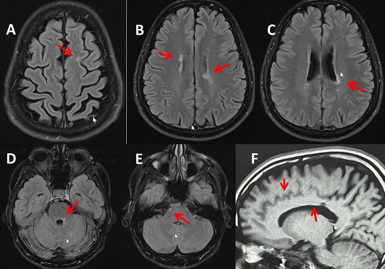
Clinical neurological examination revealed brisk patella and Achilles tendon reflexes, but no crossed adductors. Plantar responses were mute bilaterally with 2 to 3 beats of non-sustained clonus bilaterally. There was mildly reduced vibration sensation bilaterally at great toes but intact proprioception. Cerebral magnetic resonance imaging three months after onset of the sensory disturbances and visual discomfort showed multiple, nonspecific, non-expansible T2 and fluid attenuated inversion recovery hyperintense and T1 hypointense lesions involving the juxta-cortical, subcortical, periventricular white matter, the right lateral pons, and the left superior cerebellar peduncle, which did not enhance (Figure 1). Juxta-cortical involvement was noted in the left superior frontal gyrus. There was no involvement of the corpus callosum, no infarction, haemorrhage, mass, or abnormal enhancement. Magnetic resonance imaging with contrast agent of the entire spine was normal. Cerebrospinal fluid examinations revealed elevated kappa free light chains of 0.5070 mg/dl (n, <0.1 mg/dL) and 8 (n, <3) oligoclonal bands on the oligoclonal bands assay. Ophthalmologic examination by slit lamp and optical coherence tomography were normal, but the pupillary reflex was slowed and there was a right side afferent pupillary defect (1+. Aquaporin-4 antibodies, myelin oligodendrocyte glycoprotein antibodies, and neuromyelitis optica antibodies were negative. Hepatitis B surface antibodies were markedly elevated. John Cunningham virus antibodies were positive and the index was increased to 2.78 (n, <0.4). The quantiferon test was negative. Visually evoked potentials were not recorded yet. Since the patient met the revised McDonalds criteria (two flares, temporal and spatial dissemination, positive oligoclonal bands), multiple sclerosis with a positive Uhthoff phenomenon (symptoms occur with exercise or heat, which is common with demyelinating lesions)12 was diagnosed with an expanded disability status scale of 1.5, a disease activity score of 8, and secondary prophylaxis with ocrelizumab was initiated.
The case presented is of interest because it concerns a new-onset multiple sclerosis, presumably triggered by SARS-CoV-2 vaccination with BNT162b2, with a first flare 48 days after the second dose and a second flare 11 months after the third shot. The diagnosis multiple sclerosis was made using the revised McDonald criteria. The patient showed temporal dispersion (initial flair with visual impairment presumably due to optic neuritis, urinary urgency, sensory disturbances) and magnetic resonance imaging showed a juxta-cortical lesion, several periventricular lesions and two infratentorial lesions. In addition, the oligoclonal bands were positive and the free kappa light chains in the cerebrospinal fluid were significantly increased. The patient also met the Barkhof criteria with one juxtacortical lesion, two infratentorial lesions, and positive oligoclonal bands.
Differential diagnoses that were excluded include neuromyelitis optica spectrum disorders and myelon oligodendrocyte glycoprotein-associated disease but also acute viral encephalitis, immune encephalitis, acute disseminated encephalomyelitis, acute hemorrhagic necrotising encephalitis, acute necrotising encephalopathy, posterior reversible encephalopathy syndrome, cerebral vasculitis, vascular malformation, anterior ischemic optic neuropathy, antiphospholipid syndrome, sarcoidosis, congenital leucodystrophy, Fabry disease, vitamin-B12 deficiency, ischemic stroke, Susac syndrome, human immunodeficiency virus infection, Borreliosis, syphilis, fibromyalgia, Behcet disease, lupus erythematodes, Sjögren syndrome, cerebral neoplasms, and migraine. Neuromyelitis optica spectrum disorders and myelon oligodendrocyte glycoprotein-associated disease were excluded because spinal magnetic resonance imaging did not show a spinal cord lesion and because aquaporin-4 antibodies, myelin oligodendrocyte glycoprotein antibodies, and neuromyelitis optica-associated antibodies were negative. Viral encephalitis was excluded because there was no enhancing lesion on magnetic resonance imaging and because the viral panel for various viruses was negative. Immune encephalitis was excluded because there was no enhancing lesion on magnetic resonance imaging and because immune encephalitis-associated antibodies were negative. Acute disseminated encephalomyelitis was excluded because the spinal magnetic resonance imaging was normal. Acute hemorrhagic, necrotising encephalitis was excluded because there was no indication for bleeding. Acute necrotising encephalopathy was excluded because there were no lesions that could be interpreted as necrosis.
Although the latency between vaccination and onset of the first and second relapses were quite long (first flair: 48 days after second BNT162b2 dose, second flair: 11 months after third BNT162b2 dose), a causal relationship cannot be excluded. Arguments for of a causal relationship are that it is known that infections, including SARS-CoV-2,13 and vaccinations can trigger the occurrence of multiple sclerosis,7 that there was a temporal connection between vaccination and the occurrence of multiple sclerosis, especially after the second BNT162b2 dose, that the SARS-CoV-2 vaccination favours the development of autoimmune disease in general3 and triggers autoimmune-inflammatory reactions,14 and that several cases of SARS-CoV-2 vaccination associated multiple sclerosis have been reported previously.6–11 However, there are also studies that showed that SARS-CoV-2 vaccinations are safe and neither increase the prevalence of multiple sclerosis nor trigger relapses of the disease.15
A further argument in favour for a causal relation is that also several cases in which SARS-CoV-2 vaccination triggered the development of myelin oligodendrocyte glycoprotein antibody disease respectively neuromyelitis optica spectrum disorders have been reported.16 New-onset myelin oligodendrocyte glycoprotein antibody disease has been reported in a 46 year-old female who experienced reduction in visual acuity in her left eye approximately two weeks after the first dose of an RNA-based SARS-CoV-2 vaccine.16 Orbital magnetic resonance imaging showed mild enhancement of the retrobulbar fat surrounding the optic nerves and optic nerve hyperintensity and enhancement involving the infraorbital and orbital apex with sparing of the prechiasmatic optic nerves, consistent with acute bilateral optic neuritis.16 Cerebrospinal fluid analysis only revealed increased myelin basic protein. Serological analysis for aquaporin-4 antibodies was negative but antibodies against myelin oligodendrocyte glycoprotein returned positive with a titter of 1:1000 (n, 1:20).16 Based on these findings myelin oligodendrocyte glycoprotein antibody disease was diagnosed and intravenous steroids followed by plasmapheresis were given with a beneficial effect. The addition of rituximab, however, resulted in numerous relapses, which is why she was transitioned to monthly intravenous immunoglobulins.16 A second case concerns a previously healthy 23-year-old woman who presented with dizziness, vomiting, and headache starting 33 days after the second dose of the BNT162b2 vaccine.17 Magnetic resonance imaging showed hyperintensity around the fourth ventricle on fluid-attenuated inversion recovery images, which enhanced on T1 sequences with gadolinium.17 Clinical neurologic examination revealed bilateral horizontal nystagmus and mild ataxia in the finger-nose test.17 Cerebrospinal fluid analysis revealed pleocytosis of 48 cells/microL, positive oligoclonal bands but was otherwise normal.17 Serological tests revealed the presence of myelin oligodendrocyte glycoprotein antibodies in a live cell-based assay.17 The patient benefited significantly from oral and intravenous prednisolone.17 Jarius et al. reported a single 67 year-old male with myelin oligodendrocyte glycoprotein antibody disease that occurred ten days after the second BNT162b3 dose.18 The patient presented clinically with impaired vision on the left eye and pain on eye movements.18 Workup revealed swelling and enhancement of the left optic nerve, suggesting optic neuritis, prolonged left P100 latency, mild pleocytosis, and increased serum antibodies against myelin oligodendrocyte glycoprotein.18 Administration of methyl-prednisolone resulted in complete recovery.
The pathophysiology underlying the development of multiple sclerosis after SARS-CoV-2 vaccination remains elusive, but it can be speculated that the immune response against the vaccine or an adjuvant may also attack neuronal structures, the myelin sheath, or the cellular immune system. Generally, mRNA-based anti-SARS-CoV-2 vaccines carry two components. The first is a pathogen-specific antigen (immunogen synthesising the SARS-CoV-2 spike protein) against which neutralising antibodies and T cells are directed.6 The second is an adjuvant for the intrinsic properties to be recognised by pattern recognition receptors.6 The adjuvant stimulates the innate immune response and provides the second signal and pro-inflammatory cytokines to initiate the adaptive response.6 Accordingly, several mechanisms can be proposed to explain COVID-19 vaccination-induced demyelinating CNS disease. First, vaccines that stimulate the innate immune response through the adjuvant and create an inflammatory cytokine environment, may activate preformed, self-reactive T and B cells, in a process known as bystander activation. Bystander activation may develop in the early immune response and therefore may be involved in acute demyelinating events a few days after vaccination with mRNA vaccines.6 A second pathophysiologic mechanism to explain demyelination after SARS-CoV-2 vaccination could be molecular mimicry,1 Molecular mimicry is characterised by vaccine-derived antigens which mimic self-molecules that could stimulate cross-reactive T cells.6 A third mechanism could be epitope spreading.6 Epitope spreading is characterised by an immune response that reacts also against different epitopes of the same or other proteins after initial activation of antigen-specific T cells against a dominant epitope.6 All these three mechanisms involve a cell-mediated as well as humoral adaptive immune response.6
The subtle APD 1+ on the right eye and sluggish pupil movement with limited constriction on both eyes in the index patient was suspected due to a process in the Edinger-Westphal nucleus involving the right side more than left. The efferent branch was interpreted as unaffected, but limited in response. The “slow” ability to focus near was suspected due to post-inflammatory neurologic deficit involving multiple areas. Ophthalmologists interpreted the findings as definitely suspicious for multiple sclerosis but in the patient’s age bracket more likely to be a COVID vaccine immune reaction. They recommended return for formal visual field testing and dilated exam for completeness, high resolution MRI in the Edinger-Westphal nucleus region and an orbital magnetic resonance imaging looking for optic tract inflammation.
In summary, this case shows that SARS-CoV-2 vaccination can occasionally trigger new-onset multiple sclerosis, that the clinical presentation of SARS-CoV-2 vaccination-induced multiple sclerosis does not differ from multiple sclerosis due to other triggers, that the latency between vaccination and onset of multiple sclerosis manifestations is longer than with other neurological disorders, and that the severity of relapses may increase with the number of booster vaccinations administered. Although rare, neurologists should be aware of the likely event in which SARS-CoV-2 vaccination triggers new-onset multiple sclerosis in patients with no prior immunological disease or compromised immune system.
| Views | Downloads | |
|---|---|---|
| F1000Research | - | - |
|
PubMed Central
Data from PMC are received and updated monthly.
|
- | - |
Provide sufficient details of any financial or non-financial competing interests to enable users to assess whether your comments might lead a reasonable person to question your impartiality. Consider the following examples, but note that this is not an exhaustive list:
Sign up for content alerts and receive a weekly or monthly email with all newly published articles
Already registered? Sign in
The email address should be the one you originally registered with F1000.
You registered with F1000 via Google, so we cannot reset your password.
To sign in, please click here.
If you still need help with your Google account password, please click here.
You registered with F1000 via Facebook, so we cannot reset your password.
To sign in, please click here.
If you still need help with your Facebook account password, please click here.
If your email address is registered with us, we will email you instructions to reset your password.
If you think you should have received this email but it has not arrived, please check your spam filters and/or contact for further assistance.
Comments on this article Comments (0)