Keywords
Dermatochalasis; Upper eyelid blepharoplasty; Blepharoptosis; Botulinum toxin; Blepharoplasty Outcomes Evaluation (BOE); Case report.
This article is included in the Eye Health gateway.
Dermatochalasis of the upper eyelids is a frequent age-related condition that may impair vision and pose a major aesthetic concern. Although both surgical and nonsurgical treatments are available, improper or repeated administration of nonsurgical approaches, such as botulinum toxin (BT) injections, can lead to functional issues, including eyelid heaviness, ocular fatigue, and patient dissatisfaction. We report the case of a 45-year-old Syrian man with bilateral upper eyelid dermatochalasis who had received multiple periocular BT injections over two years. Despite initial improvement, he developed recurrent skin redundancy, ocular heaviness, fatigue, headaches, and aesthetic dissatisfaction. Clinical examination revealed bilateral eyelid skin redundancy and superior visual field restriction with preserved levator function. Bilateral upper eyelid blepharoplasty was performed, involving careful excision of redundant skin, removal of excessive preaponeurotic fat likely accentuated by prior BT treatment and partial excision of the orbicularis oculi muscle, while preserving the levator apparatus and orbital septum integrity. The procedure was completed without complications. Postoperatively, the patient experienced complete resolution of eyelid heaviness and subjective improvement in the visual field, as well as a more defined eyelid contour and improved symmetry. Patient satisfaction, measured using the Blepharoplasty Outcomes Evaluation (BOE) questionnaire, increased from 25% preoperatively to 96.8% at three and six months postoperatively. This case demonstrates that upper eyelid blepharoplasty remains the gold standard for managing dermatochalasis, particularly when nonsurgical interventions have failed or produced functional complications. The procedure can successfully restore eyelid function, improve visual performance, and achieve high aesthetic satisfaction, highlighting the importance of proper patient selection and timely surgical intervention in the management of upper eyelid aging.
Dermatochalasis; Upper eyelid blepharoplasty; Blepharoptosis; Botulinum toxin; Blepharoplasty Outcomes Evaluation (BOE); Case report.
Aging of the upper face commonly leads to a sensation of eyelid heaviness, resulting from brow descent, redundant upper eyelid skin, or both.1 Upper eyelid blepharoplasty, or eyelid lift, corrects redundant skin and differs from brow lift surgery, which elevates the eyebrow; in selected cases, both procedures may be combined to optimize functional and aesthetic outcomes.2,3 Age-related morphological changes, including elastolysis, collagen remodeling, and fat redistribution, contribute to dermatochalasis, which can cause aesthetic concerns, visual field obstruction, and psychological distress.4,5 Management requires careful evaluation and individualized treatment planning by surgeons experienced in periocular surgery. Available treatments range from nonsurgical approaches, such as botulinum toxin (BT) injections and energy-based devices, to definitive surgical correction via blepharoplasty.6,7 Although nonsurgical interventions are minimally invasive, repeated or improperly administered periocular BT injections can lead to functional disturbances such as eyelid heaviness, ptosis, ocular fatigue, and aesthetic dissatisfaction, ultimately necessitating surgical intervention.8–11
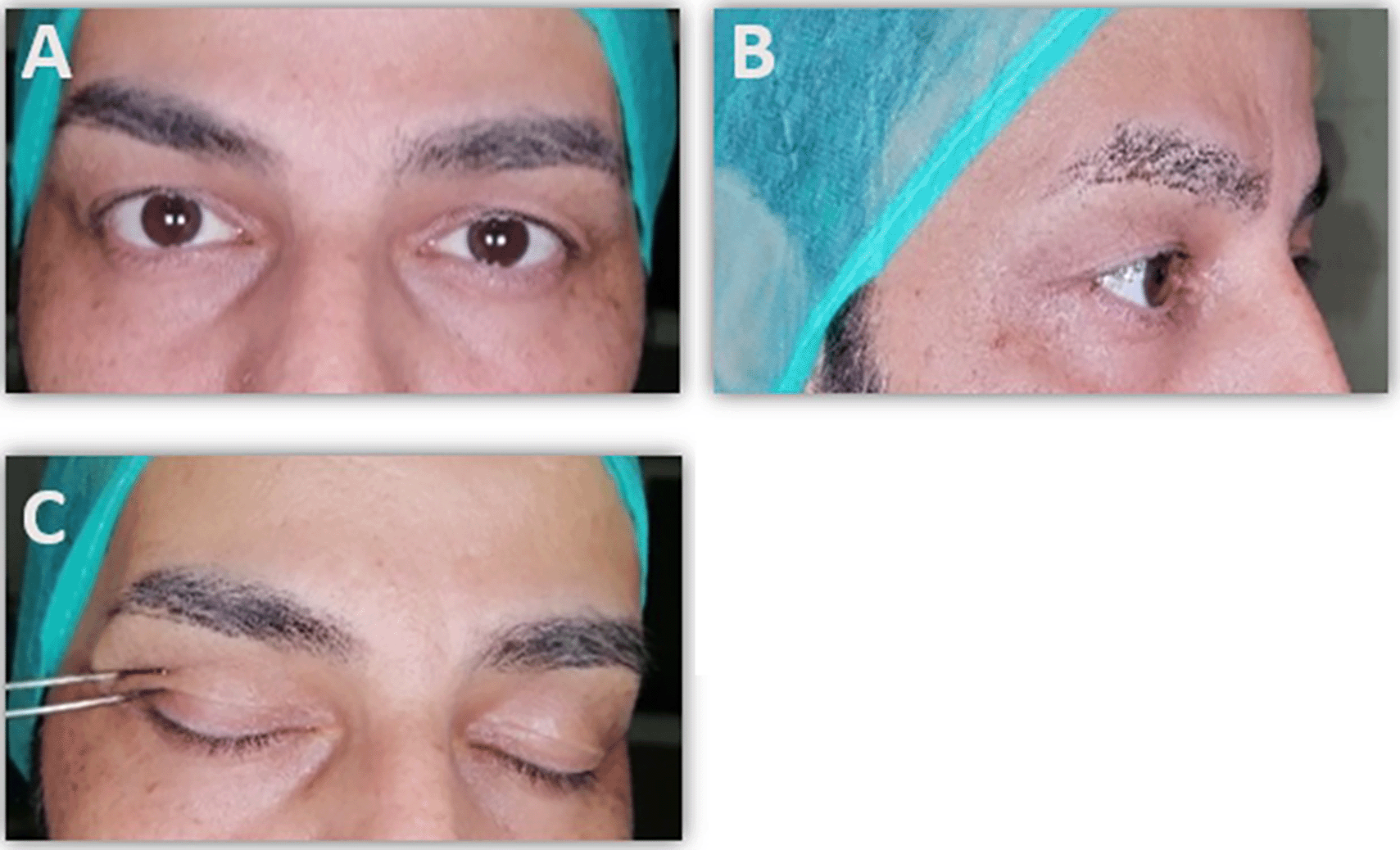
A 45-year-old Syrian man presented to the Department of Oral and Maxillofacial Surgery, Damascus University, in 2024 with complaints of bilateral upper eyelid dermatochalasis and a two-year history of repeated periocular botulinum toxin (BT) injections (two sessions every three months). While initial improvement was noted, he experienced recurrence of eyelid skin redundancy accompanied by headaches, ocular heaviness, and functional discomfort. Clinical examination revealed bilateral upper eyelid skin redundancy with mild lateral hooding and intact levator function. Superior visual field restriction was documented. Given the persistence of functional impairment and aesthetic dissatisfaction, bilateral upper eyelid blepharoplasty was planned. Intraoperatively, excessive preaponeurotic fat was identified and carefully excised along with redundant skin, while preserving the levator apparatus and orbital septum. A small segment of the orbicularis oculi muscle was also removed to enhance eyelid contour. The procedure was completed without intraoperative complications. Postoperatively, the patient reported complete resolution of eyelid heaviness and subjective improvement in the visual field (Figure 1). Aesthetic results were favorable, demonstrating improved eyelid contour and symmetry. Patient-reported satisfaction, measured using the Blepharoplasty Outcomes Evaluation (BOE) questionnaire, increased from 25% preoperatively to 96.8% at both three and six months follow-up.

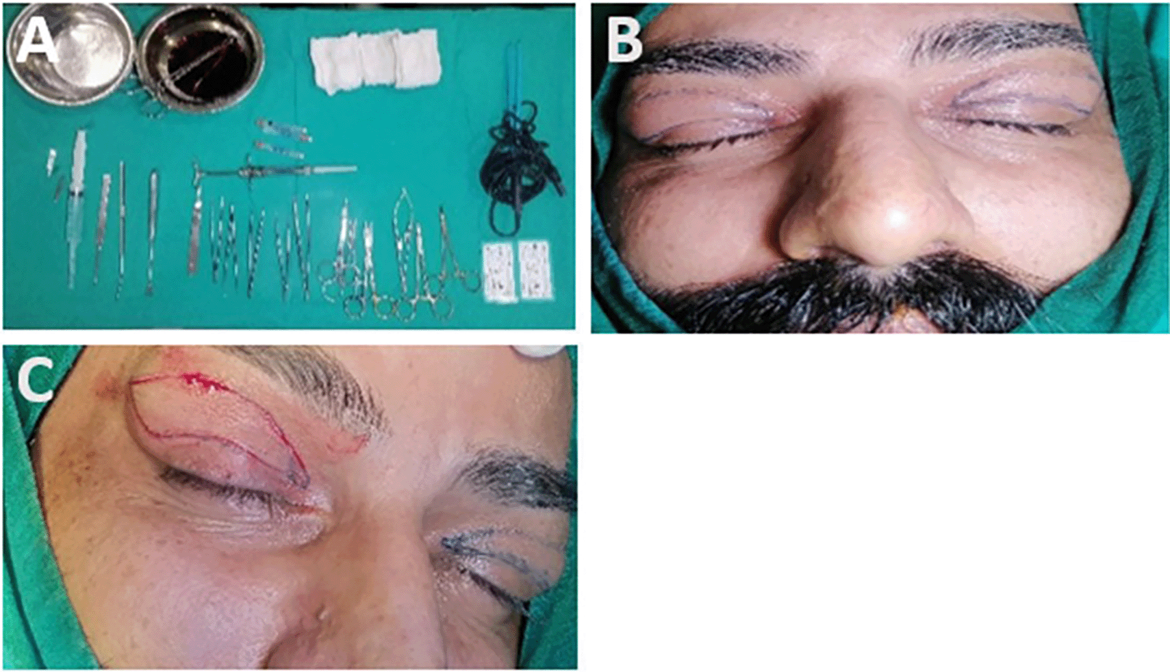
Preoperative marking: With the patient seated upright in neutral gaze, the lower margin of skin excision was delineated along the natural upper eyelid crease. In cases of indistinct or elevated creases, the central incision was marked 8–10 mm from the lid margin in women and 6–8 mm in men. The lateral limit was extended obliquely from the lateral canthus toward the lateral brow.
Anesthesia and incision: Local anesthesia was administered using 20 mg/mL mepivacaine with adrenaline.12 The skin incision followed the natural eyelid crease, and redundant skin was carefully excised using fine scissors. Hemostasis was meticulously maintained to minimize postoperative edema and ecchymosis.
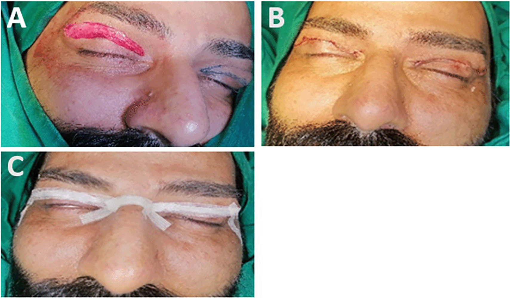
Fat and muscle management: Preaponeurotic fat from the medial and central compartments was either excised or repositioned as required. A small segment of the orbicularis oculi muscle was removed to refine eyelid contour. Care was taken to preserve at least 20 mm of vertical lid height to maintain normal eyelid closure.13
Closure: The wound edges were approximated with interrupted 6-0 polypropylene sutures placed within the natural lid crease for optimal scar concealment. The incision was reinforced with Steri-Strips to support wound healing (Figures 2–4).


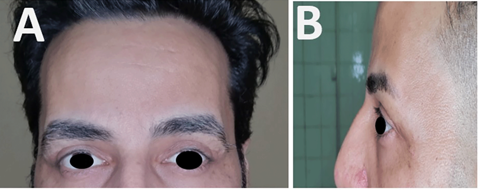
The patient experienced an uneventful recovery. Sutures were removed at one week, and superficial ecchymosis resolved within 10 days. No complications were observed, including hematoma, wound dehiscence, abnormal scarring, or overcorrection. Aesthetic results were favorable, with a well-defined supratarsal crease, appropriate pretarsal show, and a smooth lid–cheek junction. The upper eyelid margin rested approximately 2 mm below the superior limbus, and the lower eyelid aligned with the inferior limbus. Functionally, the patient reported complete resolution of eyelid heaviness and marked improvement in the superior visual field (Figures 5–7). Patient satisfaction, evaluated using the Blepharoplasty Outcomes Evaluation (BOE) questionnaire, improved from 25% preoperatively to 96.8% at six months (Figure 8).


The upper eyelid is composed of skin, subcutaneous areolar tissue, striated muscle, submuscular areolar tissue, tarsi, and the orbital septum. With aging, these structures undergo progressive morphological changes, which may be further accentuated in patients receiving repeated botulinum toxin (BT) injections for periorbital rejuvenation.14 Dermatochalasis, characterized by redundant upper eyelid skin, is a hallmark of periorbital aging and may impair ocular function through lateral hooding and superolateral visual field obstruction.15 Age-related elastolysis, collagen remodeling, loss of ground substance, and dermal thinning contribute to decreased skin elasticity, while gravitational forces and diminished subcutaneous support exacerbate tissue redundancy.16,17 Fat redistribution and weakening of connective tissue further influence the periorbital–cheek transition, intensifying the clinical presentation.14,18
BT injections have gained popularity for upper-face rejuvenation because of their rapid and reproducible effects in reducing dynamic wrinkles, particularly in the glabella, forehead, and crow’s feet regions.19,20 Although generally safe, BT administration can cause complications, including blepharoptosis, eyebrow asymmetry, headache, diplopia, lagophthalmos, ectropion, and xerophthalmia. Improper technique or excessive dosing can exacerbate eyelid heaviness, lateral hooding, and visual field obstruction.21–23
In this case, despite multiple BT treatments, the patient developed persistent dermatochalasis and functional symptoms that required surgical correction. Upper eyelid blepharoplasty successfully addressed both functional and aesthetic concerns, with intraoperative excision of redundant skin, removal of preaponeurotic fat, and partial excision of the orbicularis oculi muscle performed while preserving the levator apparatus and orbital septum. Careful hemostasis minimized postoperative edema and ecchymosis. Postoperatively, the patient reported resolution of heaviness, improvement in visual field, and high satisfaction, with BOE scores increasing from 25% preoperatively to 96.8% at six months.
This case underscores that while BT is a valuable nonsurgical tool, it does not substitute for surgery in patients with significant dermatochalasis or fat herniation. Blepharoplasty remains the gold standard for restoring eyelid function and contour. Optimal results require precise intraoperative assessment, preservation of critical anatomical structures, and meticulous surgical technique, as demonstrated in this case and supported by previous studies.
Repeated botulinum toxin injections may provide temporary periorbital rejuvenation but can lead to functional complications, particularly in patients with underlying dermatochalasis. Upper eyelid blepharoplasty offers a definitive solution, effectively restoring eyelid function, contour, and symmetry. In this case, meticulous excision of redundant skin, careful fat management, and preservation of key anatomical structures resulted in complete symptom resolution and high patient satisfaction. This report underscores the limitations of nonsurgical interventions and reinforces blepharoplasty as the gold standard for comprehensive management of upper eyelid aging, even in patients with a history of prior botulinum toxin treatment.
Written informed consent was obtained from the patient for the publication of their clinical details and images. The patient has seen the manuscript and agreed to its publication.
Ethical approval was provided by the Biomedical Research Ethics Committee (DN-020624-234) on 2 June 2024.
Clinical and intraoperative photographs were captured using a high-resolution mobile phone camera under standardized lighting conditions.
No underlying data were generated or analyzed in this study.
Surgical instruments and materials
A comprehensive set of clinical examination tools and surgical site preparation instruments were utilized. These included sterile gauze, alcohol swabs, and 10% povidone–iodine solution for skin antisepsis. Preoperative planning tools comprised a millimetric paper scale, surgical marking pen, and a Biacolis device for skin elasticity measurement.
For the surgical procedure, the following instruments were used: anesthesia suction syringe, blade holder (No. 3) with No. 15 blades, periosteal elevators (various sizes), retractors (Finger and Aufricht), needle holder, surgical scissors, forceps, dental mirror, and sterile gauze. Local anesthesia was achieved with 2% lidocaine containing epinephrine 1:80,000 delivered via 27-gauge needles. A catheter and 0.9% sodium chloride solution were used for intraoperative irrigation. Wound closure was performed with interrupted 0.6 polypropylene sutures, and wound support was reinforced with Steri-Strips.
Therapeutic agents
Postoperative management included topical dimethicone cream, silicone gel, and placebo as part of the comparative assessment.
The authors would like to thank Damascus University and AL-Mouwasat Hospital for their support and contributions to this work.
| Views | Downloads | |
|---|---|---|
| F1000Research | - | - |
|
PubMed Central
Data from PMC are received and updated monthly.
|
- | - |
Provide sufficient details of any financial or non-financial competing interests to enable users to assess whether your comments might lead a reasonable person to question your impartiality. Consider the following examples, but note that this is not an exhaustive list:
Sign up for content alerts and receive a weekly or monthly email with all newly published articles
Already registered? Sign in
The email address should be the one you originally registered with F1000.
You registered with F1000 via Google, so we cannot reset your password.
To sign in, please click here.
If you still need help with your Google account password, please click here.
You registered with F1000 via Facebook, so we cannot reset your password.
To sign in, please click here.
If you still need help with your Facebook account password, please click here.
If your email address is registered with us, we will email you instructions to reset your password.
If you think you should have received this email but it has not arrived, please check your spam filters and/or contact for further assistance.
Comments on this article Comments (0)