Keywords
Chronic kidney disease, estimated glomerular filtration rate, diabetic kidney disease, sulfated glycosaminoglycan, type 2 diabetes mellitus
Chronic kidney disease, estimated glomerular filtration rate, diabetic kidney disease, sulfated glycosaminoglycan, type 2 diabetes mellitus
Chronic hyperglycemia in diabetes mellitus contributes to an increased risk of kidney damage in the form of diabetic kidney disease (DKD), as a result of microvascular complications.1 Half of T2DM patients experience the incidence of DKD marked clinically by a progressive increase in urine albumin excretion along with blood pressure and a decrease in estimated Glomerular Filtration Rate (eGFR).2 Abnormal kidney function is defined as chronic kidney disease (CKD), where following the Kidney Disease Improving Global Outcomes (KDIGO) standard, the risk is further stratified based on the eGFR and urinary albumin-to-creatinine ratio (UACR) value.3 T2DM patients with CKD are at an increased risk of death compared to non-CKD T2DM patients.4
DKD is a silent disease affecting T2DM patients long before they show any symptoms. A cohort study by Klessens et al.5 revealed that DKD patients are diagnosed late when looking at an increased UACR because the glomerulus and tubulointerstitial are already damaged. Moreover, glomerular damage may occur even in the normoalbuminuric state or before the onset of microalbuminuria,6,7 thus restricting the predictability of UACR. As for eGFR, its decrease in diabetic complications is often preceded by hemodynamic changes in the glomerulus.8 This condition is termed renal hyperfiltration, where patients’ eGFR values slightly increase before progressively declining. Nevertheless, identifying renal hyperfiltration through eGFR has disadvantages. When compared with directly measured GFR (mGFR), eGFR cannot identify hyperfiltration conditions in patients with mGFR-confirmed hyperfiltration.9 Moreover, eGFR consistently underestimates mGFR when its value is above 90 ml/min per 1.73 m2, making it less reliable because renal hyperfiltration occurs in patients with seemingly low risk of CKD.10 Emphasizing the importance of glomerular hyperfiltration as a precursor of kidney function deterioration, it may be advantageous to rely on other biological markers that could indicate early signs of glomerular damage.
The endothelial cells lining the renal glomerulus undergo morphological changes caused by sustained high blood glucose levels.11,12 These cells function as mechanosensory and filter albumin barriers, and have a thick glycocalyx layer that consists of proteoglycans, glycoproteins, and glycolipids.13,14 Chronic hyperglycemia causes hemodynamic and metabolic changes such as increased glomerular capillary pressure and the formation of advanced glycation end products (AGE), which can cause the glycocalyx to shed into its components.8,15,16 Breakdown of these components causes endothelial dysfunction that is characterized by the presence of glycocalyx constituents, such as glycosaminoglycans and syndecan-1.13,14 Previous studies have suggested that the endothelial glycocalyx is sensitive to slight hemodynamic disturbances and prone to further develop endothelial injuries.17 Therefore, we hypothesized that glycocalyx breakdown of glomerular endothelial cells in the form of sulfated glycosaminoglycans (sGAG) could be the hallmark of DKD development. This retrospective multicenter study aimed to explore the potential of sGAG as specific and early biological markers for diabetic kidney disease.
Sample collection was conducted at the Pasar Minggu Primary Health Center from February 17, 2021, after ethical approval by the local ethics committee, Faculty of Medicine, Universitas Indonesia, Dr. Cipto Mangunkusumo Hospital (KET-49/UN2.F1/ETIK/PPM.00.02/2021), and approval from the South Jakarta City Health Department Administration. We also collected patient samples from the Depok Jaya Primary Health Center on February 10, 2022, after ethical approval (KET-26/UN2.F1/ETIK/PPM.00.02/2021) and approval from the Depok City Health Department Administration.
Written informed consent was obtained from participants after the study was approved by the ethics committee and city health department administration. For participants with difficulty reading, informed consent was read, and their verbal answers were recorded in the form through researchers’ handwriting. Validated questionnaires used in previous studies18–20 were administered to collect sociodemographic data, anthropometric data, and medication history. The authors had access to information that could identify individual participants during data collection.
Patients included in the study met the following inclusion criteria: 1) outpatients in Pasar Minggu and Depok Jaya Primary Health Center and diagnosed with type 2 diabetes mellitus; 2) patients who were older or at least 36 years old; 3) patients fasted for 8 hours the night before urine and blood collection; and 4) patients taking metformin and metformin-glimepiride combination to minimize medication variation bias. The exclusion criteria for this study were: 1) pregnant and breastfeeding women; 2) patients whose blood and urine samples could not be collected; 3) lysed blood cells during collection and analysis; 4) patients with severe anemia and/or received transfusion; and 5) patients with cardiovascular disease.
Sample collection at the Pasar Minggu Primary Health Center began on February 17, 2021, and lasted until March 31, 2021. Samples from the Depok Jaya Primary Health Center were collected on February 10, 2022, and lasted until March 28, 2022. Recruited participants were given a 50 ml urine pot for their first morning urine collection. Participants were asked to return on the appointed day for pooled blood sampling and return the urine pot. One-half ml aliquoted of fresh urine samples were used to measure UACR. Urine samples were then stored directly in a cool box during transit and subsequently at −20oC until sGAG analysis. Blood collection was performed by a certified phlebotomist from Prodia Laboratory, a nationally accredited clinical laboratory. Ten ml of serum from the median cubital vein was stored at −80oC until serum creatinine analysis.
HbA1c data were obtained from participants' finger pricks using AfinionTM HbA1c (1116795 Abbott, Norway) and Alere AfinionTM AS100 Analyzer (Alere Technologies, Norway).
Data extracted from the questionnaires included sex, age, fasting blood glucose status, weight, height, systole and diastole, disease duration, lifestyle status, comorbidities, and medication history. Body mass index (BMI) was calculated as follows: weight (kg)/ (height (m))2.
Serum creatinine and estimated Glomerular Filtration Rate (eGFR)
Serum creatinine measurement was carried out by Prodia using the Jaffe kinetic method where 100 ml samples were added to 2.0 ml mixtures of 0.5 M NaOH and saturated picric acid. The mixture was immediately mixed. After 20 seconds, absorbance (A0) was measured spectrophotometrically at 515 nm. The absorbance (At) was measured after 1 minute. The changes in absorbance (At − A0) were calculated. Calibration curves were obtained from the standard creatinine added to the serum of healthy subjects at concentrations of 0.20, 0.40, 0.81, 1.01, 2.02 and 4.04 mg/dl. The serum creatinine concentration was fitted using the calibration curve of the standard creatinine. The eGFR value was calculated from the serum creatinine concentration using the CKD-EPI equation to achieve the highest accuracy among the other equations.21
Urinary albumin-creatinine ratio (UACR)
UACR was measured using an AfinionTM ACR (1116781 Abbott, Norway) and Alere AfinionTM AS100 Analyzer (Alere Technologies, Norway). Aliquoted urine in a 1.5 ml tube and the ACR test cartridge were allowed to stand at room temperature. The supernatant was collected directly into the capillary tube provided in the cartridge. The cartridge was placed in the analyzer for the ACR reading.
Urinary sulfated glycosaminoglycans (sGAG)
Urinary sGAG were measured using a BlyscanTM Sulfated Glycosaminoglycan Assay Kit (B3000 Biocolor Ltd., UK). The urine was thawed before analysis. Room-temperature urine was flash centrifuged at 10000 rpm to precipitate the insoluble part as it interfered with the assay.22 All measurements were performed in duplicate. Urine (100 μl) was transferred to a new 1.5 ml microtube. Blyscan reagent (1.0 ml) was added and mixed manually for some time by slowly inverting the tube. The tubes were then placed in a mechanical shaker for gentle shaking (200 rpm) for 30 minutes to allow the sGAG-Blyscan complex to form. The mixture was centrifuged at 12000 rpm for 1 hour to precipitate the complex and ensure that the precipitate was firmly attached to the bottom of the tube to minimize the loss of GAG in the urine. The contents of the tubes were carefully drained by slowly inverting them. The remaining droplets were removed by carefully tapping the tube side. Irremovable droplets were allowed to remain in the tube to avoid loss of precipitate. The dissociation reagent (0.5 ml) was added and vortexed for a few seconds. After the complex had dissolved, the solution was centrifuged at 12000 rpm for 5 min to remove the bubbles. A 200 μl sample was transferred to a 96-well microplate. The sGAG content was measured spectrophotometrically at 656 nm.
The calibration curve for the measurement of sGAG was prepared from the reference standard at concentrations of 10, 20, 30, 40 and 50 μg/ml. Deionized water (100 μl) was used as the reagent blank.
Data23 were analyzed and visualized using RStudio (R Foundation for Statistical Computing, Vienna, Austria) and SPSS v.28 (IBM, USA). Statistical p-value of < 0.05 was considered significant. Kolmogorov-Smirnov test with Lilliefors significance correction was performed to determine normality. Parametric data are expressed as mean ± standard deviation (SD), and non-parametric data are expressed as median (minimum-maximum). Categorical variables were compared using the chi-squared test. An independent t-test was used to analyze parametric data to compare the two groups. The Mann-Whitney U test and Kruskal-Wallis H test were used to analyze non-parametric data between two and three groups, respectively. Correlation analysis between eGFR and sGAG was performed using the Spearman Rank Correlation. Binomial logistic regression was conducted to determine whether sGAG independently predicted CKD risk.
In total, 207 participants were included in this study. Study subjects were grouped based on the KDIGO definition of CKD risk,3,24,25 codifying both eGFR and UACR values. While KDIGO originally defined CKD risk as low, moderate, and high risk, in this study, subjects were stratified into two groups: low-risk (eGFR ≥ 60 ml/min per 1.73 m2 and UACR < 30 mg/g) and moderate-to-high-risk (eGFR and UACR outside of the low-risk criteria). The moderate-risk and high-risk groups were combined owing to the small sample size at high risk, which could interfere with the analysis. Even though there is still no definitive threshold available for the renal hyperfiltration phase to date,8 low-risk groups were considered as those with a hyperfiltration phase. The moderate-to-high-risk group represented the control group with decreased kidney function.
Table 1 shows the demographic characteristics and laboratory values of the study subjects, comprising 123 (24 males) and 84 (28 males) participants. The proportion of female subjects in all groups was significantly higher than the proportion of men (p = 0.036), accounting for more than 70% of participants across all groups. The HbA1c level was significantly higher (p < 0.001) in the decreased kidney function group (9.2%) than in the low-risk (7.8%). According to the existing theory,26 an increased HbA1c level is a risk factor for decreased eGFR. There were no significant differences between the remaining variables. In addition, the participants had a high median BMI. The majority of participants had comorbidities, with hypertension and cholesterol being highlighted, as the two comorbidities are closely related to increased pressure in the glomerular tuft.27
| Clinical characteristics | Group | p-value | ||
|---|---|---|---|---|
| Low-Risk (n = 123) | Moderate-to-High-Risk (n = 84) | |||
| Sex (%) | Male | 24 (19.51) | 28 (33.33) | 0.037 a * |
| Female | 99 (80.49) | 56 (66.67) | ||
| Age (years) | 59.5 ± 7.95 | 59.4 ± 9.02 | 0.935d | |
| BMI (kg/m2) | 26.0 (17.1-50) | 26.5 (20.7-42.6) | 0.999e | |
| Regular exercise (%) | Routine | 66 (53.66) | 35 (41.67) | 0.120 a |
| Not routine | 57 (46.34) | 49 (58.33) | ||
| Smoking status (%) | No | 116 (94.31) | 79 (94.05) | 1b |
| Yes | 7 (5.69) | 5 (5.95) | ||
| DM duration (years) | 5 (0-26) | 6 (0-30) | 0.074 e | |
| Systole (mmHg) | 130 (100-180) | 130 (100-182) | 0.258e | |
| Diastole (mmHg) | 78 (54-109) | 80 (55-115) | 0.186 e | |
| Comorbidities (%) | Hypertension | 34 (27.64) | 26 (30.95) | 0.729c |
| Hypercholesterolemia | 29 (23.58) | 18 (21.43) | ||
| Others | 41 (33.33) | 31 (36.90) | ||
| None | 19 (15.45) | 9 (10.72) | ||
| HbA1c (%) | 7.8 (6-15) | 9.2 (5.9-15) | < 0.001 e * | |
| sGAG (mg/g Cr) | 1.99 (0-17) | 2.45 (0.4-13.5) | 0.013 e * | |
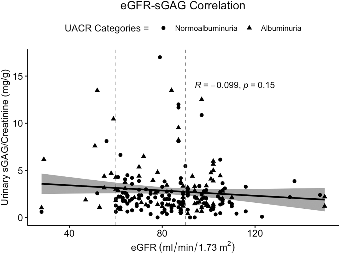
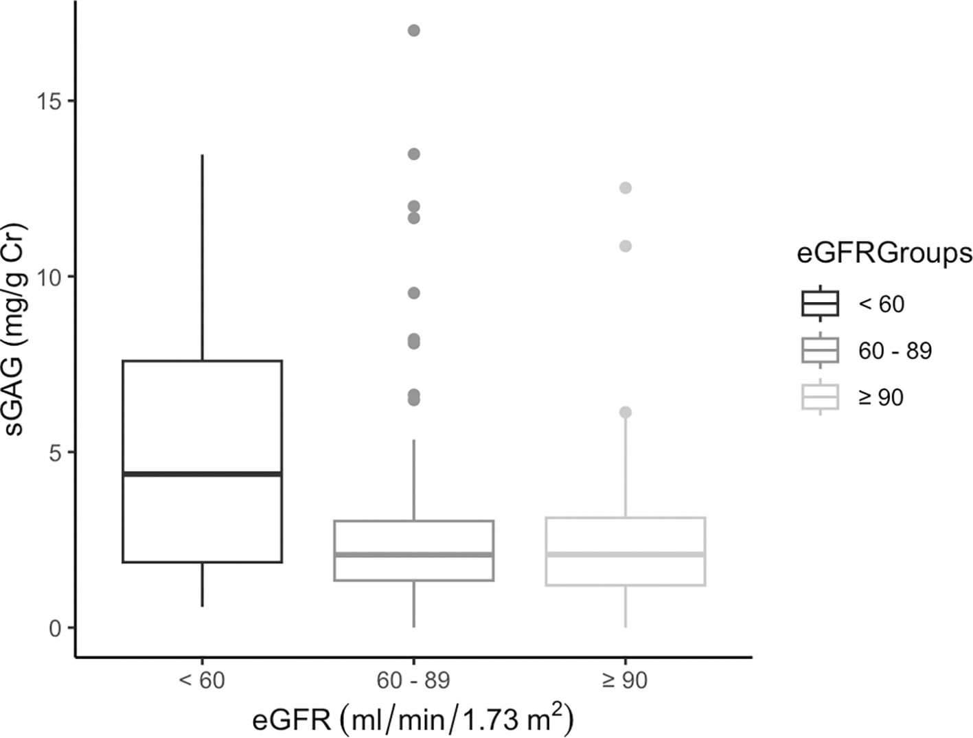
Unexpectedly, sGAG showed a significant increase in the moderate-to-high-risk control group (p = 0.013), with a median value of 2.45 mg/g Cr, differing by 0.46 in the low-risk group, showing that the glycocalyx breaks down more significantly in people with vague symptoms of DKD. Even so, when sGAG was plotted against eGFR, it showed a very low correlation and a statistically insignificant inverse relationship (r = −0.099; p = 0.15) (Figure 1). When comparing sGAG concentration in three different eGFR categories as per KDIGO recommendation (<60, 60 – 89, and ≥90 ml/min per 1.73 m2), the result confirmed that glycocalyx breakdown is more pronounced in subjects with decreased kidney function (Figure 2).
However, to confirm whether sGAG is an independent variable for CKD risk, multivariate analysis was conducted for variables with p < 0.25 in Table 1. Sex, regular exercise, DM duration, diastole, HbA1c level, and sGAG level were included in the model. However, since age and BMI are known to be associated with renal function decline, these two variables were also included. Based on the logistic regression analysis results ( Table 2), sGAG were no longer significantly associated with CKD risk after adjustment for confounders (OR 1.109; 95% CI 0.978-1.258; p = 1.08). Therefore, in this study, we found that the sGAG level was not an independent predictor of CKD risk. We noticed that HbA1c and DM duration were confounders for sGAG, indicating that when the variables were input into the model, sGAG lost their significance.
| OR (95% Confidence Interval) | p-value | |
|---|---|---|
| Crude Model | ||
| sGAG (mg/g Cr) | 1.140 (1.016-1.278) | 0.025 * |
| Adjusted Model | ||
| sGAG (mg/g Cr) | 1.109 (0.978-1.258) | 0.108 |
| Age (≥60 years) | 1.321 (0.671-2.601) | 0.420 |
| Sex (Female) | 0.376 (0.181-.0782) | 0.009 * |
| BMI (Obesity) | 2.050 (1.023-4.109) | 0.043 * |
| DM duration (≥5 years) | 2.082 (1.050-4.129) | 0.036 * |
| Regular exercise (Routine) | 0.681 (0.364-1.276) | 0.231 |
| HbA1c | 1.501 (1.236-1.822) | <0.001 * |
| Diastole | 1.022 (0.988-1.057) | 0.200 |
Our study attempted to elucidate the clinical value of urinary sGAG level by comparing it with different stages of CKD, determined by eGFR and UACR levels according to the KDIGO classification of CKD risk.3
Using consecutive sampling techniques from the two study sites, two-thirds of our subjects were female. This number was expected because females are more prone to insulin resistance, with estrogen concentration playing a role as a predisposing factor for type 2 diabetes mellitus.28,29 This study conforms with another study analyzing predictors of CKD in T2DM patients, where it was found that disease duration contributed up to 50% risk of eGFR decline to below 60 ml/min per 1.73 m2.30
HbA1c levels were also significantly higher in the group with decreased renal function. HbA1c forms when glucose binds to the N-terminal of the beta chain of hemoglobin within red blood cells, usually serving as an indicator of glycemic exposure over the previous 2-3 months. In line with our results, a study found an increasing trend of HbA1c levels along with CKD progression,31 proving that uncontrolled glycemic index is one of the main drivers of diabetes complications. However, interpreting HbA1c results can be challenging when there are changes in erythrocyte turnover, which is a common occurrence in CKD even in its early stages.32
We found significantly higher levels of urinary sGAG in the moderate-to high-risk group than in the low-risk group. This finding, together with a propensity for a negative correlation such that the higher the urinary sGAG concentration, the lower the eGFR, is consistent with an earlier study that examined blood endothelial glycocalyx levels in a group of people with chronic kidney disease. One study reported that those with chronic renal failure had higher amounts of dysfunctional serum endothelial glycocalyx syndecan-1 and hyaluronan compared to a group of healthy patients.33 Another study revealed that lower eGFR with a median of 31 (10 – 59) ml/min per 1.73 m2 had higher serum levels of glycocalyx damage (syndecan-1 and hyaluronan) and endothelial dysfunction (von Willebrand Factor; vWF and vascular cell adhesion molecule; VCAM-1) markers compared to healthy controls.34 Regarding the specific evaluation of urinary sGAG concentration in the diabetic patient group, results by Popławska-Kita et al.35 were consistent with our findings, showing that the level of urinary sGAG is higher in patients with diabetic kidney disease than in diabetic patients without microvascular complications.
The increase in the incidence of DKD was multifactorial. In addition to glomerular injury, oxidative stress and inflammation contribute to the pathogenesis of DKD. Swaminathan et al. and Yu, Song, and Li noted that Reactive Oxygen Species (ROS) and Reactive Nitrogen Species (RNS) are increased in hyperglycemia, lysing the glycocalyx into its components through syndecan dissolvation and matrix metalloproteinase activation, among others.36,37 The cleavage of protein and glycan components interferes with the functional integrity of the glomerular endothelial cell surface layer,38 causing its components to circulate freely. Indeed, the breakdown of the glycocalyx into its components could predict the incidence of early DKD.37 However, the presence of a free circulating glycocalyx, such as hyaluronic acid, is eliminated in less than 5 minutes through an extrarenal mechanism, such as by the liver.39 This implies that even though shedding may occur in advance, the glycocalyx is not directly useful as a marker in urine. Similar to eGFR and UACR, sGAG could only indicate the progressive decline of the kidney in DKD, especially when the kidney has entered a state of endothelial dysfunction.
Leppeda and De Muro extensively investigated the associations between urinary glycosaminoglycans and complications of type 1 and 2 diabetes. They found a strong association between altered sGAG excretion and hyperglycemic conditions by performing studies in type 1 diabetes patients with high HbA1c levels.40 Impaired metabolism and degradation of renal endothelial sGAG were hypothesized to be part of the pathogenesis of diabetic nephropathy.41 This is in line with our findings, in which hyperglycemic situations represented by uncontrolled HbA1c were the strongest confounder for sGAG together with DM duration. When we input them into the model, the sGAG loses its significance. As HbA1c reflects the state of blood glucose for the preceding 3 months, uncontrolled HbA1c directly affects kidney function through well-known mechanisms, such as the generation of ROS and other inflammatory markers.42
In conclusion, we concur with previous research on a possible early diagnostic tool for renal function deterioration in patients with diabetes. Urinary sGAG have been shown to be elevated in patients with moderate to high risk of CKD; however, after confounder adjustments, we found that sGAG are not an independent variable for KDIGO CKD risk. Further research should be conducted to identify its utility in detecting diabetic nephropathy in a large number of normoalbuminuric patients, as sGAG might be more accurate than UACR in detecting early glomerular basement membrane structural changes.
This study had several limitations. It was not designed to be a diagnostic study and has no power to explain a diagnostic standpoint; however, we aimed to open a path for a specific biomarker with clear clinical utility in diagnosing early renal function reduction. Moreover, the eGFR and UACR values are derived from single-point-in-time, slightly defying the persistence requirement when defining CKD, where, as per KDIGO, abnormalities should be present for more than 3 months. Doing this will also help in finding the right study subjects with a hyperfiltration phase that can better reflect the broader purpose of this study.
However, this study has several strengths. We gathered a relatively large sample size (n = 207), which allowed our results to reflect the heterogeneity of the population. We analyzed each sample closely using the same instruments and conditions according to predefined standard protocols. As biomarkers tend to be unstable during long-term storage, we performed the entire analysis within 8 weeks of sample collection. For measurements of UACR and sGAG, we used commercial kits that provide a predetermined assay protocol, minimizing the variance in handling. Additionally, the samples were stored at −20°C to maintain stability. Setting aside the aim of this study, HbA1c and UACR measurements are rarely performed in primary health centers in Indonesia because of economic concerns. Thus, the results of both variables obtained from this research could hopefully inform physicians and assist them in clinical decision-making for their patients.
The significant difference observed in urinary sGAG levels between moderate-to-high-risk and low-risk patients suggests that glycocalyx layer breakdown is one of the most important mechanisms in DKD. However, HbA1c, duration of DM, obesity, and female sex interfere with the breakdown mechanism in the initial stage of DKD.
Ethical approval was obtained prior to conducting this study in each location. This study was approved by the local ethics committee of the Faculty of Medicine, Universitas Indonesia and Dr. Cipto Mangunkusumo Hospital. The first approval was granted to perform this study in Pasar Minggu Primary Health Center (KET-49/UN2.F1/ETIK/PPM.00.02/2021). As for data collection at the Depok Jaya Primary Health Center, ethical approval was KET-26/UN2.F1/ETIK/PPM.00.02/2021.
| Views | Downloads | |
|---|---|---|
| F1000Research | - | - |
|
PubMed Central
Data from PMC are received and updated monthly.
|
- | - |
Provide sufficient details of any financial or non-financial competing interests to enable users to assess whether your comments might lead a reasonable person to question your impartiality. Consider the following examples, but note that this is not an exhaustive list:
Sign up for content alerts and receive a weekly or monthly email with all newly published articles
Already registered? Sign in
The email address should be the one you originally registered with F1000.
You registered with F1000 via Google, so we cannot reset your password.
To sign in, please click here.
If you still need help with your Google account password, please click here.
You registered with F1000 via Facebook, so we cannot reset your password.
To sign in, please click here.
If you still need help with your Facebook account password, please click here.
If your email address is registered with us, we will email you instructions to reset your password.
If you think you should have received this email but it has not arrived, please check your spam filters and/or contact for further assistance.
Comments on this article Comments (0)