Keywords
Case report, Tuberculum Sella Meningioma, Endoscopic Endonasal Approach, Remote Intracranial Haemorrhage
This article is included in the Oncology gateway.
This case report describes an exceedingly rare complication of endoscopic endonasal approach (EEA) surgery. We reported a delayed remote combined supratentorial intracerebral and subdural haemorrhage following the resection of a tuberculum sella meningioma. This report aims to analyse the pathophysiology and discuss the management of this critical complication, which is seldom documented in the literature. We present the case of a 54-year-old female with a WHO Grade I tuberculum sella meningioma. She underwent a complete (Simpson Grade I) resection via an endoscopic endonasal transsphenoidal approach. Her initial postoperative recovery was unremarkable for four days. On the fifth postoperative day, the patient experienced an acute decline in consciousness. An emergency non-contrast head computed tomography (CT) scan revealed a remote left parietal intracerebral haemorrhage (ICH) of approximately 20cc, associated with an acute left frontoparietal subdural haematoma (SDH), causing a significant midline shift. Despite the severity of the radiological findings, the patient was managed successfully with non-operative medical therapy. She made a full clinical recovery, and a three-month follow-up magnetic resonance imaging (MRI) confirmed complete resolution of the haematomas with no evidence of residual tumour or underlying vascular malformation. The clinical timeline and radiological pattern strongly suggest a venous aetiology. The most plausible mechanism is a cascade initiated by an occult cerebrospinal fluid (CSF) leak, leading to intracranial hypotension, cerebral ptosis, and the subsequent rupture of a cortical bridging vein. This case underscores the need for a high index of suspicion for remote intracranial haemorrhage (RIH) in any patient with delayed neurological deterioration after transsphenoidal surgery. Furthermore, it demonstrates that this life-threatening complication can often be managed successfully with conservative therapy.
Case report, Tuberculum Sella Meningioma, Endoscopic Endonasal Approach, Remote Intracranial Haemorrhage
The endoscopic endonasal approach (EEA) has revolutionised the surgical management of anterior skull base lesions.1 For pathologies such as pituitary adenoma and tuberculum sella meningiomas, this approach provides a direct, minimally invasive corridor that avoids the brain retraction associated with traditional transcranial methods. Despite these advantages, however, the EEA carries a unique risk profile, including cerebrospinal fluid (CSF) leakage and neurovascular injury.2,3
A particularly devastating, though infrequent, complication of any neurosurgical procedure is postoperative intracranial haemorrhage.4 While bleeding at the operative site is most common, a far rarer phenomenon is Remote Intracranial Haemorrhage (RIH), defined as bleeding anatomically distinct from the surgical bed.5
This report details a highly unusual case of a delayed, combined supratentorial intracerebral haemorrhage (ICH) and subdural haematoma (SDH) five days after an EEA for a tuberculum sella meningioma. This specific constellation of features—a supratentorial location, a combined parenchymal and subdural pattern, and a delayed onset following a purely endonasal procedure—is exceptionally rare. Therefore, the purpose of this report is to provide a detailed account of this event, analyse the underlying pathophysiological mechanisms, and discuss the critical implications for diagnosis and management.
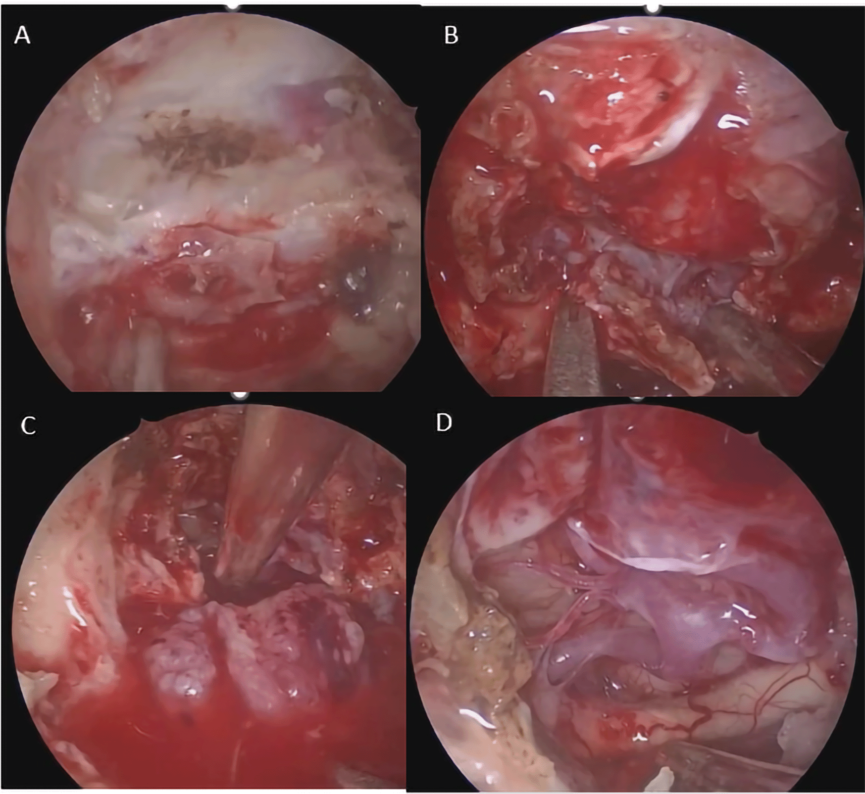
A 54-year-old female presented with a two-month history of progressively blurred vision and a right temporal hemianopia, superimposed on a two-year history of chronic headaches. Her medical history was negative for arterial hypertension, bleeding disorders or anticoagulant use. The timeline of the case presentation is shown in Figure 1. Hormonal status was within normal limit. Preoperative magnetic resonance imaging (MRI) revealed a 1.91 × 2.01 × 1.82 cm solid, globular mass that avidly enhanced with gadolinium and showed a dural tail attached to the tuberculum sella ( Figure 2). She underwent a Simpson Grade I resection of the tumour via a endoscopic endonasal transsphenoidal approach ( Figure 3). The surgery was unremarkable and histopathology confirmed a Meningothelial Meningioma, WHO Grade I. The immediate postoperative period (days one to four) was uneventful; the patient was conscious and alert with improving vision. The patient showed minimal CSF leakage from the nostril postoperatively and some polyuria adequately manage with desmopressin tablet 0.1 mg. However, on the postoperative day five, the patient was found to be drowsy (Glasgow Coma Scale score of 13).

T2 weighted image showed isointense mass at tuberculum without any remarkable surrounding oedema.

A. Tuberculum on the anterior part of sellar floor was exposed and drilled. B. U shaped durotomy was performed exposing the tumor attached to the duramater. C. Tumor was being removed. Tumor was grayish red, easy to bleed with rubbery consistency and not suctionable. Detachment of tumor from surrounding sturctures. D. Neurovascular structures revealed including anterior communicating artery, Anterior cerebral artery (A1 and A2), recurrent artery of Heubner and Optic Nerve. There was no active bleeding seen after tumor removal.
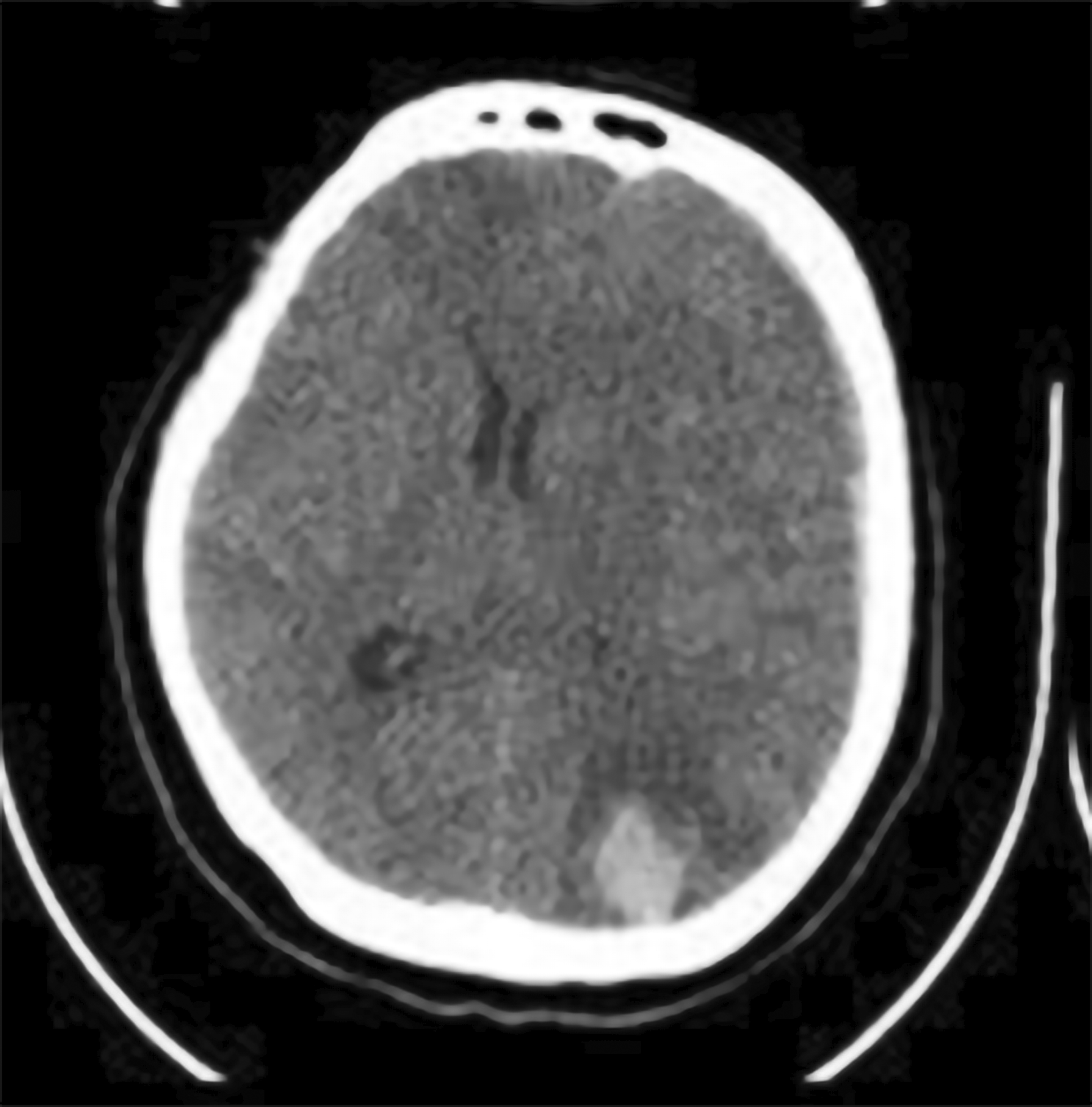
An emergency non-contrast head CT scan was performed immediately following the neurological decline. This revealed a remote haemorrhagic event, distant from the original operative site. The findings included a 20cc left parietal intracerebral haematoma and a concomitant acute left frontoparietal subdural haematoma. The combined mass effect resulted in a midline shift of over 5 mm ( Figure 4).

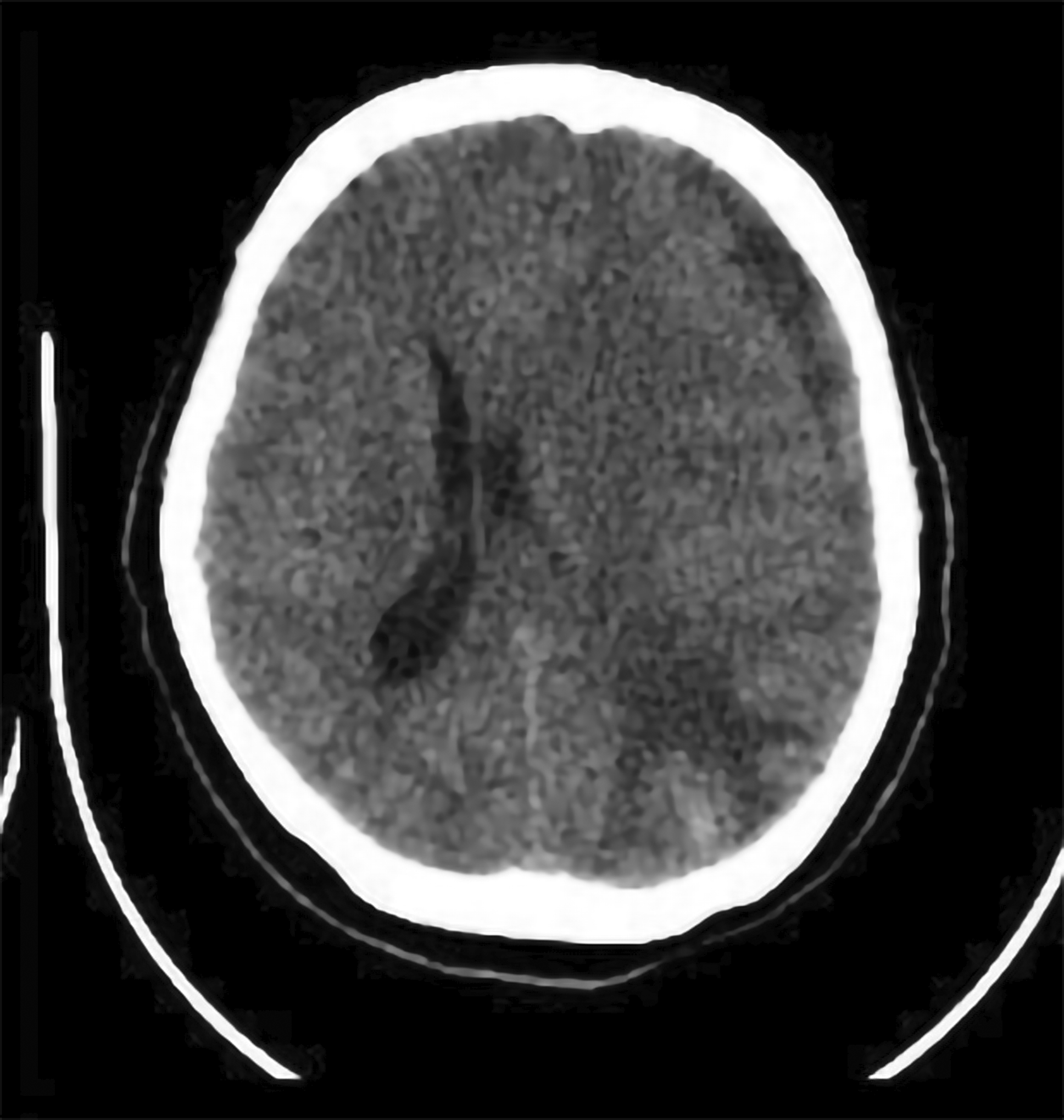
Medical therapy to control intracranial pressure (ICP) was initiated, consisting of intravenous mannitol and head-of-bed elevation. Following this conservative management, her level of consciousness progressively improved over the next three days. However, the patient suddenly became aphasic and could not understand any command on day 13th postoperatively ( Figure 5). Another head CT scan was performed and revealed partially resolved ICH at the parietal lobe with slight additional thickness of subacute SDH and midline shift to the right. Conservative management with mannitol and tranexamic acid was continued. The patient showed clinical improvement after 2 days and discharged from hospital at day 18th postoperatively. The three-month postoperative MRI confirmed gross total resection of the meningioma and complete resolution of the remote haemorrhage, without evidence of any vascular malformation ( Figure 6).

This case represents a rare and clinically significant variant of RIH. While haemorrhage is a known neurosurgical complication, it most often occurs at the operative site, with RIH only accounts for less than 1%.6 RIH is a distinct entity, with the few existing reports after EEA primarily describing extradural haematomas or subdural hematoma.7–10 The occurrence of a delayed, combined supratentorial ICH and SDH after an endonasal procedure is, to our knowledge, an exceptionally rare event.
Consequently, a unifying mechanistic cascade provides the most compelling explanation for the findings in our case. The dominant pathophysiological theory for RIH centres on the loss of CSF volume and subsequent intracranial hypotension. The proposed sequence begins with a small, occult CSF leak from the dural repair site. This gradual egress of CSF over several days leads to intracranial hypotension, causing the brain to sag caudally (cerebral ptosis).11 This downward displacement, in turn, exerts tractional force on the cortical bridging veins that anchor the cerebrum to the dural sinuses. Ultimately, this tension can lead to the tearing of a bridging vein, resulting in haemorrhage.12,13
Several key features of this case provide strong corroborative suggestion for this CSF hypovolaemia cascade. First, the five-day delay between surgery and the haemorrhagic event is a critical temporal clue. This delayed presentation argues against inadequate intraoperative haemostasis and instead points towards a more indolent process, such as a slow CSF leak.14 Second, the radiological finding of a combined ICH and SDH provides a clear anatomical signature for a ruptured cortical bridging vein. A tear in such a vein as it crosses the subdural space would lead to an SDH, and retrograde propagation of the tear to the pial surface would cause an associated ICH.15
Furthermore, the patient’s transient hypertension at the time of deterioration was likely a contributing factor rather than the primary cause. In a scenario where the bridging veins were already under critical tension from cerebral ptosis, a transient surge in venous pressure could have provided the final stress needed to induce rupture.15 The differential diagnosis also includes the haemorrhagic transformation of an ischemic infarct, a ruptured cryptic vascular malformation, and delayed cerebral vasospasm16; however, the clinical and radiological findings make these alternatives highly unlikely.
Ultimately, choosing non-operative management for this patient was successful and offers additional diagnostic validation. The approach to treating spontaneous intracerebral haemorrhage (ICH) continues to be debated, as studies present mixed outcomes when comparing surgical intervention to conservative treatment.17 However, surgical management was primarily reserved for significant hematomas (>30cc) accompanied by worsening consciousness, often occurring in relatively younger patients.18 Conservative treatment for small intracerebral haemorrhages can be effective, particularly in patients with a GCS score of 13 or higher, or with hematoma volume under 30 mL.19
In conclusion, delayed, remote, combined supratentorial ICH and SDH is a rare but potentially devastating complication of the EEA. The evidence presented in this case points unequivocally to a venous-origin haemorrhage, most plausibly initiated by occult CSF egress leading to intracranial hypotension and the rupture of a cortical bridging vein. This report delivers a critical message: clinicians must maintain a high index of suspicion for RIH in any patient with delayed neurological deterioration following transsphenoidal surgery. Prompt diagnosis and an understanding of the underlying venous mechanism are paramount, as this life-threatening complication can often be managed successfully with conservative therapy.
Written informed consent was obtained from the patient for the publication of this case report and any accompanying images.
Repository: CARE checklist for ‘Remote Intracerebral Haemorrhage after Endoscopic Transsphenoidal Surgery for Tuberculum Sella Meningioma: A Case Report’. https://doi.org/10.6084/m9.figshare.29987188.20
Data are available under the terms of the Creative Commons Zero “No rights reserved” data waiver (CC0 1.0 Public domain dedication).
| Views | Downloads | |
|---|---|---|
| F1000Research | - | - |
|
PubMed Central
Data from PMC are received and updated monthly.
|
- | - |
Is the background of the case’s history and progression described in sufficient detail?
Yes
Are enough details provided of any physical examination and diagnostic tests, treatment given and outcomes?
Partly
Is sufficient discussion included of the importance of the findings and their relevance to future understanding of disease processes, diagnosis or treatment?
Partly
Is the case presented with sufficient detail to be useful for other practitioners?
Partly
Competing Interests: No competing interests were disclosed.
Reviewer Expertise: Neurosurgery, Neurovascular, Interventional, Neuroscience, Research
Is the background of the case’s history and progression described in sufficient detail?
Yes
Are enough details provided of any physical examination and diagnostic tests, treatment given and outcomes?
Yes
Is sufficient discussion included of the importance of the findings and their relevance to future understanding of disease processes, diagnosis or treatment?
Yes
Is the case presented with sufficient detail to be useful for other practitioners?
Yes
Competing Interests: No competing interests were disclosed.
Reviewer Expertise: Neurooncology
Alongside their report, reviewers assign a status to the article:
| Invited Reviewers | ||
|---|---|---|
| 1 | 2 | |
|
Version 1 15 Oct 25 |
read | read |
Provide sufficient details of any financial or non-financial competing interests to enable users to assess whether your comments might lead a reasonable person to question your impartiality. Consider the following examples, but note that this is not an exhaustive list:
Sign up for content alerts and receive a weekly or monthly email with all newly published articles
Already registered? Sign in
The email address should be the one you originally registered with F1000.
You registered with F1000 via Google, so we cannot reset your password.
To sign in, please click here.
If you still need help with your Google account password, please click here.
You registered with F1000 via Facebook, so we cannot reset your password.
To sign in, please click here.
If you still need help with your Facebook account password, please click here.
If your email address is registered with us, we will email you instructions to reset your password.
If you think you should have received this email but it has not arrived, please check your spam filters and/or contact for further assistance.
Comments on this article Comments (0)