Keywords
Colorectal cancer; Cutaneous metastasis; Skin metastasis; Rectosigmoid adenocarcinoma; Case report
This article is included in the Oncology gateway.
Colorectal cancer; Cutaneous metastasis; Skin metastasis; Rectosigmoid adenocarcinoma; Case report
Cutaneous metastases from colorectal cancer (CRC) are rare, with reported incidences of 0.7–4% among metastatic cases. Their presence generally reflects advanced disease and poor prognosis. Typical metastatic sites include the liver, lungs, and peritoneum; skin involvement is far less common.
We present an unusual case of isolated cutaneous recurrence revealing systemic relapse after curative treatment for a rectosigmoid junction adenocarcinoma.
A 42-year-old man with no prior medical history underwent surgery for a stenosing adenocarcinoma of the rectosigmoid junction.
Initial locoregional and distant staging was negative. He underwent low anterior resection with stapled colorectal anastomosis and had an uncomplicated postoperative recovery.
Histopathological examination of the resected specimen showed a moderately differentiated adenocarcinoma, staged as ypT3N1b. The patient received eight cycles of adjuvant capecitabine (Xeloda), completed 18 months after surgery.
A follow-up CT scan performed three months after completion of chemotherapy showed no abnormalities. A completion colonoscopy, performed because a full preoperative evaluation had not been possible, was also normal.
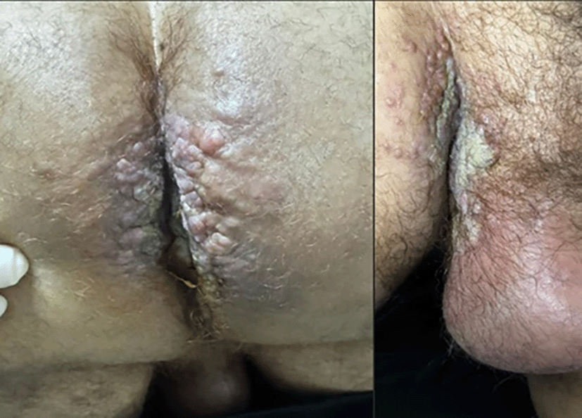
Approximately five months later, the patient presented with newly developed pruritic skin lesions. Clinical examination revealed erythematous eczematous plaques in the right inguinal fold, gluteal region, and perianal area ( Figure 1). He was otherwise in good general condition (ECOG 0). Digital rectal examination was unremarkable, and serum tumor markers (CEA, CA 19-9) were within normal limits.
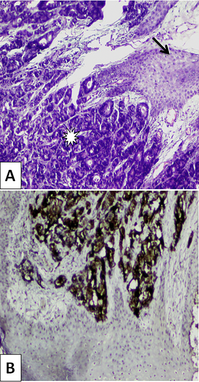
A punch biopsy of the skin lesion confirmed metastatic adenocarcinoma consistent with colorectal origin (CK20-positive, CK7-negative, CDX2-positive) ( Figure 2A,B).
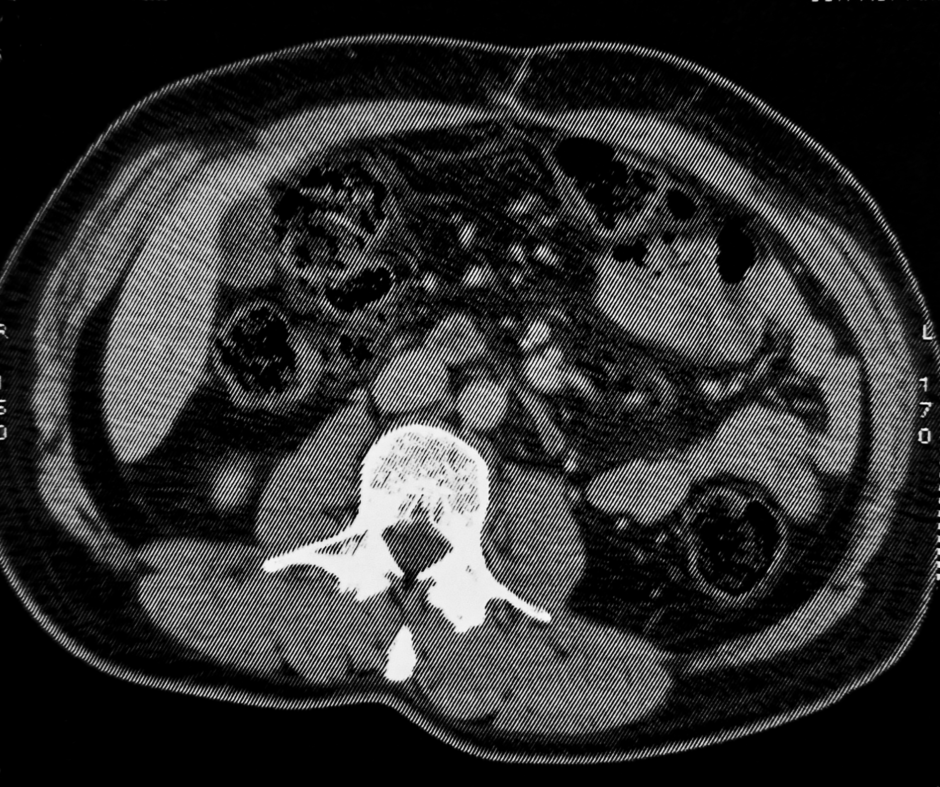
A thoraco-abdomino-pelvic CT scan revealed new, non-necrotic lymphadenopathy at the aortic bifurcation (22 × 9 mm) and in both internal iliac chains ( Figure 3).
The case was discussed in a multidisciplinary tumor board. The presence of cutaneous metastases and retroperitoneal lymph node involvement led to classification as unresectable stage IV disease. The patient was referred for palliative systemic chemotherapy, with RAS, BRAF, and MSI testing planned to guide targeted or immunotherapeutic options.
Cutaneous metastases from CRC are uncommon but clinically significant indicators of systemic dissemination. The median time from initial diagnosis to skin involvement ranges from 18–30 months, and median survival after diagnosis of skin metastasis is approximately 8 months.1–3
Skin metastases typically involve the abdominal wall, perineal, or perianal regions, often following surgical scars or areas of venous/lymphatic drainage from the primary tumor.1,2 In our patient, the inguinal and perianal lesions likely reflected pelvic lymphatic spread from the rectosigmoid area.
Cutaneous metastases may present as nodules, plaques, or inflammatory-like or eczematous lesions that mimic benign dermatoses, delaying diagnosis.3–5
Histopathology with immunohistochemistry (CK20+, CK7–, and CDX2+) is essential for confirming colorectal origin. Tumor markers such as CEA and CA 19-9 may remain normal, limiting their diagnostic value in isolation.
Treatment is mainly palliative and depends on molecular profile and disease extent:
• RAS/BRAF wild-type tumors: Chemotherapy (FOLFOX or FOLFIRI) plus anti-EGFR agents (cetuximab or panitumumab).
• MSI-high/dMMR tumors: Immune checkpoint inhibitors (pembrolizumab, nivolumab ± ipilimumab).
• RAS/BRAF-mutant or MSS/pMMR tumors: Chemotherapy combined with anti-angiogenic therapy (e.g. bevacizumab).
For symptomatic relief, surgical excision or palliative radiotherapy can alleviate pain, bleeding, or ulceration, while topical or systemic antipruritic agents may improve comfort.
Cutaneous metastases from colorectal cancer are rare and often signal systemic relapse. Awareness of this entity is essential, as lesions may mimic benign dermatologic conditions and occur even when tumor markers and imaging appear normal. Early biopsy and multidisciplinary evaluation are critical for accurate diagnosis and management.
Systemic therapy guided by molecular profiling remains the mainstay of treatment, while novel immunotherapy–anti-angiogenic combinations show promise in refractory microsatellite-stable disease. Continued reporting of such cases will aid in refining prognostic understanding and therapeutic strategies.
| Views | Downloads | |
|---|---|---|
| F1000Research | - | - |
|
PubMed Central
Data from PMC are received and updated monthly.
|
- | - |
Provide sufficient details of any financial or non-financial competing interests to enable users to assess whether your comments might lead a reasonable person to question your impartiality. Consider the following examples, but note that this is not an exhaustive list:
Sign up for content alerts and receive a weekly or monthly email with all newly published articles
Already registered? Sign in
The email address should be the one you originally registered with F1000.
You registered with F1000 via Google, so we cannot reset your password.
To sign in, please click here.
If you still need help with your Google account password, please click here.
You registered with F1000 via Facebook, so we cannot reset your password.
To sign in, please click here.
If you still need help with your Facebook account password, please click here.
If your email address is registered with us, we will email you instructions to reset your password.
If you think you should have received this email but it has not arrived, please check your spam filters and/or contact for further assistance.
Comments on this article Comments (0)