Keywords
Adrenal pseudocyst, retroperitoneal mass, Adrenalectomy, Case report
Adrenal pseudocyst, retroperitoneal mass, Adrenalectomy, Case report
Adrenal pseudocysts are rare, non-functional lesions accounting for a small fraction of adrenal incidentalomas. They are typically asymptomatic and discovered incidentally during imaging studies or autopsy. Their pathogenesis remains unclear and they are often mistaken for other retroperitoneal cystic masses. When large, adrenal pseudocysts can cause compressive symptoms involving adjacent organs. Despite advances in imaging modalities, diagnosis often remains uncertain preoperatively, with histopathology required for confirmation. This case describes a giant adrenal pseudocyst presenting with dysuria and flank pain, illustrating its clinical, radiological, and pathological features. This report complies with the CARE Checklist guidelines.11
A 70-year-old man with no significant medical history presented with a three-month history of left flank pain, upper abdominal pressure, and dysuria. He denied trauma, fever, hematuria, or weight loss, and had no personal or family history of malignancy or endocrine disorders. Physical examination revealed a firm, painless mass in the left upper quadrant. Laboratory investigations, including renal function, adrenal hormonal profile, and complete blood count, were normal.
Contrast-enhanced abdominal CT ( Figure 1) showed a large, well-circumscribed, left retroperitoneal cystic mass (15 cm) displacing the spleen superiorly and kidney inferiorly. The left adrenal gland appeared displaced but morphologically normal. Mild left pyelocaliceal dilatation suggested ureteropelvic junction compression. No solid or enhancing components were seen. Possible diagnoses included liquefied adrenal hematoma or cystic lymphangioma.
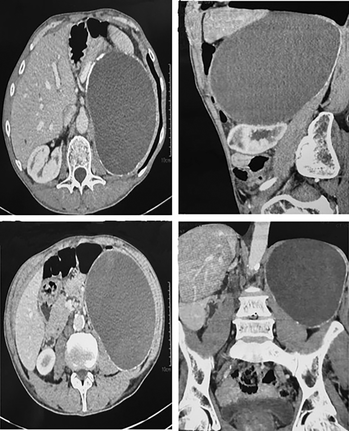
Mild left pyelocaliceal dilatation is visible, suggesting ureteropelvic junction compression.
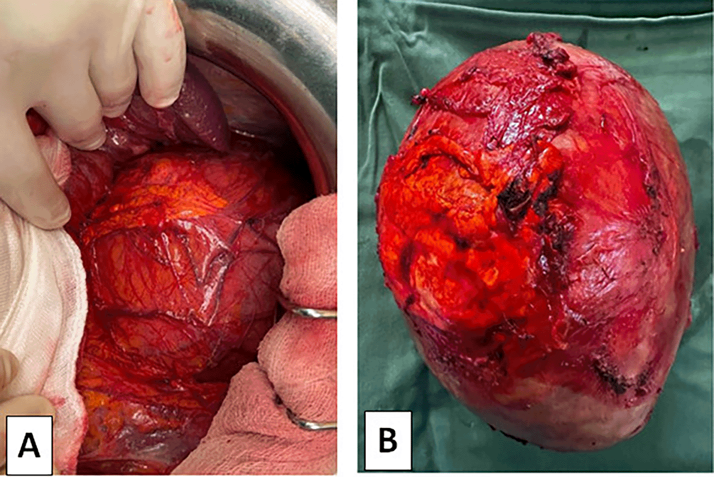
Surgical exploration via midline laparotomy revealed a large retroperitoneal cystic mass adherent to the adrenal gland and adjacent organs ( Figure 2A). Complete resection with left adrenalectomy was performed ( Figure 2B). The postoperative course was uneventful, and the patient was discharged on day five.
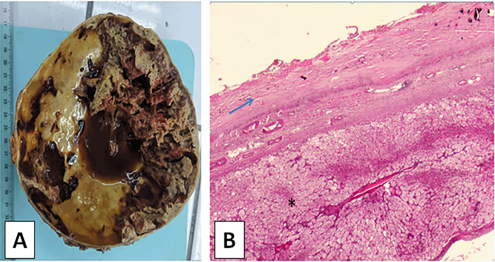
Gross examination ( Figure 3A) showed a unilocular cyst measuring 14 × 11 × 7 cm with a thick fibrous wall and hemorrhagic content. Microscopy ( Figure 3B) revealed a fibrous wall with calcifications, chronic inflammation, cholesterol clefts, and multinucleated giant cells, but no epithelial lining—confirming an adrenal pseudocyst. Adjacent adrenal parenchyma was compressed but otherwise normal.
Adrenal pseudocysts are rare,1 accounting for about 5–7% of adrenal incidentalomas.2 They lack epithelial lining and are thought to arise from prior hemorrhage, infection, or degeneration.3 Most are asymptomatic and found incidentally, but those exceeding 10 cm can cause symptoms due to mass effect.1,3–5
In this case, the pseudocyst compressed the urinary tract, explaining the dysuria. Similar presentations have been described with ureteral or gastrointestinal compression.6,7 CT imaging aids localization but cannot reliably distinguish benign from malignant cystic lesions.8 Differential diagnoses include adrenal hemorrhage, cystic lymphangioma, pancreatic pseudocyst, and cystic adrenal neoplasms.9
Histology remains the diagnostic gold standard. The absence of epithelial lining and presence of cholesterol clefts and giant cells confirmed the pseudocyst.10 Although most adrenal cysts are benign, up to 7% may harbor malignancy.2,3,10 Management depends on size, symptoms, hormonal activity, and malignancy risk. Lesions fewer than 5 cm can be monitored; larger, symptomatic, or uncertain lesions require surgical excision.3,9 Laparoscopic adrenalectomy is suitable for small lesions, while open surgery is preferred for giant or suspicious masses, as in this case.
Adrenal pseudocyst is a rare entity, particularly when reaching giant size. Surgical intervention is essential for symptomatic or uncertain cases to relieve symptoms and establish a definitive diagnosis. Given the non-specific radiological and clinical characteristics, histopathological examination remains crucial.
The patient expressed satisfaction with the outcome and was relieved by the resolution of his symptoms. He appreciated the clarity of communication and quality of care throughout diagnosis and treatment.
This report highlights a rare presentation of a giant adrenal pseudocyst revealed by atypical urinary symptoms, which is scarcely described in the literature. A key strength is the detailed radiological and histopathological documentation. A limitation is the short follow-up period and lack of long-term outcome data.
| Views | Downloads | |
|---|---|---|
| F1000Research | - | - |
|
PubMed Central
Data from PMC are received and updated monthly.
|
- | - |
Provide sufficient details of any financial or non-financial competing interests to enable users to assess whether your comments might lead a reasonable person to question your impartiality. Consider the following examples, but note that this is not an exhaustive list:
Sign up for content alerts and receive a weekly or monthly email with all newly published articles
Already registered? Sign in
The email address should be the one you originally registered with F1000.
You registered with F1000 via Google, so we cannot reset your password.
To sign in, please click here.
If you still need help with your Google account password, please click here.
You registered with F1000 via Facebook, so we cannot reset your password.
To sign in, please click here.
If you still need help with your Facebook account password, please click here.
If your email address is registered with us, we will email you instructions to reset your password.
If you think you should have received this email but it has not arrived, please check your spam filters and/or contact for further assistance.
Comments on this article Comments (0)