Keywords
Osteochondral fracture, Femoral condyle, Pediatric fracture, Arthrotomy, Case report
Osteochondral fractures (OCF)s of the femoral condyle are rare in pediatric patients but can have significant implications if missed or left untreated. Surgical fixation is often recommended, particularly in young, to restore joint congruency and function.
We report the case of a 14-year-old boy who presented with isolated weight-bearing OCF of the lateral femoral condyle following a sports injury. Magnetic resonance imaging (MRI) revealed a 30.4 mm osteochondral fragment confirmed by arthroscopy but unsuitable for percutaneous management due to its size and location. Open reduction and internal fixation were performed using headless compression screws, a technique reserved for those cases where based on sufficient detached fragment bone to facilitate secure fixation and bone-to-bone healing.
This case underscores the importance of prompt diagnosis and individualized treatment to prevent long-term joint damage, contributing valuable insights into the management of pediatric femoral osteochondral fractures.
Osteochondral fracture, Femoral condyle, Pediatric fracture, Arthrotomy, Case report
While osteochondral fractures OCFs have long been recognized, their precise incidence in the pediatric population is not well-established. OCFs around the knee. Typically results from either acute trauma or repetitive microtrauma, such as torsional injuries or patellar dislocations. Radiographic diagnosis can be difficult due to the potentially thin bony component of the osteochondral fragment.1 Despite numerous publications describing various surgical techniques for OCFs, a consensus on the optimal approach remains elusive. Historically, surgical management often involved fragment removal unless sufficient subchondral bone was present for anchoring.2
Here, we report good radiological and excellent functional outcomes at 18 months postoperatively following fragment fixation of a lateral femoral condyle OCF in a pediatric patient.
Patient information: A 14-year-old boy with no significant medical history presented to the emergency department with left knee pain and swelling following a sport injury.
Clinical findings: Initial examination of the left knee revealed swelling, pain, and a positive patellar tap, with no skin lesions observed. Three days post-injury, the patient continued to experience the same symptoms, with increased severity of knee pain.
Timeline of current episode: In May 2023, following a sports-related injury with an unknown mechanism, a child presented with a closed left knee trauma characterized by functional impairment, pain, and swelling, but no skin lesions. Initial radiographs were unremarkable, and the patient was treated symptomatically. Three days later, the child returned with persistent symptoms and increased pain. MRI revealed an osteochondral fracture. Surgical intervention occurred seven days post-injury, initially planned arthroscopically. However, due to the lesion’s inaccessibility, an open approach with ORIF using two headless compression screws was performed via a lateral parapatellar incision. Post-operatively, the patient followed a three-month rehabilitation program, including 45 days of non-weight-bearing. At 18 months follow-up, the patient demonstrated excellent results with full range of motion and good quadriceps strength.
Diagnostic assessment: Initial physical examination revealed a painful knee with functional impairment but no deformity or skin lesions. Initial radiographs did not demonstrate any osseous abnormalities ( Figure 1).
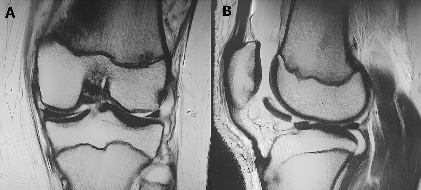
Due to persistent symptoms, MRI was performed, which revealed a 30.4mm osteochondral fracture ( Figure 2).

A key diagnostic challenge in such cases is considering the possibility of an osteochondral lesion, especially when initial radiographs are negative, as these fractures can be subtle or even invisible on plain films if the bony component is small.1
Diagnosis: The final diagnosis was an osteochondral fracture of the lateral femoral condyle. Other diagnoses considered, prior to MRI confirmation, included meniscal tear and ligament sprain.
Therapeutic interventions: The patient underwent arthroscopy to visualize the osteochondral fragment and assess for associated ligamentous or meniscal injuries. Arthroscopy confirmed the osteochondral defect and the absence of associated injuries ( Figure 3).

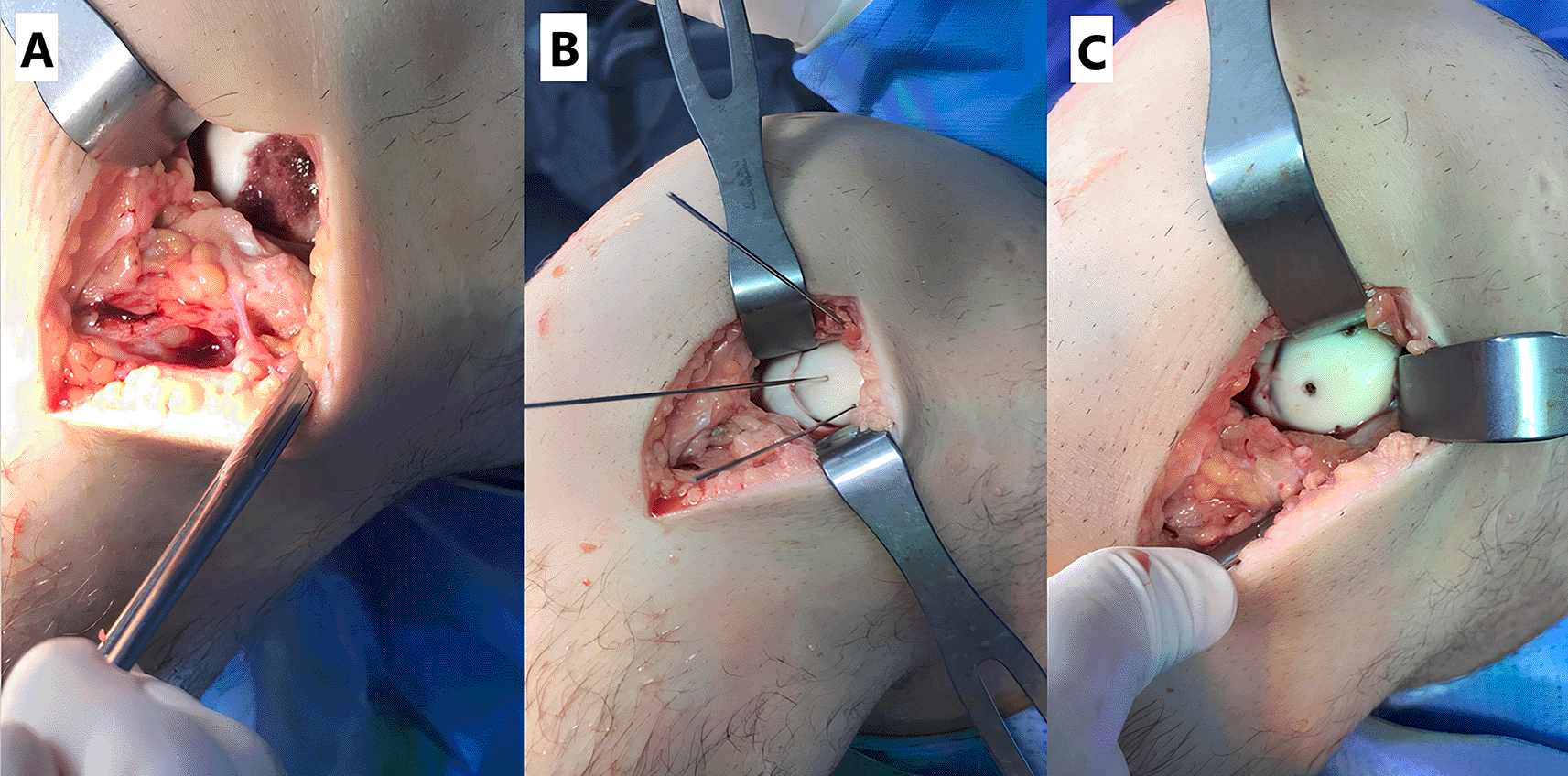
However, due to the size and location of the fragment, arthroscopic fixation was deemed infeasible. The procedure was converted to an open approach via a lateral parapatellar incision. Open reduction and internal fixation (ORIF) was then performed using two headless compression screws ( Figure 4).

Postoperatively, the patient began immediate mobilization with a non-weight-bearing protocol for 45 days, followed by continued physiotherapy for a total of three months.
Follow-up and outcome of interventions: At 18 months post-operatively, the patient demonstrated excellent clinical results with a pain-free, stable, and non-effused knee. Full range of motion was achieved. Radiographs also confirmed excellent results with no evidence of arthritic changes ( Figure 5).
The patient returned to sports.
Patient perspective: “After my knee injury, I was worried I wouldn’t be able to play sports again. The initial pain and swelling were really bad. I was glad when the surgery was over, but the recovery was tough. But it was worth it. My knee feels great now, and I’m back playing with my friends. I’m so grateful to the doctors and physiotherapists who helped me get better.”
Traumatic OCFs of the distal femur are relatively uncommon compared to other femoral injuries. The concept of traumatic OCF was first detailed by Milgram in 1943.3 It is important to differentiate a recent fracture from osteochondritis dissecans (OCD) based on the presence of a trauma-related incident. Recent fractures typically result in fragments that are well-suited for fixation techniques.4 OCFs commonly occur on the articular surfaces of bones frequently involved in trauma, such as the glenoid, femur, patella, and talus. The mechanism of injury can vary based on lesion location. Common causes include shearing, rotational or impaction forces, and excessive tangential loading of the articular surface.5
The precise incidence of OCFs around the knee remains uncertain. Current evidence suggests a higher frequency in pediatric patients compared to adults.6–8 Generalized joint laxity and patellar dislocation are often associated with OCFs.8,9 In patellar dislocations, the contracted quadriceps can exert high pressure on the lateral femoral condyle during patellar reduction, frequently resulting in OCFs.5 Studies have shown that the osteochondral unit in adolescents has lower resistance to shear forces,10 while adults exhibit greater fracture resistance at the osteochondral junction.11
Diagnosing OCFs in pediatric patients around the knee can be particularly challenging. Small bony fragments may be overlooked, and the actual size of the cartilage component might be underestimated or misinterpreted as an accessory bone. MRI is the preferred imaging modality for evaluating osteochondral injuries,12,13 while radiographs or computed tomography (CT) scans are more useful for evaluating the bony component of the fragment.
The management of chondral injuries with tissue loss remains a significant challenge. Recent studies suggest that innovative techniques offer promising results for cartilage repair and improved clinical outcomes. However, a universally accepted treatment approach has yet to be established.14,15
Key factors influencing the management of pediatric knee osteochondral lesions include lesion location, size, stability, and symptom severity.16 While fragment fixation is generally considered the gold standard treatment for OCFs, the impact of patient age, fragment size, and the interval between injury and diagnosis on surgical success remains unclear.17 Current literature supports operative intervention for osteochondral injuries, particularly those involving weight-bearing surfaces, lesions larger than 2 cm2, and those causing mechanical symptoms.18 Surgical fixation of acute OCFs in pediatric patients has demonstrated favorable clinical and radiological outcomes,8,16,19 likely due to the superior regenerative capacity of pediatric cartilage, facilitated by bony union and chondral extensions from the fragment.7,11 Historically, fragment excision was often performed in late-diagnosed cases, treating the fragment as a loose body.6,20 However, this approach can lead to early degenerative changes in the knee due to the resulting defect.21,22 Similarly, untreated intra-articular fragments can cause further cartilage damage and arthritis. Therefore, even in neglected or late-diagnosed cases, fixation remains the preferred treatment strategy.
Bioabsorbable pins offer the advantage of postoperative MRI compatibility. While multiple pins can enhance rotational stability, they may limit compression of the lesion. Screws, conversely, provide immediate compression and, when used in multiples, rotational stability.23 Postoperatively, patients can begin rehabilitation and undergo follow-up imaging with CT or MRI.
The literature provides limited guidance on the minimum bony component size required for OCF fixation. Fabricant et al.24 reported a 90% success rate in young athletes with chondral-only shear fractures treated with bioabsorbable implants or sutures. Other studies suggest fibrin sealant or tissue glue can be effective for larger fragments lacking sufficient bone for implant fixation.25,26 Schlechter et al.16 demonstrated good outcomes with bioabsorbable fixation in OCFs with a mean lesion size of 299 mm2. Hsu et al.27 recommended headless cannulated screws for fragments larger than 3 cm2 in a pediatric case. These findings suggest that while overall osteochondral fragment size helps determine whether surgical or conservative treatment is warranted, the size of the bony component dictates the specific fixation method (headless screws, adhesives, rods, or sutures).
Untreated osteochondral defects in children can enlarge with skeletal growth. Furthermore, fibrotic tissue formation on the bony component can reduce fragment size after debridement, potentially leading to incongruity between the fragment and the defect, hindering anatomical reduction. In this case, complete anatomical reduction was achieved, resulting in excellent outcomes at 18 months postoperatively. While the literature suggests eventual removal of metallic implants,28 we are conducting annual follow-up to monitor for potential articular cartilage damage and to guide the decision regarding implant removal.
This case contributes to the limited literature on isolated, weight-bearing lateral femoral condyle OCFs in pediatric patients, demonstrating the potential for successful outcomes with careful surgical management and emphasizing the need for individualized treatment approaches.
This case highlights the diagnostic challenges associated with isolated OCFs of the weight-bearing portion of the lateral femoral condyle. The subtle radiographic findings in such fractures underscore the importance of advanced imaging, such as MRI, when clinical suspicion remains high despite normal initial radiographs. Furthermore, this case demonstrates the efficacy ORIF when arthroscopic management is not feasible due to fragment size or location. Prompt and appropriate surgical intervention can effectively restore joint congruity and facilitate optimal healing in these challenging pediatric knee injuries.
Written informed consent for publication of their clinical details and/or clinical images was obtained from the patient’s parents.
| Views | Downloads | |
|---|---|---|
| F1000Research | - | - |
|
PubMed Central
Data from PMC are received and updated monthly.
|
- | - |
Is the background of the case’s history and progression described in sufficient detail?
Yes
Are enough details provided of any physical examination and diagnostic tests, treatment given and outcomes?
Yes
Is sufficient discussion included of the importance of the findings and their relevance to future understanding of disease processes, diagnosis or treatment?
Yes
Is the case presented with sufficient detail to be useful for other practitioners?
Yes
Competing Interests: No competing interests were disclosed.
Reviewer Expertise: Sports Medicine, Cartilage Reconstruction procedures.
Alongside their report, reviewers assign a status to the article:
| Invited Reviewers | |
|---|---|
| 1 | |
|
Version 1 04 Feb 25 |
read |
Provide sufficient details of any financial or non-financial competing interests to enable users to assess whether your comments might lead a reasonable person to question your impartiality. Consider the following examples, but note that this is not an exhaustive list:
Sign up for content alerts and receive a weekly or monthly email with all newly published articles
Already registered? Sign in
The email address should be the one you originally registered with F1000.
You registered with F1000 via Google, so we cannot reset your password.
To sign in, please click here.
If you still need help with your Google account password, please click here.
You registered with F1000 via Facebook, so we cannot reset your password.
To sign in, please click here.
If you still need help with your Facebook account password, please click here.
If your email address is registered with us, we will email you instructions to reset your password.
If you think you should have received this email but it has not arrived, please check your spam filters and/or contact for further assistance.
Comments on this article Comments (0)