Keywords
Ultrasound; Doppler;Leiomyomas;Adenomyosis;AUB.
This article is included in the Global Public Health gateway.
This article is included in the Manipal Academy of Higher Education gateway.
Ultrasound; Doppler;Leiomyomas;Adenomyosis;AUB.
Abnormal uterine bleeding (AUB) is a common problem among women in the reproductive age group.1 It accounts for approximately 70% of the pathology in perimenopausal and post-menopausal women. AUB may occur because of hormonal imbalances or as a result of benign or malignant lesions.2
Benign myometrial lesions are common in women of reproductive age and can cause significant morbidity.3 The most common causes of uterine bleeding are adenomyosis and uterine leiomyoma. Leiomyomas are benign masses of uterine smooth muscles that can be surgically removed. Adenomyosis is an ill-defined lesion in which endometrial glands and stroma are interspersed with the myometrium, making complete removal through surgery challenging.4 Therefore, accurate preoperative diagnosis of these two conditions is critical. Imaging techniques play a crucial role in distinguishing leiomyomas from adenomyosis.
Histopathology, considered a corner stone in confirming the diagnosis, remains a gold standard.4
Transabdominal/transvaginal sonography is often performed and is planned as a first line imaging modality. The ability to globally visualise the pelvic structures and organs combined with colour Doppler greatly enhances diagnostic ability.5
This study aimed to differentiate clinically suspected cases of uterine Leiomyoma and Adenomyosis using grey-scale 2D ultrasonography with color Doppler to determine its accuracy and confirm histopathological findings.
Owing to similar clinical signs and symptoms, misdiagnosing one in favour of the other is not uncommon. The organic cause responsible for menstrual disorders should be ruled out to provide a final cure for a broad range of gynecological diseases and preoperative diagnosis of these two conditions is vital for patients wishing to retain their fertility.
Study design: Observational Study.
Study period: August 2018 to August 2020.
Study setting: This study was carried out in tertiary care centres attached to the Kasturba Medical College, Mangaluru, Government Lady Goschen Hospital, and KMC Hospital Attavar.
Study population: Women reporting with complaint of Abnormal uterine bleeding in pre and perimenopausal age and clinically suspected cases of uterine Leiomyoma and Adenomyosis were selected.
Zα = 1.96 at 95% confidence level
Zβ = 1.28 at 90% power, the sample size calculated as 60.
The study included 60 patients with a history of Abnormal Uterine bleeding. Clinically suspected cases of Uterine Leiomyoma and Adenomyosis were included. Institutional ethical board clearance was also obtained. Informed written consent was obtained after explaining the study details in the language that was best understood by the study group.
Pre-operative assessment was performed by detailed history taking with respect to age, symptoms, parity, menstrual pattern, other coexisting medical conditions, gynaecological complaints, treatment taken prior to this visit and relevant family history. General and pelvic examinations and relevant investigations were performed. The clinical diagnosis was made based on the chief complaints and clinical examination. The study subjects underwent ultrasonography to confirm the clinical diagnosis.
The equipment used for transabdominal sonographic imaging was GELOGIQ S7, GES8, GEVOLUSON EXPERT730, PHILLIPS HD7, SAMSUNG 70A using a 2-5 MHz curvilinear and 6-12 MHz transvaginal transducer. Images were evaluated with respect to morphology. Sonographic images were recorded and stored for archiving purposes. A treatment plan was suggested based on radiological diagnosis.
After obtaining fitness, surgery was performed individualizing the approach for each patient. Myomectomy or total hysterectomy was performed according to standard surgical protocols. The specimens were subjected to histopathological evaluation. The final diagnosis was made based on the histological diagnosis. The collected data were subjected to statistical analyses. The clinical diagnosis, ultrasonographic findings, and histopathological reports were compared.
With this background, this study aimed to establish the accuracy of ultrasonography in diagnosing uterine leiomyoma and adenomyosis.
This is an observational clinical study. The Statistical Package for Social Sciences (SPSS) for Windows, Version 22.0 released 2013. Armonk, NY: IBM Corp.
Chi square test was used for comparison.
Independent student “t” test to compare the mean values of Doppler parameters.
Sensitivity and specificity analysis was done to estimate the accuracy of USG in differentiating Leiomyoma and Adenomyosis.
Significance level was set at P value of <0.05.
This study included 60 patients with a history of AUB and clinically suspected uterine leiomyoma and Adenomyosis ( Table 1).
| Variable | Category | Number of Leiomyoma Cases (n) | % | Number of Adenomyosis Cases (n) | % |
|---|---|---|---|---|---|
| Age | 21-30 Years | 2 | 3.33 | 0 | 0.00 |
| 31– 40 Years | 7 | 11.67 | 3 | 5.00 | |
| 41– 50 Years | 31 | 51.67 | 13 | 21.67 | |
| 51-60 Years | 1 | 1.67 | 3 | 5.00 | |
| Total | 41 | 68.33 | 19 | 31.67 |
The study patients were in the age range of 28–53 years. The maximum number of patients was 41 – 50 years (n=44, 73.3%). The mean age was 44.45 yrs.
The maximum incidence of fibroids and adenomyosis was found to between 41-50 yrs. We found 31 (51.67%) cases of leiomyoma and 13 (21.67%) cases of Adenomyosis within 41-51 yrs range. When compared to other age categories, both conditions were found to be comparatively less prevalent especially in those aged <30 years and > 51 years.
More uterine leiomyomas n= 28 (46.67%) and adenomyosis n= 18(30%) were found in multiparas. Eight cases (13.33%) of leiomyoma were observed in the primiparous group followed by five cases (8.33%) in the nulligravida group. In the case of adenomyosis, one case (1.67%) was observed in primiparous patients and none in Nulligravida ( Table 2).
| Variable | Category | Leiomyoma (n) | % | Adenomyosis (n) | % |
|---|---|---|---|---|---|
| Parity | Nulligravida | 5 | 8.33 | 0 | |
| Primiparous | 8 | 13.33 | 1 | 1.67 | |
| Multiparous | 28 | 46.67 | 18 | 30.00 | |
| Total | 41 | 68.33 | 19 | 31.67 |
The most common clinical symptom among the study patients was heavy menstrual bleeding 50 (83.3%), followed by pelvic pain n= 42 (70%), and dysmenorrhea n =36(60%). Near equal distribution of mass per abdomen and Urinary Tract complaints were observed in 15 (25%) and 14 (23.3%) cases respectively ( Table 3).
| Variable | Category | Number of patients (n) | % |
|---|---|---|---|
| Clinical Symptoms | Heavy Menstrual Bleeding | 50 | 83.3% |
| Chronic pelvic pain | 42 | 70.0% | |
| Dysmenorrhea | 36 | 60.0% | |
| Mass per abdomen | 15 | 25.0% | |
| Urinary Tract Complaint | 14 | 23.3% |
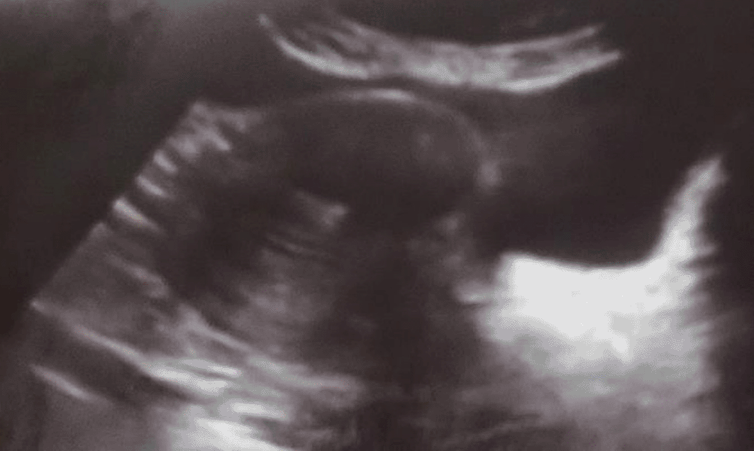
Out of 60 cases, 41 were suspected clinically as leiomyomas. USG detected leiomyoma in 36 cases (60.0%) ( Figure 1). Nineteen cases were suspected clinically as Adenomyosis, out of which USG detected 15 cases (25%) to be adenomyosis. USG also detected nine cases which were categorized as other conditions. (Note: Other conditions are pathologies other than Leiomyoma and Adenomyosis.)
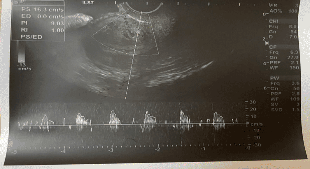
While imaging patients with leiomyomas using USG and color Doppler, peripheral vascularity was observed in 39 cases (95.12%). The morphological features of USG findings with well-defined margins were present more in 39 cases (95.12%) followed by absence of cystic space 31 (75.61%) and least observed was edge shadowing 5 (12.20%) ( Table 4).
Imaging adenomyosis using USG with color Doppler central vascularity accounted for 18 (94.70%) ( Figure 2). The morphological features of USG findings accounted for a more ill-defined margin 19(100.0%), near equal distribution of globular uterus 18 (94.70%), uterine wall thickening 16 (84.20%) and least observed was hypoechoic linear striations 3(15.80%) ( Table 5).
Total abdominal hysterectomy was the most common type of surgery performed than myomectomy.
The distribution of the type of surgery performed among the study patients revealed that total abdominal hysterectomy was done in 40 cases (66.67%) and myomectomy in 20 cases (33.33%) ( Table 6).
| Age | Abdominal Hysterectomy | Myomectomy | ||
|---|---|---|---|---|
| Cases | % | Cases | % | |
| 21-30 Years | 1 | 2.50 | 1 | 5.00 |
| 31-40 Years | 5 | 12.50 | 5 | 25.00 |
| 41-50 Years | 31 | 77.50 | 13 | 65.00 |
| 51-60 Years | 3 | 7.50 | 1 | 5.00 |
| Total | 40 | 20 | ||
Both surgical procedures were found to be performed in the age range of 41-50 years.
Histopathological features of leiomyoma revealed more specimens with smooth muscle whorls 36 (97.3%), followed by pseudo capsule 30 (81.1%), Dense Connective tissue 27 (73.0%) and hyalinization 26 (70.3%) ( Table 7).
Histopathological features of adenomyosis revealed specimens with equal distribution of endometrial glands and smooth muscle hyperplasia in 15 cases (100%) each followed by disruption of the endometrium and myometrium in 12 cases (80.0%) ( Table 8).
The test results demonstrated that the mean PI scores among adenomyosis cases [1.722 ± 0.205] were significantly higher than those in leiomyoma cases [0.994 ± 0.190] at P<0.001 ( Table 9).
Similarly, the mean RI scores among adenomyosis cases [1.450 ± 0.293] was significantly higher than that compared to leiomyoma cases [0.679 ± 0.147] at P<0.001.
In contrast the mean Vmax scores among leiomyoma cases [10.836 ± 1.448] were significantly higher than those of adenomyosis cases [7.280 ± 1.864] at P<0.001. This difference was Statistically Significant.
Out of the 60 cases, clinical diagnosis over estimated 9 cases as follows:
However, Imaging was correctly performed in up to 51 cases (83.33%)-36 cases of Leiomyoma and 15 cases of adenomyosis.
The test results demonstrated that the clinical diagnosis was strongly correlated with USG findings P <0.001was considered statistically significant.
Histopathological diagnosis unveiled the falsified USG diagnosis in 9 cases and diagnosed all 52 (86.66%) cases appropriately. However, imaging was correctly performed in 51 (85.0%) cases of Leiomyoma and Adenomyosis.
The comparison analysis test results demonstrated that the USG findings had a very strong correlation with histopathological diagnosis P < 0.001 which was found to be statistically significant ( Table 10).
| USG findings | Histopathological diagnosis | χ2 Value | P-Value | |||||
|---|---|---|---|---|---|---|---|---|
| Leiomyoma | Adenomyosis | Other pathology | ||||||
| n | % | n | % | n | % | |||
| Leiomyoma | 36 | 100.0% | 0 | 0.0% | 0 | 0.0% | 111.89 | <0.001* |
| Adenomyosis | 0 | 0.0% | 15 | 100% | 0 | 0.0% | ||
| Other pathology | 1 | 11.1% | 0 | 0.0% | 8 | 88.9% | ||
Accuracy was calculated using the area under the receiver operating characteristics (ROC) curve. An area of 1 denotes a perfect test indicating that USG findings can be a good test to accurately detect leiomyoma ( Figure 3 and Table 11).
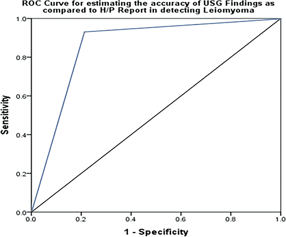
An area of 1 denotes a perfect test indicating that USG findings can be a good test to accurately detect leiomyoma.
| Area under curve | Std Error | P-Value | 95% CI | |
|---|---|---|---|---|
| Lower | Upper | |||
| 0.85 | 0.13 | 0.02* | 0.59 | 1.00 |
Accuracy was calculated using the area under the receiver operating characteristics (ROC) curve. An area of 1 denotes a perfect test indicating that USG findings would be a good test to accurately detect Adenomyosis ( Figure 4 and Table 12).

An area of 1 denotes a perfect test indicating that USG findings would be a good test to accurately detect adenomyosis.
AUB is a common clinical symptom. It encompasses bleeding due to systemic causes, pathology of the genital tract and dysfunctional uterine bleeding.6
Myometrial lesions form various groups among which leiomyoma (benign smooth muscle tumour) is the most common followed by adenomyosis, leiomyosarcoma, endometrial stromal tumours, secondary tumours, and vascular lesions. Leiomyoma is a benign neoplasm that affects women of reproductive age. They are observed clinically in 20-30% of women above 30 years of age. Large tumours give rise to diffuse enlargement of the uterus with irregular uterine contours, which may be related to infertility.7
Ultrasonography is a dependable method for diagnosing tumours, supplemented with Doppler mode to provide additional hemodynamic information.8 In obstetrics and gynaecology the pulsatility index (PI) is presently the preferred Doppler index due to its sensitivity to change in waveform appearance and it analyzes the entire cardiac cycle. Doppler is a principle supplement to ultrasonography playing a vital role in the assessment of uterine leiomyomas, pelvic hormone replacement therapy, infertility, arteriovenous malformations, etc.8
The vascularity of the leiomyoma and flow patterns were evaluated using Doppler. Velocimetry is of practical use for the management of uterine leiomyomas. Ultrasonography of arteries is useful in observing leiomyomas in response to therapy, distinguishing Adenomyosis from Leiomyomas, and evaluating tumor size changes in response to uterine artery embolization. The degree of vascularity reflects the growth pattern of the tumour and increase the bleeding risk during surgery.
The leiomyoma vascular pattern showed multiple vessels located peripherally surrounding the tumour and the vessels were longer than 1 cm when only a few vessels were close to the tumor. Focal Adenomyosis intramural signals seen without peripheral vessels, and less than 1 cm long if peripheral vessels are present.8
This was an observational study of 60 patients of reproductive age group. Women with history of heavy menstrual bleeding and clinically suspected case of Leiomyoma and Adenomyosis were included. All study patients were subjected to ultrasonography. Depending upon the treatment suggested surgery was carried out individualising the approach to each patient. Myomectomy or transabdominal hysterectomy was performed with standard surgical protocols. Ultimate diagnosis was made on the basis of histology report. Histopathological report of the specimens was collected and maintained as a record. The collected data were subjected to statistical analysis. Finally, Clinical diagnosis, Ultrasonographic findings and Histopathological reports were compared.
In the present study the age of the patients ranged from 21 to 53 years. The majority of patients were predominantly aged 4150years. Majority of patients were of the perimenopausal group contributing to 73.3% of total sample size. A similar finding was reported by Kumari G et al. with age > 40 years.9 Supria et al.10 found maximum patients in 40-49 years (40.22%).
A wide age range was observed in all the study samples. The increased incidence of AUB in this age group may be due to their climacteric period, prior to which changes occur in ovarian-pituitary function.10
The present study revealed that the maximum number of multiparous patients was 46(76.7%). Lithingo Lotha et al.11 (76.7%) and Dinesh et al.12 (79.3%) observed the maximum number of patients in the multiparous group.
In this study the parity distribution among women ranged from 0-4. The maximum number of leiomyoma cases was observed in women with a parity score of 2(48.7%) and adenomyosis cases in women with a parity score of 3 (52.63%). However, 12.40% of leiomyoma cases were also found in nulliparous women. This could be related to a family history of the disease.
Singh et al.13 found the maximum number of cases was 44.15% in para 3. A study by Mohamed Ranaei14 found that adenomyosis is common in multiparous women.
This age distribution may be because the effects are hypothesized to occur as a result of a decrease in menstrual cycle changes in levels of ovarian hormones and growth factors, and a reduction in oestrogen receptor levels in myometrial tissue.15
In a study by Gupta et al., the authors found heavy menstrual bleeding was present in 97.83% of subjects, 60.87% had chronic pelvic pain, 93.48% had dysmenorrhea, 43.48% had dyspareunia, and 84.78% had a uterine size of ≥8 weeks on clinical examination.16
As per FIGO The causes of AUB have been broadly classified into structural/organic causes and non-structural/functional causes.17 Menorrhagia is a subjective complaint evidenced by women as the heaviness of their period. AUB presents with different patterns of bleeding.
In the present study a greater number of cases of leiomyoma were clinically suspected than adenomyosis. Among the 60 patients, 41 (68.3%) Leiomyoma cases and Adenomyosis19 (31.7%) was observed.
As the endometrium is a dynamic and hormonally sensitive and responsive tissue, it continually experiences variations throughout reproductive life; thus it is vulnerable to pathological lesions and manifests as different symptoms17 Unopposed estrogen stimulation may lead to endometrial proliferation and hyperplasia, which may cause menorrhagia.
In the present study the most common clinical presentation was heavy menstrual bleeding (83.3%) followed by chronic pelvic pain. The study patients presented with a combination of clinical symptoms, including dysmenorrhea and mass per abdomen.
Adenomyosis may be asymptomatic but is also a structural cause of AUB. Adenomyosis is extremely variable and exact etiology may be hyper-estrogenism, endometriosis, pregnancy, and prior uterine surgeries.17
In the present study, using color Doppler imaging in patients with leiomyomas, peripheral vascularity was observed in 39 cases (95.12%).The morphological features of leiomyoma revealed maximum cases with well defined margins which accounted for 39 cases (95.12%) edge shadowing accounted for 5 cases(12.20%), and using several sonographic morphological and color Doppler imaging was able to correctly diagnose 85% of the total cases.
Other studies have observed higher blood velocity and lower PI and RI in uterine arteries with uterine fibroids. Fibroids significantly change the velocity flow of blood in the arteries and PI Values < 1.0 in fibrooids.18
In the present study color Doppler central vascularity was observed in 18 (94.73%) cases, and the morphological features of USG accounted for adenomyosis with ill-defined margins in 19 cases (100%) globular uterus in 18 cases (94.70%), and hypoechoic linear striations in 3 cases (15.80%). The contribution of various sonographic morphological features and color Doppler imaging correctly diagnosed 36 cases (60%) of leiomyoma and 15 (25%) cases of adenomyosis. However imaging was accurately performed in 51 cases (85%) in the total sample.
The presence of myometrial cysts, deformed or heterogeneous echo texture, and scanty foci of abnormal myometrium observed by TAS characterize adenomyosis. The presence of ill-defined myometrial heterogeneity is a prognostic ultrasound feature in adenomyosis. The absence of a distinct margin and morphological features of linear striations appears to have high specificity of sonography in evaluating adenomyosis.19
In our study, the test results demonstrated the validity of USG findings in the detection of leiomyoma in patients as compared to histopathological methods, with a P value of 0.001 which was statistically significant.
In the diagnosis of submucosal fibroids and small fibroids (<5 mm) TVS is more sensitive, but they recommend that TAS and TVS should be performed in conjunction.20
MUSA criteria have enhanced the potential of diagnosing adenomyosis using ultrasound.21
The receiver operating characteristic curve (ROC) is a plot of the true positive rate against the false positive rate for the different possible cut-off points of a diagnostic test.
ROC Curve demonstrated area under curve value of 0.85 (95% CI 0.59-1.00) with a significance of P=0.02. An area of 1 represented a perfect test. This indicate that USG findings can be a good test for accurately detecting leiomyoma.
The ROC Curve demonstrated an area under the curve value of 0.88 (95% CI 0.68-1.00) with a significance of P=0.01 indicating that USG findings would be a good test to accurately detect cases of adenomyosis. An area of 1 represents a perfect test.
This study revealed a USG Sensitivity of 97% and a positive predictive value of 97% for leiomyoma and 100% sensitivity and positive predictive value for adenomyosis. Doppler velocimetry revealed that a lower pulsatility index and resistive index were highly suggestive of leiomyoma, and vice versa in cases of adenomyosis. Histopathology correlated with ultrasound and colour Doppler velocimetry findings in identifying and differentiating clinically suspected cases of Leiomyoma and Adenomyosis.
The study was approved by Intsitutional Ethics Committee of Kasturba Medical College, Mangaluru (IEC KMC MLR 09-18/240) on 19th September 2018. Informed written consent was obtained after explaining the study details in the language that was best understood by the study group.
Repository name- Mendeley: TITLE-ROLE OF ULTRASOUND WITH DOPPLER IN DIFFERENTIATING CLINICALLY SUSPECTED CASES OF LEIOMYOMA AND ADENOMYOSIS OF UTERUS. https://doi.org/10.17632/y4c6bk9h2m.2.22
This project contains the following underlying data:
Data is available under the terms of the CC0 1.0
| Views | Downloads | |
|---|---|---|
| F1000Research | - | - |
|
PubMed Central
Data from PMC are received and updated monthly.
|
- | - |
Provide sufficient details of any financial or non-financial competing interests to enable users to assess whether your comments might lead a reasonable person to question your impartiality. Consider the following examples, but note that this is not an exhaustive list:
Sign up for content alerts and receive a weekly or monthly email with all newly published articles
Already registered? Sign in
The email address should be the one you originally registered with F1000.
You registered with F1000 via Google, so we cannot reset your password.
To sign in, please click here.
If you still need help with your Google account password, please click here.
You registered with F1000 via Facebook, so we cannot reset your password.
To sign in, please click here.
If you still need help with your Facebook account password, please click here.
If your email address is registered with us, we will email you instructions to reset your password.
If you think you should have received this email but it has not arrived, please check your spam filters and/or contact for further assistance.
Comments on this article Comments (0)