Keywords
percutaneous nephrolithotripsy, model, surgery training, Op-simulation
percutaneous nephrolithotripsy, model, surgery training, Op-simulation
In response to the single round of peer review, we have revised the manuscript as follows:
• Methods – Model preparation:
We now specify that an off‑label culinary gelatin without a reported Bloom rating was used and clarify that Psyllium Plantago was not incorporated because of limited availability. To ensure anatomical kidney angulation, we describe the newly adopted two‑stage pouring technique (gelatin partially set before kidney placement, then topped up).
• Figures:
A new Figure 6 has been added showing real‑time ultrasound images of needle insertion into the simulated collecting system, as requested, to illustrate image quality and puncture trajectory. All subsequent figures have been renumbered accordingly.
• Discussion – Limitations:
We acknowledge that only novice urologists evaluated the phantom; expert validation was not feasible within current resources. This limitation is now explicitly stated, and we outline plans for expert assessment in future work.
Title, abstract, author list, tables, data set, and supplementary files remain unchanged.
See the authors' detailed response to the review by Enrique Pulido-Contreras
Urolithiasis affects around 0.1–19.1% of the total population in Asia.1 However, throughout the course of time, the frequency and incidence of the disease have shifted in a variety of nations or areas as a result of differences in socioeconomic standing as well as geographic locations.1 Over the course of the past few decades, the frequency and incidence of urolithiasis have skyrocketed across the majority of the nations in Asia.1 One of the most performed treatments for urolithiasis is percutaneous nephrolithotomy (PCNL).2
For percutaneous nephrolithotripsy (PCNL) to be as effective as possible and to reduce any potential morbidity, it is imperative that the practitioner have sufficient training. Currently, training in PCNL is offered all over the globe via organized residency and fellowship programs. Individual theoretical and practical training courses are also offered to supplement this kind of education. However, there is some debate over the usefulness of the shorter training programs.3
Surgical simulations are commonly performed in training for various urological procedures.4–6 However, commercially printed organ models and surgical simulators which use synthetic materials are typically lacking in anatomical details and often fabricated from materials with different sensations from the target organ.4 Meanwhile, the use of animal tissues such as pig kidneys is usually cheaper but carries potential animal ethical tissues.2
This study aims to develop a low-cost kidney transplantation surgery training model using both animal parts and synthetic materials.
The ethical approval to conduct this study was issued by the research ethic committee of the Faculty of Medicine, Universitas Indonesia with ethical clearance letter number KET-909/UN2.F1/ETIK/PPM.00.03/2022. Written informed consent was obtained from the urologists participating in this study. They were provided with a structured questionnaire to evaluate the training model’s realism, functionality, and overall effectiveness, using a Likert scale for scoring.
This study is designed to develop and evaluate a low-cost PCNL training model using synthetic materials (silicone for kidney and gelatine jelly for the interstitial space) for both urologists and urology residents in developing countries such as Indonesia.
3D printing of the kidney and calyceal system mould
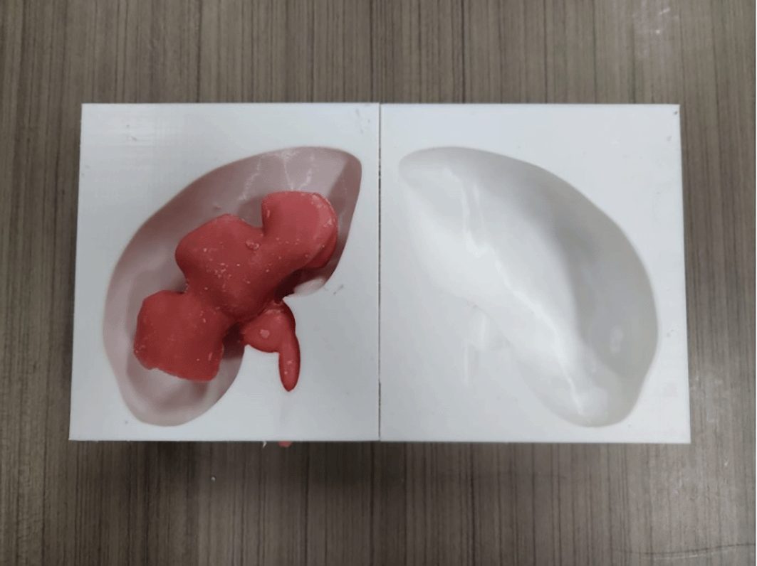
The kidney mould model was printed using a 3D-printer with Polylactid Acid (PLA) filament. The mould was separated into two interconnecting parts. Printing of a two-part mould took 25 hours and 288 grams of PLA filament. An adult kidney with hydronephrosis was scanned using computed tomography (CT) imaging. The DICOM file was deconstructed in InVesalius 3.0.0 (Centro de Tecnologia da Informação Renato Archer, Brazil, freely available on the web) and exported as STL (stereolithography) file. The file was then converted and stitched using a freely-available plug-in “Autodesk Mesh Mixer”. The STL file was printed out on a commercially available 3D printer (Ender-3 Pro, Creality 3D, China). Both the model of the kidney and calyceal system were printed as the initial mould ( Figure 1).
Calyceal system moulding using wax
Much like the kidney mould, the calyceal system was acquired from the same adult kidney with hydronephrosis scanned using CT imaging, the DICOM file was converted into STL file using InVesalius 3.0.0. STL refinement was done using Autodesk Mesh Mixer. The STL file of the calyceal system was printed with the same 3D printer. Printing of the calyceal system took 6 hours and 44 grams of PLA filament. The printed calyceal system was then casted in 400 mL of silicone RTV 52 with catalyst ratio of 1 to 100. In order to create a negative mould for further replication, wax casting of the calyceal system was done using 100 mL of commercially available paraffin wax ( Figure 2).

Silicone kidney model assembly
Following the development of both the 3D printed kidney mould and the wax calyceal system, the kidney was created using RTV52 silicone rubber that was poured into the mould with the wax calyceal system sandwiched between the two part mould. A total of 350 mL RTV52 silicone rubber was used to create one kidney with a catalyst ratio of 1 to 100. Following the curing time of the silicone, the silicone kidney was demoulded form the master mould and was boiled in water (80° C or 176° F) for 15 minutes to melt the wax calyceal system within the silicone kidney, leaving a hollow space inside the silicone kidney model.
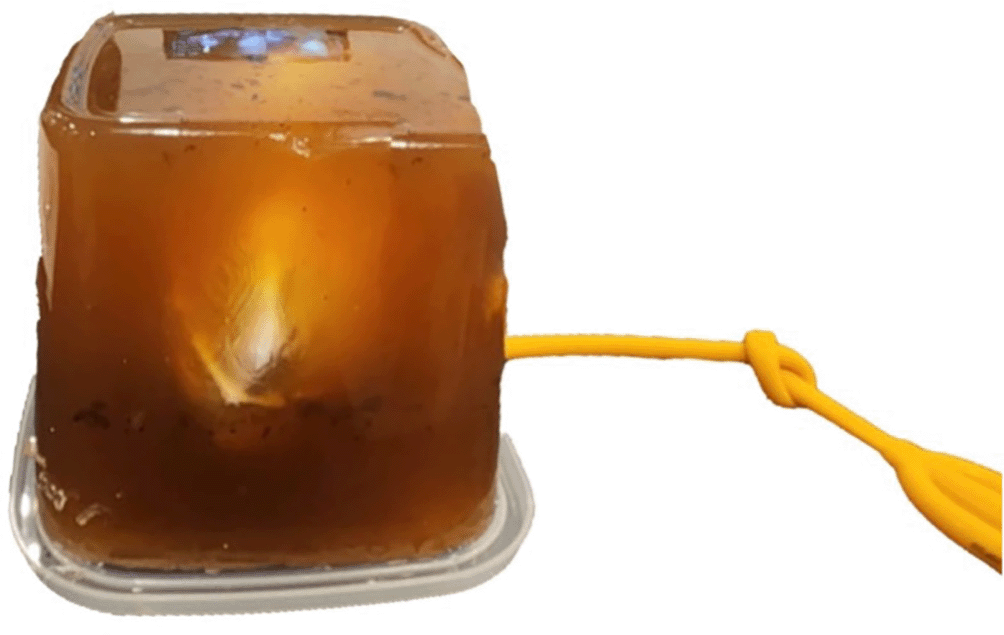
Artificial stones made from commercially available camphor were inserted to facilitate nephrolithotripsy training. The kidney was sealed using a three-way foley catheter 24 Fr (Rusch®, Teleflex, USA) balloon to create a water sealed environment and simulate the closed urinary system. The foley catheter was clamped after adding tap water into the calyceal system ( Figure 3).

A 300 mL of gelatin medium was prepared from commercially available gelatin powder (culinary grade, no Bloom rating provided) mixed with water in a 1:9 ratio. The gelatine powder was added while boiling and stirring the water to avoid clumps. To ensure anatomical kidney angulation, the gelatin was poured in two stages. The first half was allowed to partially set before placing the kidney in its desired position, then the top half was poured afterward. The silicone kidney was submerged in a gelatine medium with a minimum depth of 7 cm to simulate the environment for PCNL procedure. The final model was then put into a chiller with a temperature of 4°C (40°F) for 6 hours to set the gelatine medium. The final model was then taken out from the chiller shortly before use ( Figure 4).
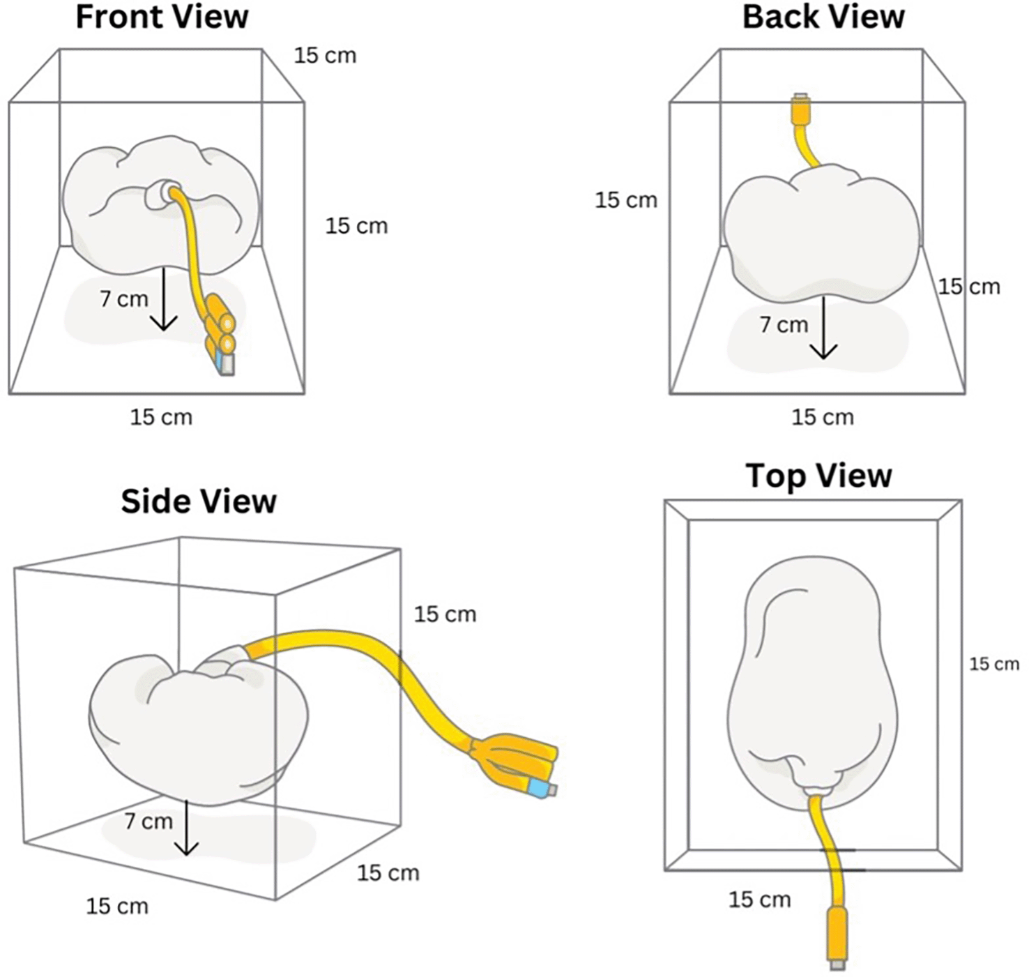
The details of the surgical technique used in our training have been described in other publications.7 Initially, an ultrasound convex ultrasound transducer with 3-5 megahertz (Mhz) frequency and needle-insertion bracket was used assess the calyceal system and identify route of insertion. Moreover, the distance between the outer part of the model (representing the skin) and the calyceal system was measured. A gravity bag of saline was hanged 60 cm above the model and connected to the foley catheter to simulate passive retrograde filling. The percutaneous renal access is established using a 17.5-gauge, echogenic, renal access needle.
Once the needle has reached the collecting system, removal of the needle stylet will facilitate visualization of normal saline (representing urine). The needle stylet is then withdrawn to allow the insertion of J-tip, superstiff, guidewire through the access needle. The guidewire was inserted until secured in place within the calyceal system.
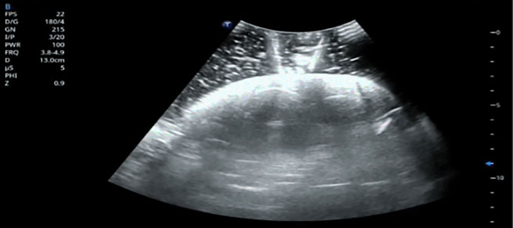
Following the guidewire insertion, a series of fascial dilator is passed over the guidewire until the measured distance to create a working tract. A percutaneous access sheath is then carefully advanced until the efflux of the normal saline is seen at the outer tip. Nephroscopy for stone removal and nephrolithotripsy are then done as per usual standard fluoroscopy-guided PCNL. An ultrasound image showing the needle entering the simulated calyceal system has been added ( Figure 6) to demonstrate the real-time visualization during ultrasound-guided access. Although silicone provides adequate tactile sensation, one of its drawbacks is that ultrasound imaging cannot visualize the calyces effectively due to its acoustic properties.
The evaluation of our model was done through an anonymous response to a questionnaire developed by Ali et al (2020).8 The questionnaire consisted of 8 primary questions with a Likert scale of 1 (strongly disagree) to 10 (strongly agree). A higher score represented a better perception from the respondent. Moreover, an additional comment and suggestion section was placed following the questionnaire. The complete contents of the questionnaire can be found in Table 1.
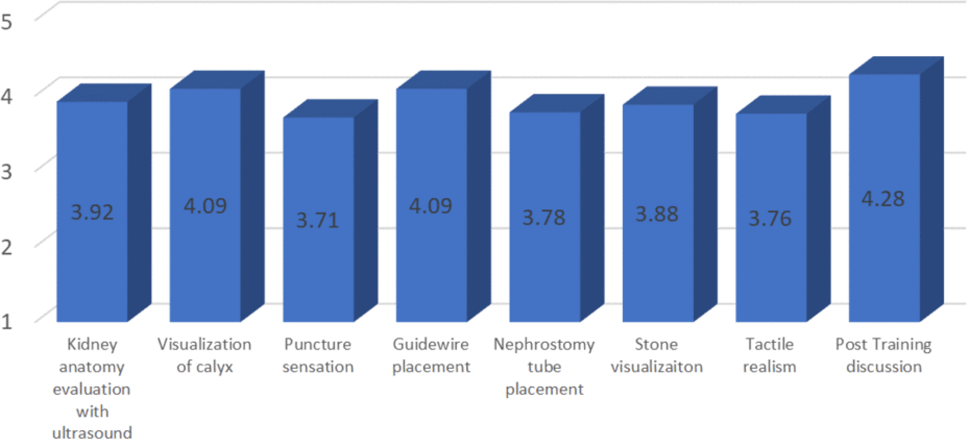
During the study, a total of 21 urologists with no experience of performing US-guided PCNL in Indonesia participated in the training and anonymously gave the evaluation of our model. The questionnaire used can be found in Table 1, whereas the result of the perception of the model could be found in Table 2.
Based on the evaluation, there were highly positive results among participants (average score of kidney anatomy evaluation using ultrasound 3.92/5, visualization of calyx 4.08/5, puncture sensation 3.71/5, guidewire placement 4.09/5, nephrostomy tube placement 3.78/5, Stone visualization 3.88/5, Tactile realism 3.76/5 and Post training discussion 4.28/5. The question with the lowest average score was about puncture sensation. However, there was no additional comment or suggestion regarding the issue.
In this study, we have developed an affordable non-biological model using with similar characteristics to the kidney for percutaneous nephrolithotomy (PCNL) training. Based on the post-training evaluation of our model, a high satisfactory rate was obtained from every participant in our study.
The results of our study was in line with other PCNL training model created in other studies.8,9 Although traditionally PCNL training consists of dry lab training using commercially available bench model and wet lab training on real patients, the cost of traditional model and the safety of the patients remain as an issue. Moreover, training using animal models such as porcine kidney may not fully represent the calyceal system in human, while training using virtual reality (VR) simulation model is currently very limited in developing countries such as Indonesia.10
For surgical teaching systems, there are a variety of possibilities, including synthetic models, virtual reality simulations, animal parts, and complete animals.2 Synthetic models are frequently selected for training in several teaching hospitals and educational institutions due to their nature as the comparatively affordable, simply available, and repeatedly reusable model. When executing the procedure, it is, however, similarly deficient in actual tactile input. On the other hand, whole animals and reality simulations are expensive and challenging to obtain.2 To offer a comparable experience while still keeping the cost of the model low, we thus sought to use an affordable synthetic model with realistic consistency.
The hands-on training using animal model and live surgery is considered useful in changing the practices and techniques in PCNL, even for attending surgeons.3 We hypothesized that the hands-on training using non-biological model would also provide similar experience and changes to the current practice of the urologists in developing countries.
One limitation of this study is the lack of expert urologist validation prior to involving novices. Due to logistical constraints, only urologists without prior PCNL experience participated. Future studies are planned to include validation by experienced PCNL practitioners to enhance model reliability.
Currently, the fluoroscopy guidance for PCNL is the most commonly used imaging modality for the surgery, starting from obtaining renal access to performing stone extraction. However, radiation exposure is an important issue for patients with urolithiasis as it tends to form repeatedly during a lifetime.11 Thus, the rising use of ultrasound-guided PCNL is deemed as an alternative with fewer risk of radiation exposure.12 There are only limited options of training model for ultrasound-guided PCNL. Therefore, we hope that the development of our training model to be beneficial in improving the learning curve of ultrasound-guided PCNL.
The limitation of this study is the lack of comparison between the developed model and a similar model. Moreover, there is no objective measurement of the models’ characteristics, such as consistency, plasticity, and tensile strength.
An affordable model utilizing 3D printed mould, silicone, and gelatine jelly is a feasible option for ultrasound guided PCNL training among urologists in developing countries. The utilization of 3D printing and silicone moulding will be beneficial in reducing the cost of surgery model while preserving the realistic tactile feedback.
The ethical approval to conduct this study was issued by the Research Ethics Committee of the Faculty of Medicine, Universitas Indonesia, with ethical clearance letter number KET-909/UN2.F1/ETIK/PPM.00.03/2022, dated 22 September 2022. Written informed consent was obtained from the subjects participating in the study. This study complies with the Declaration of Helsinki and was performed according to ethics committee approval.
RAH: Conceptualization, Data curation, Formal analysis, Investigation, Resources, Software, Methodology.
PB: Conceptualization, Data curation, Formal analysis, Investigation, Project administration, Resources, Software, Validation, Methodology, Writing, Supervision, Funding acquisition.
NR: Conceptualization, Formal analysis, Investigation, Project administration, Software, Validation, Methodology, Writing, Supervision, Funding acquisition.
IW: Conceptualization, Data curation, Formal analysis, Investigation, Project administration, Writing, Supervision, Funding acquisition.
GRS: Conceptualization, Data curation, Formal analysis, Investigation, Project administration, Writing, Supervision, Funding acquisition.
WA: Formal analysis, Investigation, Software, Validation, Methodology, Writing, Supervision, Funding acquisition.
MRD: Conceptualization, Data curation, Formal analysis, Investigation, Writing, Supervision
KY: Conceptualization, Data curation, Formal analysis, Investigation, Validation, Methodology, Writing.
JPS: Conceptualization, Investigation, Project administration, Resources, Validation, Writing.
All authors has approved the final version of the manuscript.
| Views | Downloads | |
|---|---|---|
| F1000Research | - | - |
|
PubMed Central
Data from PMC are received and updated monthly.
|
- | - |
Provide sufficient details of any financial or non-financial competing interests to enable users to assess whether your comments might lead a reasonable person to question your impartiality. Consider the following examples, but note that this is not an exhaustive list:
Sign up for content alerts and receive a weekly or monthly email with all newly published articles
Already registered? Sign in
The email address should be the one you originally registered with F1000.
You registered with F1000 via Google, so we cannot reset your password.
To sign in, please click here.
If you still need help with your Google account password, please click here.
You registered with F1000 via Facebook, so we cannot reset your password.
To sign in, please click here.
If you still need help with your Facebook account password, please click here.
If your email address is registered with us, we will email you instructions to reset your password.
If you think you should have received this email but it has not arrived, please check your spam filters and/or contact for further assistance.
Comments on this article Comments (0)