Keywords
Lung Cancer, Pancoast-Tobias Syndrome, Colonic Metastasis, Non-Small Cell Lung Cancer.
This article is included in the Oncology gateway.
This article is included in the Rare diseases collection.
Lung cancer remains one of the most prevalent malignancies worldwide, with a high mortality rate. However, Pancoast tumors, a rare subset of non-small cell lung cancer (NSCLC), represent an uncommon clinical presentation. While the liver, bones, brain, and adrenal glands are the most frequent metastatic sites of lung cancer, gastrointestinal involvement, particularly colonic metastasis, is exceedingly rare. Herein, we present the case of a 61-year-old man diagnosed with Pancoast-Tobias syndrome, initially manifesting through colonic metastasis.
Lung Cancer, Pancoast-Tobias Syndrome, Colonic Metastasis, Non-Small Cell Lung Cancer.
Lung cancer is a prevalent malignant tumor with a high mortality rate, ranging from 18% to 23%.1 Approximately half of lung cancer cases present with distant metastases at diagnosis, with mortality rates exceeding 50% in these patients.2 The most common metastatic sites include the lungs, liver, bones, brain, and adrenal glands.3,4 In contrast, colonic metastasis from lung cancer is exceptionally rare,3 with an incidence ranging from 0.2% to 1.7%.5
Among lung cancer subtypes, squamous cell carcinoma is the most frequently reported histological type associated with colonic metastasis, whereas adenocarcinoma rarely spreads to the colon or rectum.6 Given its rarity, distinguishing primary colonic neoplasms from metastatic lung cancer remains challenging.
Herein, we present a case of Pancoast-Tobias syndrome (PTS) initially manifesting as colonic metastasis and anemia, highlighting an unusual metastatic presentation of lung cancer.
A 61-year-old man with a history of chronic alcoholism and heavy smoking (60 pack-years) presented to our hospital with asthenia, anorexia, and significant weight loss, accompanied by anemia-related symptoms such as dyspnea and palpitations. He denied any gastrointestinal bleeding, digestive symptoms, or respiratory complaints. However, he reported back and right shoulder pain.
On examination, mucocutaneous pallor was the primary clinical finding. Abdominal, cardiovascular, and pulmonary examinations were unremarkable, and no lymphadenopathy was detected. Laboratory tests revealed severe iron deficiency anemia (hemoglobin: 2 g/dL), while renal and liver function tests were within normal limits. The patient received blood transfusion and intravenous iron supplementation, leading to an improvement in hemoglobin and ferritin levels.
An upper gastrointestinal endoscopy revealed no abnormalities, whereas lower gastrointestinal endoscopy identified a large ulcerated mass in the sigmoid colon. Histopathological examination confirmed a poorly differentiated adenocarcinoma ( Figure 1).

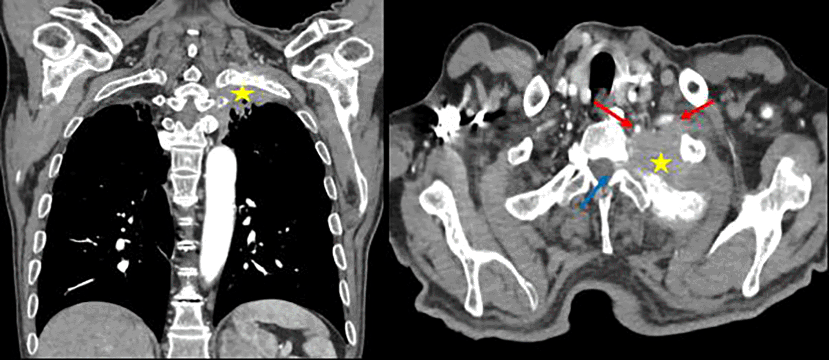
For tumor staging, a contrast-enhanced computed tomography (CT) scan of the chest, abdomen, and pelvis was performed. Coronal and axial contrast-enhanced thoracic CT scans (parenchymal and bone windows) revealed mediastinal lymphadenopathy and a left apical mass with heterogeneous enhancement. The lesion invaded the posterior arch of the second rib with lytic destruction and extended into the D1-D2 lateral foramen, showing proximity to the ipsilateral subclavian and vertebral arteries ( Figure 2). These findings were consistent with PTS.

It invades the posterior arch of the second rib with lytic lesions and extends towards the lateral foramen D1-D2 (blue arrow). The process shows close association with the ipsilateral subclavian and vertebral arteries (red arrow).
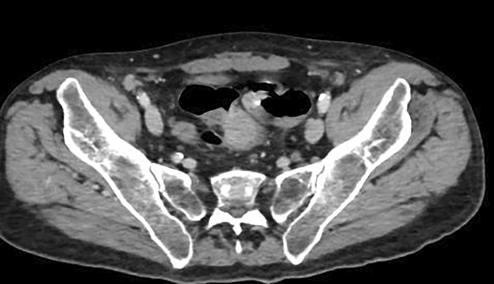
Abdominal and pelvic CT scans demonstrated an irregular circumferential thickening of the sigmoid colon wall, suspicious for malignancy, along with perilesional lymphadenopathy ( Figure 3), an irregular right retroperitoneal nodule suggestive of carcinoma, and bilateral adrenal nodules concerning for metastatic disease.

A percutaneous biopsy of the lung mass was performed, and histopathological analysis confirmed pulmonary adenocarcinoma with positive thyroid transcription factor-1 (TTF-1) staining ( Figure 4). Given the suspicion of lung carcinoma with colonic metastasis, immunohistochemical analysis of the colonic biopsy for TTF-1 was conducted and returned positive, confirming colonic metastasis of pulmonary adenocarcinoma ( Figure 5).


The patient was enrolled in oncologic care and initiated on systemic chemotherapy.
Pancoast-Tobias syndrome is a rare presentation of lung cancer, accounting for 3% to 5% of all cases.7 Smoking is the primary risk factor, and the disease predominantly affects men in their sixth decade of life,7 which aligns with our case, as the patient was a 61-year-old smoker. Clinically, this tumor subtype is characterized by shoulder and arm pain, as observed in our patient. Radiological findings typically include an upper lobe mass with pleural invasion and rib infiltration. The histological type in our case was adenocarcinoma, which is consistent with the literature, as adenocarcinoma is the most commonly reported cause of PTS.8
Lung cancer is one of the most prevalent malignancies worldwide, with nearly half of cases presenting with distant metastases at diagnosis, regardless of the primary histological type. The most common metastatic sites include the brain, lungs, bones, liver, adrenal glands, and lymph nodes.1 Our patient had bone, peritoneal, adrenal gland, and lymph node metastases.
However, less common metastatic sites have also been reported in the literature. In this case, we focused on colonic metastasis, a rare manifestation of lung cancer. The incidence of colonic metastases is estimated at 12% in autopsy studies, whereas symptomatic cases are exceedingly rare (0.1%).9,10 The reported incidence varies depending on the diagnostic method used (digestive endoscopy, surgical specimens, or autopsy).11 Gastrointestinal metastases from lung cancer can manifest with anemia, gastrointestinal bleeding, or bowel obstruction.12–16 In our patient, metastatic lung adenocarcinoma was revealed by iron deficiency anemia, a presentation similar to that reported by Vasa Jevremovic et al. in a 71-year-old female patient who presented with iron deficiency anemia. Digestive endoscopy confirmed adenocarcinoma in the transverse colon, and chest CT revealed a left upper lobe mass. The histological subtype was also adenocarcinoma.15
A literature review of 18 cases of lung cancer with gastrointestinal metastases identified intestinal obstruction (5 cases) and anemia (4 cases) as the most frequent clinical presentations. Other symptoms included abdominal pain, weight loss, and intestinal perforation. The most common metastatic sites were the small bowel (9 cases) and the stomach (5 cases), while colonic metastases were observed in only two patients. Histologically, 10 patients had large cell carcinoma, while 8 had adenocarcinoma.13 A more recent review of 34 cases of lung cancer with colonic metastases found that squamous cell carcinoma was the most common histological type, identified in 15 cases. Adenocarcinoma, as in our patient, accounted for one-third of cases, while large cell carcinoma and small cell carcinoma were less frequently observed.14
In contrast, colorectal cancer frequently metastasizes to the lungs,17 making it challenging to differentiate primary pulmonary cancer from metastatic colorectal cancer. For instance, Ana Cunha et al. reported a case of metastatic colorectal cancer presenting as a Pancoast tumor.18 Immunohistochemical staining plays a crucial role in distinguishing primary tumors in such cases. Typically, primary lung cancer is positive for TTF-1, CK7, and CK20 markers.19 In our patient, both colonic and lung biopsies tested positive for TTF-1, confirming the diagnosis of primary lung cancer with colonic metastasis.
This case report highlights a rare presentation of colonic metastasis revealing a Pancoast tumor. In patients presenting with both pulmonary and colonic tumors, a thorough evaluation of radiological features and histopathological findings, particularly immunohistochemical markers, is essential to accurately determine the primary site of malignancy.
Written informed consent for the publication of clinical details and/or images was obtained from the patient.
| Views | Downloads | |
|---|---|---|
| F1000Research | - | - |
|
PubMed Central
Data from PMC are received and updated monthly.
|
- | - |
Is the background of the case’s history and progression described in sufficient detail?
Yes
Are enough details provided of any physical examination and diagnostic tests, treatment given and outcomes?
Partly
Is sufficient discussion included of the importance of the findings and their relevance to future understanding of disease processes, diagnosis or treatment?
Partly
Is the case presented with sufficient detail to be useful for other practitioners?
Partly
References
1. Matthias Dettmer, Tae Eun Kim, Chan Kwon Jung, Eun Sun Jung, Kyo Young Lee, Chang Suk Kang, Thyroid transcription factor-1 expression in colorectal adenocarcinomas. Pathology - Research and Practice. 2011. Volume 207;11:686-690.Competing Interests: No competing interests were disclosed.
Reviewer Expertise: Lung cancer, colorectal cancer, histology.
Alongside their report, reviewers assign a status to the article:
| Invited Reviewers | |
|---|---|
| 1 | |
|
Version 1 09 Apr 25 |
read |
Provide sufficient details of any financial or non-financial competing interests to enable users to assess whether your comments might lead a reasonable person to question your impartiality. Consider the following examples, but note that this is not an exhaustive list:
Sign up for content alerts and receive a weekly or monthly email with all newly published articles
Already registered? Sign in
The email address should be the one you originally registered with F1000.
You registered with F1000 via Google, so we cannot reset your password.
To sign in, please click here.
If you still need help with your Google account password, please click here.
You registered with F1000 via Facebook, so we cannot reset your password.
To sign in, please click here.
If you still need help with your Facebook account password, please click here.
If your email address is registered with us, we will email you instructions to reset your password.
If you think you should have received this email but it has not arrived, please check your spam filters and/or contact for further assistance.
Comments on this article Comments (0)