Keywords
Gallbladder, Tuberculosis, Ant tubercular drugs, cholecystectomy
This article is included in the Cell & Molecular Biology gateway.
Gallbladder tuberculosis (GTB) is extremely rare, accounting for less than 1% of all TB cases. We present a 62-year-old woman in whom the diagnosis of GTB was made pre-operatively by percutaneous biopsy. The patient was put on antitubercular treatment for six months. The control CT scan showed an almost disappearance of the gallbladder mass leaving a discreet wall thickening. Because of the presence of stones in the gallbladder the patient was operated she had a laparoscopic cholecystectomy. Primary tuberculosis of the gallbladder is a rare and elusive condition that can be easily misdiagnosed due to its nonspecific clinical presentation. Early diagnosis and appropriate anti-tubercular therapy are crucial for successful management and prevention of further complications.
Gallbladder, Tuberculosis, Ant tubercular drugs, cholecystectomy
Tuberculosis (TB) is a common infectious disease caused by Mycobacterium tuberculosis, primarily affecting the lungs. However, it can also involve extra pulmonary sites such as the lymph nodes, bones, and abdomen. Gallbladder tuberculosis (GTB) is extremely rare, accounting for less than 1% of all TB cases. Most cases of gallbladder TB are secondary, meaning they arise from the spread of TB from other organs. Primary TB of the gallbladder is exceptionally uncommon, with very few cases reported in medical literature. In this article, we discuss a rare case of primary gallbladder tuberculosis, highlighting its clinical presentation, diagnosis, and management.
A 62-year-old woman presented with nonspecific symptoms, including right upper quadrant abdominal pain, nausea, and intermittent fever. She had no significant past medical history of tuberculosis or contact with individuals suffering from TB. Physical examination revealed mild tenderness in the right hypochondrium, but no palpable masses. Routine laboratory tests were within normal limits, except for mildly elevated liver function tests and inflammatory markers.
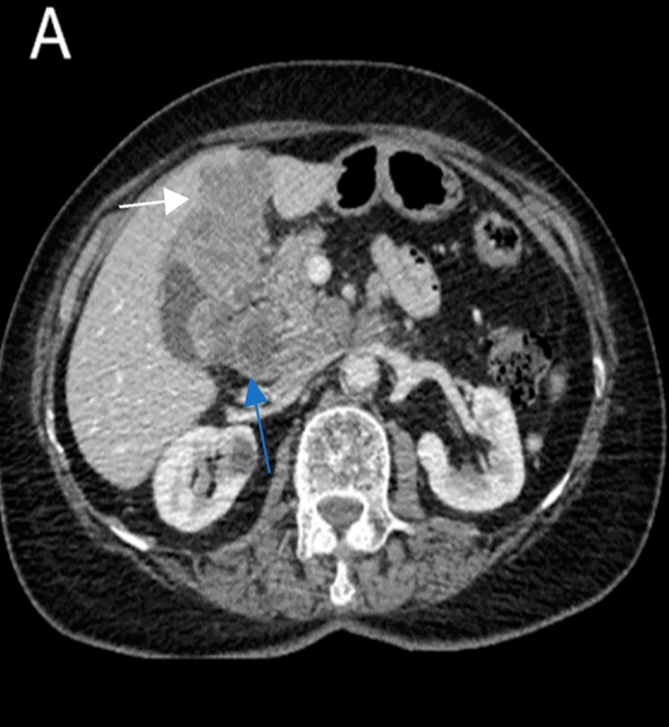
A CT scan was performed and showed a mass in the fundus of the gallbladder with irregular borders, locally advanced, invading the right colic angle, the segment IV of the liver and infiltrating the gastric wall ( Figure 1).

The patient underwent a percutaneous biopsy, and the histopathological examination revealed granulomatous inflammation with caseation necrosis, which raised suspicion for tuberculosis ( Figure 2). Subsequent staining for acid-fast bacilli (AFB) and polymerase chain reaction (PCR) testing confirmed the presence of Mycobacterium tuberculosis in the gallbladder tissue, leading to a diagnosis of primary gallbladder tuberculosis.
The patient was put on antitubercular drugs with a remarkable improvement.
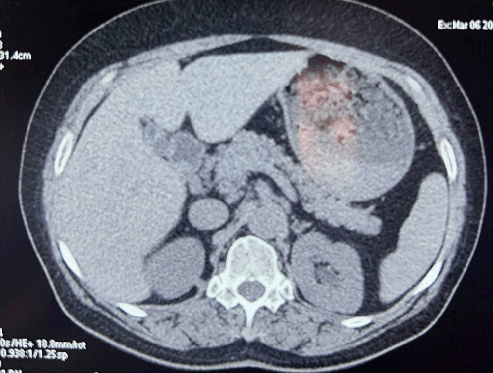
The control CT scan showed an almost disappearance of the gallbladder mass leaving a discreet wall thickening ( Figure 3).

At the end of the antitubercular treatment and because of the presence of stones in the gallbladder the patient was operated she had a laparoscopic cholecystectomy. The post-operative course was uneventful, and the patient was discharged on post-operative day one.
Histopathological examination of the specimen showed a residual granulomatous inflammation without malignancy.
Primary tuberculosis of the gallbladder remains an elusive diagnosis due to its rarity and the absence of specific clinical features. In most cases, patients present with symptoms that mimic common biliary diseases, such as chronic cholecystitis or gallbladder cancer. Symptoms like abdominal pain, nausea, vomiting, weight loss, and low-grade fever are often nonspecific and can be easily mistaken for more prevalent conditions, such as gallstones or chronic inflammation of the gallbladder. Because of these overlapping symptoms, gallbladder tuberculosis is rarely considered in the initial differential diagnosis.1,2,3
A key challenge in diagnosing gallbladder tuberculosis is the lack of characteristic imaging findings. Conventional diagnostic tools, such as ultrasound and computed tomography (CT), typically show nonspecific features like gallbladder wall thickening or the presence of gallstones.4 In some cases, a mass may be mistaken for a neoplastic lesion. However, imaging alone cannot definitively distinguish gallbladder tuberculosis from other gallbladder pathologies. In this case, ultrasound and CT scans only revealed wall thickening, without any clear indicators of tuberculosis. This underlines the difficulty in detecting TB preoperatively and the need for heightened clinical suspicion, especially in regions with a high prevalence of tuberculosis.1,4
The pathophysiology of primary gallbladder tuberculosis remains poorly understood. It is believed that gallbladder involvement usually occurs via hematogenous or lymphatic spread from a primary site, most commonly the lungs or gastrointestinal tract. However, in rare instances of primary gallbladder tuberculosis, such as in this case, no other organ involvement is detected. This raises intriguing questions about how Mycobacterium tuberculosis reaches the gallbladder without first affecting other parts of the body.3
Several theories have been proposed to explain primary gallbladder tuberculosis. One hypothesis is that latent TB bacilli could be harbored in the lymphatic system or subserosal tissues, where they remain dormant for years before reactivating in response to certain stimuli, such as immune suppression or chronic biliary inflammation.2,4 The role of the gallbladder epithelium and bile, which both possess natural resistance to bacterial infections, is another important consideration. For TB to successfully infect the gallbladder, there may be contributing factors such as bile stasis, gallstones, or pre-existing conditions that compromise the gallbladder's defenses.3,4
Histopathology remains the gold standard for diagnosing gallbladder tuberculosis. The presence of granulomatous inflammation with caseating necrosis, along with the identification of acid-fast bacilli through staining techniques, is highly suggestive of TB. In most cases, the diagnosis was only confirmed after the patient underwent a laparoscopic cholecystectomy and tissue samples were analyzed. This highlights the critical role of surgical intervention, not only for symptom relief but also for obtaining diagnostic samples in cases where tuberculosis is suspected but not confirmed through non-invasive methods.3,5 Our case is the first to confirm gallbladder tuberculosis before cholecystectomy.
Management of gallbladder tuberculosis involves a combination of surgical and medical approaches. Cholecystectomy is usually necessary to relieve symptoms and obtain tissue for definitive diagnosis.2,5 However, the cornerstone of treatment remains anti-tubercular therapy (ATT), which is administered postoperatively to eradicate the infection and prevent recurrence. A standard course of ATT, similar to that used in pulmonary TB, typically includes a combination of rifampicin, isoniazid, pyrazinamide, and ethambutol. Treatment duration is generally six months, although it may be extended in cases of drug-resistant TB or complicated disease.
Given the rarity of primary gallbladder tuberculosis, there are no established guidelines for its management, and treatment decisions are often based on case reports and expert opinion.5,7 Early diagnosis is essential to avoid complications, such as perforation of the gallbladder, peritonitis, or fistula formation. In endemic areas, physicians should maintain a high index of suspicion for tuberculosis in patients with atypical biliary presentations and unexplained histological findings [5.7].
This case demonstrates the importance of considering gallbladder tuberculosis as part of the differential diagnosis in patients presenting with chronic biliary symptoms, especially in countries where tuberculosis remains prevalent. While rare, primary gallbladder tuberculosis is treatable with timely diagnosis and appropriate anti-tubercular therapy. Further research is needed to better understand the pathogenesis of this condition and to develop more effective diagnostic strategies that could allow for earlier detection and treatment.6,8
Primary tuberculosis of the gallbladder is a rare and elusive condition that can be easily misdiagnosed due to its nonspecific clinical presentation. Diagnosis is usually established postoperatively through histological examination. This case emphasizes the importance of considering gallbladder tuberculosis in patients with chronic biliary symptoms and unexplained histopathological findings, especially in regions where TB is endemic. Early diagnosis and appropriate anti-tubercular therapy are crucial for successful management and prevention of further complications.
Written informed consent for publication of their clinical details was obtained from the patient.
| Views | Downloads | |
|---|---|---|
| F1000Research | - | - |
|
PubMed Central
Data from PMC are received and updated monthly.
|
- | - |
Provide sufficient details of any financial or non-financial competing interests to enable users to assess whether your comments might lead a reasonable person to question your impartiality. Consider the following examples, but note that this is not an exhaustive list:
Sign up for content alerts and receive a weekly or monthly email with all newly published articles
Already registered? Sign in
The email address should be the one you originally registered with F1000.
You registered with F1000 via Google, so we cannot reset your password.
To sign in, please click here.
If you still need help with your Google account password, please click here.
You registered with F1000 via Facebook, so we cannot reset your password.
To sign in, please click here.
If you still need help with your Facebook account password, please click here.
If your email address is registered with us, we will email you instructions to reset your password.
If you think you should have received this email but it has not arrived, please check your spam filters and/or contact for further assistance.
Comments on this article Comments (0)