Keywords
uterine fibroid, Leiomyoma, Hemoperitoneum, Acute abdomen, vessel rupture.
Uterine fibroids, also known as leiomyomas, are the most common benign tumors in reproductive-aged individuals. Although they may remain asymptomatic, they can occasionally lead to acute complications, including sudden hemorrhage. Spontaneous hemoperitoneum due to fibroids is extremely rare and can be life threatening.
We report the case of a 48-year-old multiparous woman with a history of hypertension who presented to the emergency department with acute abdominal pain, vomiting, and hypovolemic shock. An emergency computed tomography (CT) scan showed a large hemoperitoneum and a uterine mass measuring 10 cm. Emergency laparotomy revealed a pedunculated fundal fibroid with an actively bleeding superficial vein. Myomectomy was performed, followed by hemostatic control and abdominal washout. The patient required blood transfusion and recovered well without further complications. Histopathological examination confirmed the diagnosis of a benign leiomyoma.
Hemoperitoneum due to rupture of a superficial vein overlying a fibroid is an exceedingly uncommon but serious condition demanding prompt diagnosis and surgical management. Clinicians should consider this condition in patients with an acute abdomen and hypovolemic shock, especially in those with known fibroids. Early detection and timely surgical intervention are vital for achieving favorable outcomes.
uterine fibroid, Leiomyoma, Hemoperitoneum, Acute abdomen, vessel rupture.
Uterine fibroids are the most prevalent benign growth in women of reproductive age. They can remain symptom-free for a long time and are usually diagnosed because of chronic complications. However, acute complications due to fibroids are possible but are rare, torsion, degeneration, rupture, or hemorrhage, are rare.1,2
Intra-abdominal hemorrhage due to leiomyoma is a rare occurrence. In the literature, it is mostly reported as sporadic cases, and there have been about a hundred cases, of which 30 cases are caused by a rupture of a superficial vessel on the fibroid.3,4
It is difficult to diagnose preoperative hemoperitoneum caused by fibroids. In fact, various differential and more common diagnoses are usually suspected, including ectopic pregnancies and ruptured cysts. Clinically, the symptoms are the result of a peritoneal reaction to blood leakage, involving acute abdominal pain with signs of hypovolemia. Immediate intervention, including volume resuscitation and surgical hemorrhage control, is crucial.5
Herein, we present a rare case of massive hemoperitoneum secondary to rupture of a superficial vein overlying a pedunculated fibroid that was successfully managed with emergency surgery.
A 48-year-old woman with a medical history of high blood pressure treated with monotherapy and no prior surgical history, gravida 4, para 2, with two vaginal deliveries, presented to the emergency department with acute abdominal pain and vomiting starting 6 hours before consultation, which progressively aggravated, and the onset of vomiting and asthenia led the patient to seek urgent medical help.
The initial examination revealed diffuse abdominal tenderness, low blood pressure (70/40 mmHg), tachycardia (140 beats per minute), and normal body temperature. The patient was diagnosed with a Glasgow score of 13/15 (E3V4M6). Owing to hypovolemic shock, recourse to vasoactive drugs was necessary. The patient required vasoactive support with 2 mg/h norepinephrine. Blood tests showed a hemoglobin level of 6 g/dl, TP of 76%, and negative beta-human chorionic gonadotropin level.
An urgent CT scan was conducted, showing an extensive hemoperitoneum and a large mass dependent on the uterus measuring 100.3 × 90.4 mm ( Figures 1 and 2).

The patient was immediately transferred to the department of gynecology and obstetrics.
Immediate blood transfusion was initiated, and the patient received a total of 4 units of red blood cells with 6 units of PFC. Immediate laparotomy with midline incision was performed, and the intra-operative findings were dark clotted blood filling the abdominal cavity up to Morrison’s pouch. The uterus was normal in size, and both ovaries appeared normal.
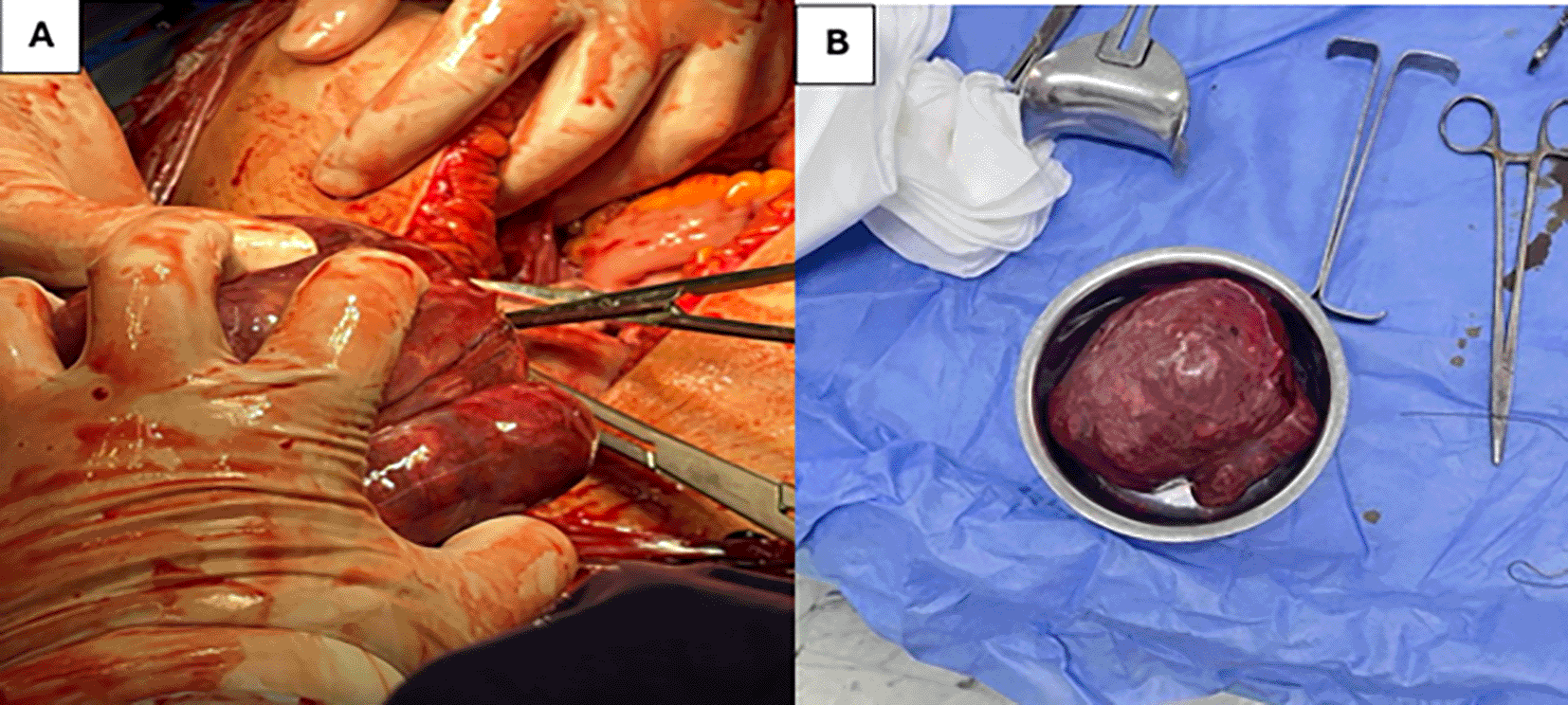
A type 7 fundal myoma measuring 10 cm was found, and the pedunculated fibroid had an actively haemorrhaging superficial posterior vein, causing abundant hemoperitoneum.
The myoma was resected, given its location and pedunculated nature, after coagulation of the bleeding vein ( Figure 3).

Then an abdominal wash and evacuation of the blood clots was done rigorously.
Postoperative care was carried out in collaboration with anesthesiology team, the patient remained hemodynamically stable, and the hemoglobin level increased to 9 g/dl. The patient was discharged after 3 days and returned after 2 weeks for check-up. Ultrasound was normal, and the final pathological examination confirmed a leiomyoma weighing 546 g and measuring 11 cm.
Patient’s perspective: The patient was deeply grateful for the quick care she received and expressed great relief after the traumatic experience that she described as a near-death experience. She expressed that, after this ordeal, she became more aware of how important it was to seek help if something did not feel right.
Uterine fibroids or leiomyomas are the most common benign uterine tumors.6,7
It is assumed that they occur in more than 70% of women at the time of menopause. It has been reported that they are clinically pronounced in 25% of women of reproductive age. According to a meta-analysis conducted in 2017, the incidence ranged between 217 and 3745 cases per 100 000 women per year, while the prevalence ranged from 4.5% to 68.6%. They are also more common among black women.7
The most affected age group was women during the reproductive period. They are very rare in the prepubertal years and usually reduce in size and fade in menopausal women.7,8
However, the pathogenesis of leiomyomas is not well elucidated. They originate from individual uterine smooth muscle cells, also called myometrial cells, the expansion of which relies on hormonal production (estrogens).8,9
This medical condition remains understudied given that many patients are asymptomatic or disregard their symptoms.10
The clinical presentation of symptomatic fibroids depends on their location and size. They can present with abnormal uterine bleeding, which may lead to anemia, painful menstruation, persistent pelvic pain or pressure, back ache, and symptoms related to tumor compression such as bowel, bladder, or sexual discomfort and complications. They can also lead to infertility and compromise obstetric outcomes (abnormal fetal positioning, preterm labor, restricted fetal growth, cesarean section, and bleeding before labor). Moreover, quality of life can be really affected by this affection and can lead to significant morbidity and mortality, especially with late diagnosis and management.10,11
Acute complications linked to leiomyomas are remarkably rare, and it is difficult to attribute a specific incidence to these complications due to their rarity. These encompass urinary retention, torsion of pedunculated fibroids, red degeneration in pregnant women, thrombosis of the mesenteric vein, thromboembolism, and sudden intraperitoneal or vaginal hemorrhage. Most of these complications lead to acute abdominal presentation and need urgent diagnostic surgery.12
Intra-peritoneal hemorrhage due to fibroids is an exceptionally rare occurrence, in fact, according to a systematic review by Lim et al., only 125 cases have been identified in English published research since 1902, and four patients succumbed to this complication. The root of this manifestation is yet to be understood; thus, it is uncommon to suspect the diagnosis based on the clinical onset of a hypovolemic choc associated with acute abdominal pain, which is often discovered intra-operatively.1,3,6
The pathogenesis of abdominal hemorrhage secondary to fibroids remains uncertain, with numerous speculations and theories proposed. Bleeding was linked to a subserosal or superficial vessel rupture or to avulsion/torsion of a pedunculated leiomyoma and rupture of a degenerated fibroid.1,2
In cases where bleeding originated from a ruptured vessel, it was mainly veinous, and even a minor vein could lead to massive abdominal hemorrhage. However, arterial bleeding was reportedly associated with high blood pressure. Ruptures can be traumatic or spontaneous in nature.4–6 In our case, there was no traumatic event reported by the patient.
Rupture of vessels overlying leiomyomas usually affects multiparous women aged 30–49 years, who have large fibroids exceeding 10 cm in diameter. Our patient met the criteria for age and parity, and had a sizable fibroid measuring 11 cm.13
Multiple speculations have been proposed regarding the spontaneous rupture of these vessels. First, elevated abdominal pressure could contribute to vessel congestion, which resulting in rupture. This situation could be caused by heavy exercise, intense sexual activity, difficult defecation, and menstruation. Second, larger leiomyomas were, allegedly, correlated with malformations and fragilization of superficial vessels due to the tension and stretching effect of a growing fibroid; thus, fibroids that exceed 10 cm in diameter are more likely to have weaker and distended vessels that may rupture. Finally, on a molecular level, recent reports have found that micro-RNAs, particularly miR-29b, play an essential role in the pathogenesis of leiomyomas, and that their downregulation is crucial for fibroid tumorigenesis. Progesterone plays a role in downregulating miR-29b and upregulating mRNAs for multiple collagens, which means that a decrease in progesterone may potentially weaken the blood vessels.5,6
The typical clinical presentation is acute abdominal and pelvic pain with signs of hypovolemic shock. A precise preoperative diagnosis of this entity is uncommon. It is usually the result of exploration of an unexplained hemoperitoneum.5,14
The management of such patients includes two crucial therapeutic components that are part of hemorrhagic shock management: the ‘titrated hypotensive resuscitation’ tactics and the ‘damage control surgery’ strategy. In fact, it is compulsory to promptly and definitively control the origin of bleeding and replace blood loss.15
Urgent surgery is the cornerstone of treatment, and the chosen procedure depends on age, hormonal status, and fertility preservation in younger patients. Hysterectomy should be performed in postmenopausal women, while myomectomy should be prioritized in younger patients wishing to preserve their fertility. If bleeding is uncontrollable, hemostatic hysterectomy should be carried out. Lately, embolization of the uterine artery has been increasingly used owing to its minimally invasive nature and quick recovery. Open surgery is the typical technique; however, some successful laparoscopic approaches have been reported. Maemura et al. reported the first successful laparoscopic myomectomy for the management of hemoperitoneum of unknown origin.4,5
Although fibroids are extremely common, they are unlikely to cause acute complications that require surgical management. Abdominal hemoperitoneum is particularly quite rare and remains underdiagnosed until surgical exploration. Thus, rupture of a superficial vessel in a fibroid should be suspected in every patient presenting with acute hemoperitoneum, especially when a history of fibroids is reported. CT scans and MRI could be helpful for diagnosing stable patients, but urgent surgery prevails as the key approach to confirm the diagnosis and control bleeding without omitting the resuscitation component.
Consent for publication: Written informed consent was obtained from the patient for publication of this case, with assurances of anonymity and confidentiality.
No data are associated with this article.
The checklist used for the development of this care guideline can be found on our data guidelines page at Zenodo. The dataset is titled “CARE Checklist for Case Report of Chayma Rjiba”, available under the CC0 license. The working DOI for the dataset is https://doi.org/10.5281/zenodo.15204318.16
| Views | Downloads | |
|---|---|---|
| F1000Research | - | - |
|
PubMed Central
Data from PMC are received and updated monthly.
|
- | - |
Provide sufficient details of any financial or non-financial competing interests to enable users to assess whether your comments might lead a reasonable person to question your impartiality. Consider the following examples, but note that this is not an exhaustive list:
Sign up for content alerts and receive a weekly or monthly email with all newly published articles
Already registered? Sign in
The email address should be the one you originally registered with F1000.
You registered with F1000 via Google, so we cannot reset your password.
To sign in, please click here.
If you still need help with your Google account password, please click here.
You registered with F1000 via Facebook, so we cannot reset your password.
To sign in, please click here.
If you still need help with your Facebook account password, please click here.
If your email address is registered with us, we will email you instructions to reset your password.
If you think you should have received this email but it has not arrived, please check your spam filters and/or contact for further assistance.
Comments on this article Comments (0)