Keywords
Thalassemia Major, Thalassemia Minor, Candida albicans, Iron Overload, Ferritin levels
This article is included in the Pathogens gateway.
Thalassemia Major, Thalassemia Minor, Candida albicans, Iron Overload, Ferritin levels
This revised article clarifies the quantitative methodology for Candida albicans levels, detailing serial dilution and cfu/ml calculation in the Methods section. It specifies thalassemia diagnosis via hemoglobin electrophoresis in the Subject Selection section. The Discussion section adds how iron overload impairs immunity via ROS and promotes C. albicans growth, and addresses limitations like small sample size (n=30 per group) and variability in transfusions and immune responses, enhancing methodological clarity and scientific depth compared to the previous version.
See the authors' detailed response to the review by Pradana Zaky Romadhon
See the authors' detailed response to the review by Anwer Faisal
Thalassemia is a hereditary condition affecting hemoglobin, the condition involves a decrease or absence in the formation of one or more globin chains in the hemoglobin tetramers, resulting in uncontrolled destruction of red blood cells and, ultimately, severe anemia.1,2 Beta-thalassemia arises from mutations that impact many stages of beta-globin protein synthesis, encompassing transcription, translation, and the stability of beta-globin production.3 One of the most prevalent genetic illnesses in humans is β-thalassemia. Around 60,000 infants worldwide are born with β-thalassemia annually, predominantly in countries of the Mediterranean but also in the Middle East, West Africa, India, and South-East Asia.4 The estimated prevalence of thalassemia carriers is 3 to 10 individuals per 100 individuals. Given a population of 200 million individuals, a birth rate of 20%, and a thalassemia carrier rate of around five percent, it is projected that there will be an annual occurrence of 2,500 kids born with thalassemia congenital illness.5 Patients with β-thalassemia often have blood transfusions and suffer from iron overload.6 Serum ferritin (SF) indicates probable iron excess.7 Serum ferritin is an effective monitoring instrument for iron overload in thalassemia major.8 In healthy individuals, the average serum ferritin (SF) levels range from 12 to 300 μg/L for men and from 12 to 150 μg/L for females.9 Serum ferritin levels exceeding 1000 μg/L are indicative of iron overload and are linked to adverse outcomes, including increased mortality and organ injury, a more significant likelihood of cardiac events, and hepatic difficulties.10 Candida albicans is a fungal infection that results in approximately 1.7 million fatalities annually on a global scale, primarily affecting those with weakened immune systems and several underlying medical problems.11 The prevalence of infectious illnesses by Candida has consistently risen since the 1970s, potentially attributed to an elevated susceptibility to opportunistic infections, advancements in clinical techniques for detecting fungi-related hospital-acquired infections, and the emergence of antifungal resistance resulting from prolonged treatment exposure.12,13 The clinical features of oral cavities in patients with thalassemia include pointed and shortened root morphology, taurodontism, and a characteristic chipmunk appearance. These individuals often have a higher caries index and display gingival hypertrophy, which suggests gingival inflammation. Additionally, these patients are susceptible to infections caused by bacteria or fungi, including C. albicans.14 Thalassemia, especially thalassemia major, is linked to a heightened incidence of C. albicans colonization in the oral cavity. This increased vulnerability is chiefly attributable to variables including iron accumulation from repeated blood transfusions, which creates a nutrient-rich environment favorable for fungal proliferation, and immunological impairment caused by the disease and its therapies.15 This study aims to examine the correlation between iron and ferritin levels and C. albicans distribution in individuals with major and minor beta-thalassemia compared with healthy controls.
The saliva and blood samples were collected between December 2023 and March 2024 from a total of sixty patients; thirty of them were individuals diagnosed with Beta Thalassemia Major, and thirty were diagnosed with Beta Thalassemia Minor, diagnosing was based on clinical history, hematological parameters, and hemoglobin electrophoresis, as per standard protocols at the Thalassemia Centers of Ebn-Albalidy and Al-Karama hospitals in Baghdad, compared with thirty apparently healthy individuals served as control. Data collected from each participant included demographic information (name, age, gender) and duration of illness. The Research Ethics Committee of the University of Baghdad College of Dentistry granted ethical permission for the study, with reference number 889 and project number 889824, on January 11, 2024. The research was conducted in conformity with the principles established in the Declaration of Helsinki. Before doing the analysis, all patient data was anonymized.
Inclusion criteria included patients with stable Beta Thalassemia Major (T Major) without other diseases or medication influences this group representing the 1st patients group, and stable Beta Thalassemia minor (T Minor) patients with no significant health changes, representing the 2nd patients group compared with apparently healthy individuals with no blood disorders or chronic illnesses affecting iron metabolism representing the control group. All participants with ages ranged between 18-60 years.
Exclusion criteria included any subject between 18 and 60 years of age but with an inability to provide informed consent or with conditions or treatments that could affect the biomarkers being studied. Indeed, pregnant or lactation women were also excluded.
Three milliliters of unstimulated saliva were obtained from each participant by the spitting method,16 and subsequently transferred to sterile tubes. The samples were centrifuged at 4000 rpm for 3 minutes to separate cellular debris. For blood samples, 5 mL were obtained through venipuncture into sterile tubes, centrifuged for 15 minutes at 3000 rpm, and the serum extracted using an automated pipette. Both serum and saliva were preserved at -20°C until later usage.
A quantitative assessment of Candida albicans levels was performed by determining the viable colony count in colony-forming units per milliliter (CFU/mL). Saliva samples from both patients and control groups were processed using a serial dilution method17 4.5 ml of nutrient broth was added to sterile tubes, and 0.5 ml of freshly collected saliva was introduced into the first tube, followed by inoculating the following tubes successively to achieve the tenfold dilutions. One hundred microliters of each dilution were distributed over a plate of Sabouraud Dextrose Agar (SDA) medium containing 1% chloramphenicol and incubated at 37°C for 24-48 hours. The colony count was calculated as cfu/ml using the formula: cfu/ml = (number of colonies × dilution factor) / volume plated (0.1 ml).
C. albicans were initially identified and explored microscopically to observe the oval or elongated yeast cells exhibiting budding and pseudohyphae. Additionally, the C. albicans inoculum was incubated in 3-5 ml of human serum. A small quantity of the grown serum was placed on a glass slide and examined under a microscope to determine the presence or absence of germ tube formation.
C. albicans were subcultured onto a chromogenic medium (CHRO Magar TM Candida media) and then incubated at 37°C for 48 to 72 hours. This technique uses substrates linked to chemical dyes to distinguish several species of Candida by the coloration of the developing colonies. For additional identification, the VITEK® 2 COMPACT was employed to accurately identify C. albicans isolates to the species level, according to the manufacturer’s guidelines (Biomerieux/France).
The method employs microparticle-enhanced immunoturbidimetry using a Thermo Scientific™ Indiko™ Clinical Chemistry Analyze (Finland). Sera samples from patients and controls were utilized and processed with microparticles coated with rabbit antibodies specific to either human ferritin or iron. The rabbit antibodies specific to human ferritin and iron were not sourced directly from live animal cells in our laboratory. They were obtained externally as part of commercial reagent kits from Thermo Scientific: the Human Ferritin Chemistry Analyzer Kit (Ref 981949, 4 × 4 mL, Indiko™) and the Human Iron Chemistry Analyzer Kit (Ref 981236, 10 × 20 mL, Konelab™, Indiko™). These kits provide pre-coated microparticles with polyclonal rabbit anti-human antibodies, validated for use with the Thermo Scientific™ Indiko™ Clinical Chemistry Analyzer. The measurement of immunocomplex formation for Ferritin is conducted by assessing absorbance variations at 700 nm, within a detection range of 0.32–20 ng/mL, iron immunocomplex formation is quantified by absorbance at 600 nm.
The statistical analyses were carried out by applying the SPSS 15 software (SPSS Inc., IL, USA, https://spssdownload.com/spss-software-version-15-free-download). It was employed the Shapiro-Wilk test, Kruskal-Wallis H test, and the Pairwise test. The p-value of less than 0.05 was considered statistically significant.
The research study examined the age-based disparities in mean values for the T Major and T Minor groups and for the control group. T Minor group demonstrated a superior mean value of age (35.533 ± 7.523) years, followed by the T Major group (23.233 ± 7.398) years in comparison with the control group as it had a somewhat elevated mean than the minor group (29.667 ± 9.517) ( Table 1).
| Control | T Major | T Minor | ||||
|---|---|---|---|---|---|---|
| N | Mean ± S.D. | N | Mean± S.D. | N | Mean ± S.D. | |
| Total | 30 | 29.667 ± 9.517 | 30 | 23.233 ± 7.398 | 30 | 35.533 ± 7.523 |
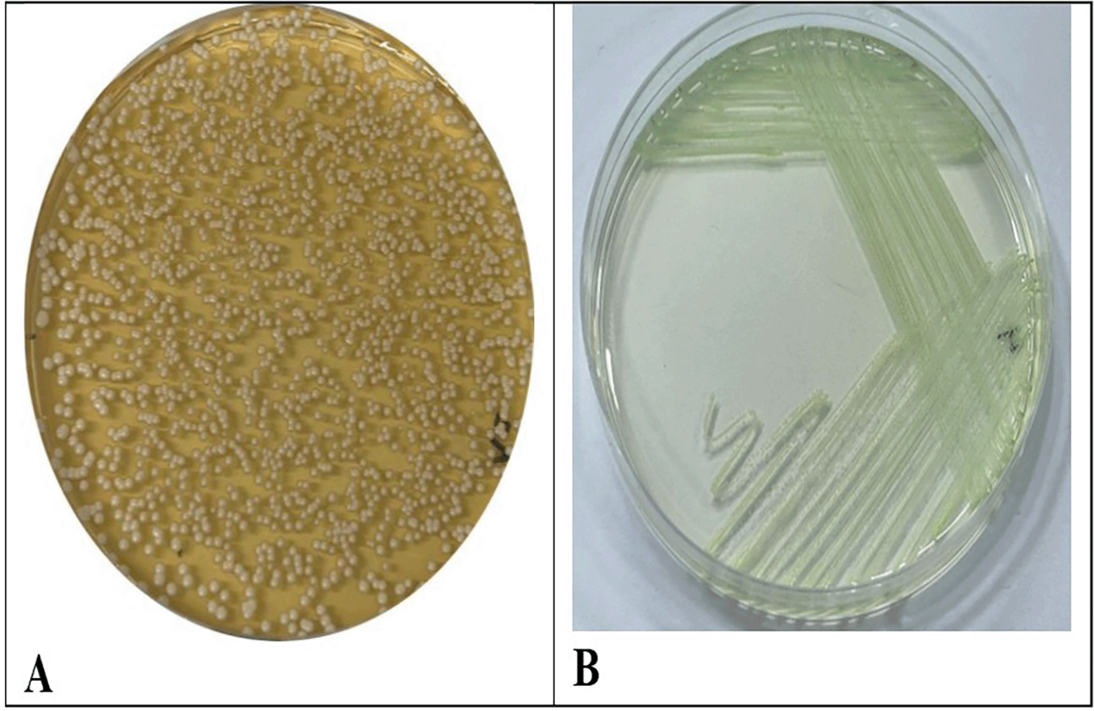
C. albicans isolates were isolated and diagnosed successfully from the saliva of patients suffering from B-thalassemia. C. albicans display distinctive colonies. When cultivated on (SDA), the colonies exhibited a smooth, creamy texture and were white to off-white in hue. They are often grown with a delicate texture. Over time, the colonies may display the development of a corrugated surface Figure 1. CHRO Magar was utilized for more accurate identification. In this instance, colonies of C. albicans display a characteristic green coloration due to the enzymatic activity of the yeast. The combination of these characteristics with microscopic identification facilitates the accurate diagnosis of C. albicans infections.
For advanced identification, all isolates were accurately identified using the VITEK 2 Compact System, with a probability of 88% to 90%, hence demonstrating the system’s efficiency in identifying C. albicans. Figure 2 and Table 2 show the distribution of C. albicans among the groups as the T Major has the highest carriage rate, as twenty-two out of thirty patients have C. albicans accounting for (73.33%) of the group. In contrast, in the T Minor, only twelve out of thirty patients carrying C. albicans represented (40%) of the group, and the control group exhibited the lowest prevalence rate with merely two individuals, constituting (6.67%) of the healthy subjects. The results indicated a markedly elevated colonization rate of C. albicans in the major group relative to the minor and control groups, respectively.

This study analyzed two biochemical parameters, Iron and ferritin, across the beta thalassemic Major and Minor patients in comparison with the control group. Descriptive statistics, comprising the median, which was computed for each parameter simultaneously with C. albicans. Statistical analysis indicated substantial differences across the groups (p < 0.001) for all parameters, as the T-Major group revealed a higher iron, ferritin, and C. albicans count when compared with the results of both T Minor and control groups, as seen in Table 3.
The T Major group for Iron demonstrated the most excellent median (270.016) in comparison to the Control (116.985) and T Minor (117.599). Likewise, for Ferritin, T Major had a significantly higher median (2783.800), greatly surpassing T Minor (77.800) and control (63.150). In addition to these biochemical markers, C. albicans levels in saliva were significantly elevated in the T Major group (1.715 x10⁴ cfu/ml) compared to the Minor and control as the median has zero value for both groups. These data demonstrate substantial increases in these parameters within the T Major group vs. other groups.
The results in Table 4 illustrated the pairwise analysis of Iron, Ferritin, and C. albicans levels among the T Major, T Minor and control groups demonstrated substantial increases in the T Major group relative to the control group for all three parameters (p < 0.001). Likewise, significant differences were noted between the T Major and T Minor groups across all parameters (p ≤ 0.005), further emphasizing the heightened levels in the T Major group. No significant differences were observed between the T Minor and control groups for the biochemical parameters, Iron and Ferritin (p = 0.748 and p = 0.851, respectively), and the difference for C. albicans was also not statistically significant between these groups (p = 0.106).
The results in Tables 5, 6, and 7 showed no significant correlations among all markers under investigation (iron, ferritin, and C. albicans levels) in both patient groups (T Major and T Minor) and the control group, except for a significant correlation noted between iron and ferritin levels in the control group.
| Parameters | C. albicans | Ferritin | |
|---|---|---|---|
| Iron (μg/dl) | r | -0.062 | 0.092 |
| p | 0.746 | 0.628 | |
| Ferritin (mg/ml) | r | -0.214 | |
| p | 0.257 |
| Parameters | C. albicans | Ferritin | |
|---|---|---|---|
| Iron (μg/dl) | r | 0.082 | 0.343 |
| p | 0.668 | 0.064 | |
| Ferritin (mg/ml) | r | 0.037 | |
| p | 0.844 |
The result of this study indicated that 73.33% of the significant thalassemia patient group had oral cavity colonization by C. albicans, a substantially greater rate compared to the minor group at 40% when compared with the control group which displayed the lowest prevalence at 6.67%. A markedly elevated mean candidal count was noted in thalassemic patients in comparison to the healthy cohort, the findings closely agree with another study conducted in Jordan, which indicated that Candida species were identified in 74% of thalassemic patients. Similarly, the present results obtained were comparable to another study conducted in Egypt, as C. albicans was isolated in 69.2% of cases, whereas 30.8% were non- C. albicans.18 The data obtained exhibited a higher elevation rate of C. albicans when compared with a local study performed in Najaf, Iraq, which evaluated samples from 50 thalassemia patients, of which only 14(28%) demonstrated positive growth for Candida species.19 One of the primary reasons that significant C. albicans have infected some minor patients is the immune problems that may have increased sensitivity to oral fungal colonization in these thalassemic patients.20 Iron overload in beta-thalassemia major generates reactive oxygen species (ROS), impairing neutrophil and macrophage function, thus weakening immune defenses against Candida albicans.21 Anemia may serve as a predisposing factor for oral fungal colonization in people with thalassemia.22 Another reason is that repeated blood transfusions lead to iron accumulation in salivary glands, resulting in the development of non-transferrin-bound iron (NTBI), which circulates in plasma and generates reactive oxygen species (ROS).23 Iron accumulation in salivary gland acini cells induces inflammation and reduces salivary output and components.24 Salivary components bolster the immune system’s response to infections caused by C. albicans by preventing the spread and protecting the mucosal epithelial barrier. Reduced salivary production in beta-thalassemia major patients can lead to dysbiosis, causing C. albicans to overgrow and adhere to the oral epithelium.25 Increased levels of iron and ferritin create an environment favorable for the growth of C. albicans in the major group.26 The findings of the current study reveal a unique biochemical and microbiological profile in the T Major group, marked by considerably increased levels of iron, ferritin, and C. albicans as compared to the T Minor and control groups. The T Major group exhibited significantly elevated iron and ferritin levels, indicating modified iron metabolism, potentially associated with inflammation or stress responses, alongside markedly increased C. albicans concentrations, indicating possible microbiome disruption or immune system changes. The pairwise analysis confirmed that these elevations were statistically significant (p < 0.001), differentiating the T Major group, from each of minor and control group. Still, no significant changes were noted between the T Minor and control groups for iron and ferritin (p = 0.748 and p = 0.851) or C. albicans (p = 0.106). The results obtained correspond with another two previous studies, one conducted in Turkey and the other in Indonesia. Both studies investigated iron and ferritin levels in saliva and serum across various groups, including thalassemia patients (major and minor) and controls. The findings of these studies indicate significantly elevated blood ferritin and iron levels in the significant thalassemia group compared to both minor and control groups.27,28 The findings of the current investigation align with those of a study performed in Pakistan involving 155 patients with T Major. The study also revealed markedly increased ferritin and iron levels in the T Major group relative to the control group.29 The increased concentrations of iron and ferritin in individuals in the T Major group are mainly due to the frequent blood transfusions necessary for the treatment of severe anemia. Each transfusion sends roughly 200 mg of iron into the body,30 and due to the absence of a natural physiological mechanism for excreting excess iron, patients progressively collect iron.31 This iron overload results in the accumulation of surplus iron in tissues and organs, evidenced by markedly increased serum ferritin levels, a crucial indicator of iron storage.32 Moreover, the inadequate erythropoiesis in beta-thalassemia significantly leads to heightened intestinal iron absorption, hence intensifying iron buildup. In the absence of sufficient chelation therapy, iron excess may escalate to dangerous levels, leading to problems such as organ damage.33 Conversely, beta-thalassemia minor is marked by less severe anemia as it resulting from a singular faulty beta-globin gene.34 Individuals with this illness generally do not necessitate blood transfusions, hence reducing the risk of iron excess from exogenous sources.35 Moreover, the extent of inefficient erythropoiesis is considerably reduced in beta-thalassemia minor, resulting in a better-controlled iron absorption and storage mechanism. As a result, their iron and ferritin levels typically stay within normal limits.36 The present work illustrated a correlation between biochemical markers and colonization by Candida albicans in thalassemia patients. The increased levels of C. albicans in individuals in the T Major group may be indirectly associated with the raised concentrations of iron and ferritin in this population.21 Iron is essential for microbial proliferation and pathogenicity, particularly in fungal infections such as C. albicans.37 In instances of iron overload, such as in beta-thalassemia major, excess free iron in the bloodstream and tissues could be served as a plentiful substrate for Candida, facilitating its growth.38 Ferritin, a principal iron storage protein, is also raised in beta-thalassemia major as a result of continuous transfusions and poor erythropoiesis.39,40 Candida might have evolved ways to extract iron from host ferritin, hence augmenting its survival and proliferation in iron-abundant settings. Conversely, patients with beta-thalassemia minor demonstrate normal or nearly normal iron and ferritin levels owing to the lack of recurrent transfusions and modest inefficient erythropoiesis.41 As a result, there is an absence of surplus iron to promote Candida proliferation, and their immune systems are often less impaired. This leads to markedly reduced amounts of C. albicans in patients with beta-thalassemia minor relative to those with beta-thalassemia major.42
The current study has a limitation that may explain the absence of significant correlations between iron, ferritin, and Candida albicans levels across the study. The relatively small sample size (n=30 per group) may have limited statistical power to detect weaker correlations. Additionally, variability in transfusion frequency, chelation therapy, and individual immune responses among T Major patients could have confounded the relationship between iron, ferritin, and fungal levels.
This study shows the heightened prevalence of C. albicans colonization in patients with thalassemia major relative to individuals with thalassemia minor and healthy controls. The results indicate that iron overload, diminished immune system impairment, and contribute to the increased vulnerability to fungal infections in thalassemia major patients. These findings underscore the significance of iron control and iron chelation therapy in averting problems, such as oral fungal infections, and indicate that consistent monitoring may enhance patient outcomes.
The Research Ethics Committee of the University of Baghdad College of Dentistry granted ethical permission for the study, with reference number 889 and project number 889824, on January 11, 2024. The study started on February 1, 2024, and was conducted in conformity with principles established in the Declaration of Helsinki. Before doing the analysis, all patient data was anonymized.
All participants provided written informed consent, as approved by the Research Ethics Committee of the University of Baghdad, College of Dentistry, which granted ethical permission for the study. The patients were fully informed about the study’s objectives, procedures, and potential benefits, and were assured of their complete freedom to participate. Consent was obtained for the collection of both blood and saliva samples.
| Views | Downloads | |
|---|---|---|
| F1000Research | - | - |
|
PubMed Central
Data from PMC are received and updated monthly.
|
- | - |
Provide sufficient details of any financial or non-financial competing interests to enable users to assess whether your comments might lead a reasonable person to question your impartiality. Consider the following examples, but note that this is not an exhaustive list:
Sign up for content alerts and receive a weekly or monthly email with all newly published articles
Already registered? Sign in
The email address should be the one you originally registered with F1000.
You registered with F1000 via Google, so we cannot reset your password.
To sign in, please click here.
If you still need help with your Google account password, please click here.
You registered with F1000 via Facebook, so we cannot reset your password.
To sign in, please click here.
If you still need help with your Facebook account password, please click here.
If your email address is registered with us, we will email you instructions to reset your password.
If you think you should have received this email but it has not arrived, please check your spam filters and/or contact for further assistance.
Comments on this article Comments (0)