Keywords
Endoscopy – surgery – duodenal duplication – duodenotomy – MRI – CT scan.
Gastrointestinal duplications are rare congenital anomalies, occurring in about 1 in 25,000 births, with duodenal duplications making up 2% to 7% of cases. Typically diagnosed in childhood, they pose diagnostic challenges in adults.
We present a case of a 14-year-old boy with mild epigastric pain, whose imaging revealed a large inter-duodeno-pancreatic cystic mass displacing nearby structures. Surgical exploration identified a cystic formation in the second part of the duodenum with a small communication to the duodenal lumen. A subtotal resection and cysto-duodenostomy were performed, leading to an uneventful postoperative recovery.
Duplication cysts, or alimentary tract duplications, are rare congenital lesions, typically found in the distal ileum but infrequently in the duodenum. Diagnosed mainly in early childhood, they may occasionally present in adulthood, manifesting as abdominal mass, obstruction, or discomfort; in some cases, they mimic choledochal cysts, complicating diagnosis. Surgical removal is the standard treatment, focusing on ensuring proper drainage while preserving biliary and pancreatic ducts.
Duodenal duplication is a rare, challenging diagnosis in adolescents; this case offers guidance for timely identification and treatment. It aims to support healthcare providers in achieving timely diagnosis and effective management in similar cases.
Endoscopy – surgery – duodenal duplication – duodenotomy – MRI – CT scan.
Duodenal duplication is an uncommon congenital anomaly with diverse and nonspecific symptoms, often challenging to diagnose, especially in adolescents.
Surgical excision is the primary treatment, with total removal being ideal, though subtotal resection or digestive bypass may be necessary in complex cases.
Duodenal duplication is a rare congenital anomaly involving the formation of a duplicated segment along the gastrointestinal tract, typically located in the small intestine, and even more rarely in the duodenum.1 These duplications are typically diagnosed in infancy or early childhood due to symptoms like abdominal pain, vomiting, or signs of obstruction; however, they may go undetected into adolescence or adulthood if symptoms are mild or absent.
The absence of specific symptoms in some cases, as well as the overlap with other more common gastrointestinal conditions, can make diagnosis challenging. Imaging studies, such as ultrasound, CT, or MRI, are essential tools for identifying the location, size, and relationship of the duplication with surrounding structures, especially when there is no clear communication with the duodenal lumen.2
In this case, we report a particularly rare occurrence of cystic duodenal duplication diagnosed in an adolescent male, who presented with mild, nonspecific symptoms. This report highlights the importance of a thorough diagnostic process and careful consideration of differential diagnoses in similar cases, offering valuable insights for healthcare providers. This report was written following the CARE guidelines.3
A 14-year-old male patient was referred to our clinic after experiencing mild vague epigastric pain for three months with no other associated symptoms. There was no history of vomiting, anorexia or change in bowl movements.
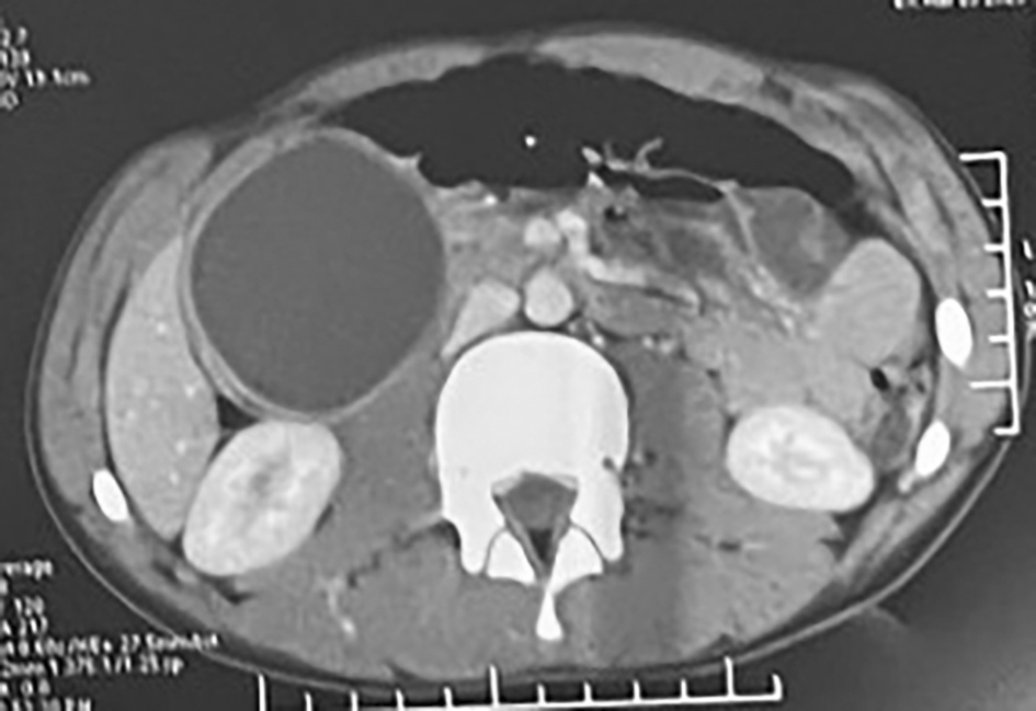
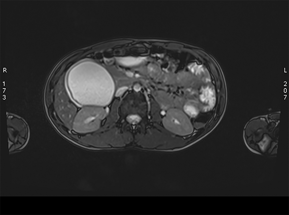
A computed tomography (CT) of the abdomen was performed, revealing a well-defined, rounded, inter-duodeno-pancreatic cystic formation measuring 65 × 55 mm and extending 75 mm in height. This mass makes contact with the inferior vena cava and the right renal vein, both of which are patent. There was no apparent communication with the main bile duct and no dilation observed in the intrahepatic or extrahepatic bile ducts ( Figure 1). To further substantiate the diagnosis a magnetic resonance imaging (MRI) showed an inter-duodeno-pancreatic cystic mass with hypo intensity on T1 and hypersignal on T2, homogenous, without apparent diffusion hyperintensity, and no evident duodenal communication. It imposes a mass effect on the cephalic pancreas and displaces the duodenum upward and outward ( Figure 2).


It imposes a mass effect on the cephalic pancreas and displaces the duodenum upward and outward.
The patient underwent an open abdominal exploration by midline incision. Upon exploration, a cystic formation involving the second duodenum (D2) was found, it was closely in contact with the right renal vein and the inferior vena cava ( Figure 3).
Due to the difficulty in identifying the boundaries of the cystic formation, an upper gastrointestinal endoscopy ( Figure 4) was performed in the operating room. A duodenotomy was then performed on the anterior side of D2, revealing a 7 cm cystic formation protruding and connecting to the inner edge of D2.
The decision was made to perform a cholecystectomy and introduce a trans cystic drain to locate the papilla. The drain was palpated inside the duplication. The latter communicates with the duodenal lumen through a small foramen ( Figure 5). Subsequently, a subtotal resection of the duplication wall was performed, and the duodenotomy was transversely closed, creating a wide internal cysto-duodenostomy.
The post operative course was uneventful.
Duplication cysts, also known as alimentary tract duplications, are congenital lesions of the gastrointestinal (GI) tract. Although the exact cause still eludes us, multiple theories have been put forward to explain its occurrence.1
In terms of epidemiology, alimentary tract duplications occur in approximately 1 in 4500 births, showing slight male predominance.4 These are typically identified in childhood, with most duplications causing symptoms during the initial two years of life. Nevertheless, instance of late presentation in adulthood are also observed.5
GI duplications are most frequently found in the distal ileum, with subsequent occurrences in the esophagus, colon, and jejunum. The duodenum accounts for a mere 2%~7% of GI duplications. 6
The diagnosis of duodenal duplication (DD) typically takes place during infancy and childhood, a meta-analysis of 47 DD cases revealed that 40,4% occurred in the pediatric period, surpassing the 30,8% found in individuals older than 20. The lowest incidence was observed in the second decade at 21,3%. 7
Diverse clinical manifestations have been recorded. Predominantly, patients manifest a discernible abdominal mass, signs of intestinal obstruction, or abdominal discomfort. In certain cases, the occurrence of ectopic gastric mucosa may precipitate ulceration, hemorrhage, and prospective perforation.8 In our case the patient was asymptomatic and the duplication was discovered while performing an abdominal CT for mild epigastric pain.
It is also important to mention that a duodenal duplication cyst may exhibit symptoms resembling a type III choledochal cyst, also referred to as choledochocele.2 This uncommon condition involves abnormalities in the distal bile duct and ampullary structures, protruding into the duodenal lumen, mimicking the appearance of a duodenal mass. Despite the challenges in distinguishing between these conditions, it is crucial for proper surgical planning.
DDs have traditionally been addressed through surgical means. The surgical approach is determined by the cyst wall’s relationship with the biliary and pancreatic drainage systems. The main objective of surgery is to ensure unimpeded cyst drainage into the duodenum while safeguarding the integrity of the biliary and pancreatic ducts. However, in cases where the proximity of DD to the pancreaticobiliary system poses challenges and risks, an endoscopic procedure emerges as a viable alternative.9,10
Duodenal duplication, a seldom-discussed congenital anomaly in adolescents, presents diverse clinical manifestations, posing a diagnostic challenge, even for expert clinicians, due to the absence of specific signs and symptoms. Here we reported a particularly rare instance of an adolescent diagnosed with cystic duodenal duplication. This case report serves as valuable guidance for healthcare providers, facilitating early diagnosis and optimal therapeutic decision-making in similar cases.
Zenodo: Duodenal Duplication: A Case Report of a Rare Gastrointestinal Anomaly, https://doi.org/10.5281/zenodo.15623271 11
This project contains the following underlying data:
CARE checklist
Data is available under Creative Commons Zero v1.0 Universal license.
Written informed consent was obtained from the patient for publication of this case report. A copy of the written consent is available for review by the editor-in-chief of this journal on request.
| Views | Downloads | |
|---|---|---|
| F1000Research | - | - |
|
PubMed Central
Data from PMC are received and updated monthly.
|
- | - |
Is the background of the case’s history and progression described in sufficient detail?
Yes
Are enough details provided of any physical examination and diagnostic tests, treatment given and outcomes?
Yes
Is sufficient discussion included of the importance of the findings and their relevance to future understanding of disease processes, diagnosis or treatment?
Yes
Is the case presented with sufficient detail to be useful for other practitioners?
Yes
Competing Interests: No competing interests were disclosed.
Reviewer Expertise: General surgery, Surgical gastroenterology
Is the background of the case’s history and progression described in sufficient detail?
Yes
Are enough details provided of any physical examination and diagnostic tests, treatment given and outcomes?
Yes
Is sufficient discussion included of the importance of the findings and their relevance to future understanding of disease processes, diagnosis or treatment?
Partly
Is the case presented with sufficient detail to be useful for other practitioners?
Partly
Competing Interests: No competing interests were disclosed.
Reviewer Expertise: Pediatric Surgery
Alongside their report, reviewers assign a status to the article:
| Invited Reviewers | ||
|---|---|---|
| 1 | 2 | |
|
Version 1 27 Jun 25 |
read | read |
Provide sufficient details of any financial or non-financial competing interests to enable users to assess whether your comments might lead a reasonable person to question your impartiality. Consider the following examples, but note that this is not an exhaustive list:
Sign up for content alerts and receive a weekly or monthly email with all newly published articles
Already registered? Sign in
The email address should be the one you originally registered with F1000.
You registered with F1000 via Google, so we cannot reset your password.
To sign in, please click here.
If you still need help with your Google account password, please click here.
You registered with F1000 via Facebook, so we cannot reset your password.
To sign in, please click here.
If you still need help with your Facebook account password, please click here.
If your email address is registered with us, we will email you instructions to reset your password.
If you think you should have received this email but it has not arrived, please check your spam filters and/or contact for further assistance.
Comments on this article Comments (0)