Keywords
Basal cell, Carcinoma, Case report, Eyelids
This article is included in the Eye Health gateway.
Basal cell, Carcinoma, Case report, Eyelids
Malignant lesions are approximately 13.1% of eyelid tumors.1 Basal cell carcinoma (BCC) is the most common type of cancer.2 Risk factors, such as exposure to UV light, were identified. It is the main contributor to the development of these malignant lesions.1,2 Intracranial extension requires efficient emergency treatment with safe margin surgical excision.3 We present the case of a patient with primary lower eyelid basocell carcinoma discovered during routine examination.
A 57-year male, presented to the ophthalmology department with optical correction. He had no medical history and skin prototype III. The patient was a farmer with chronic sun exposure.
Ophthalmological examination of the right eye revealed an ulcerative budding lower eyelid lesion measuring 1*0.5 centimeter, with some telangiectasia and pigmentation ( Figure 1).
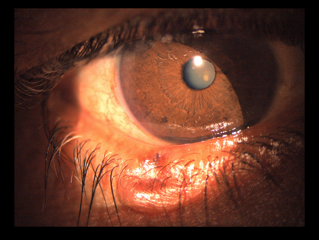
It was indurated on palpation and fixed to deeper tissues. The remaining ophthalmological examinations showed no abnormalities. The patient reported that it had evolved over 3 months and was painless. No history of trauma was found.
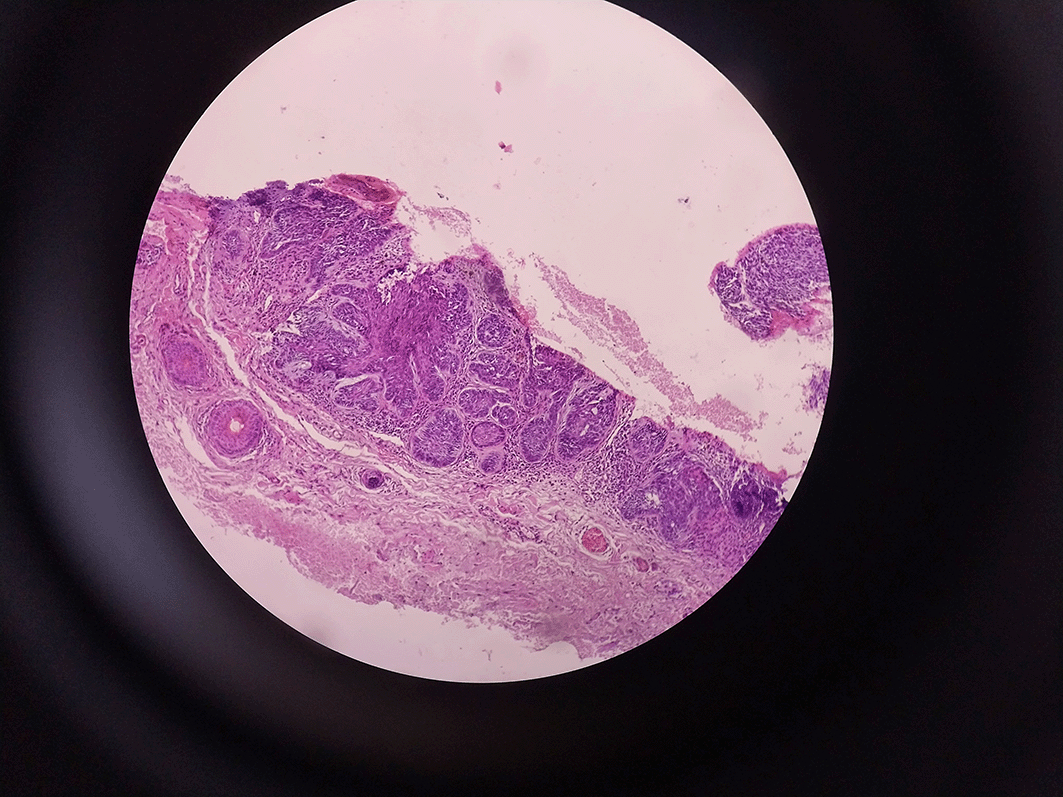
The patient underwent biopsy of the lesion. Histological examination revealed malignant epithelial proliferation consisting of solid cell masses bordered by malignant basaloid tumor cells, confirming the diagnosis of basal cell carcinoma. The stroma is dotted with inflammatory cells containing melanin deposits ( Figure 2).
This case highlights the importance of careful ophthalmological examination of all ocular structures during routine examination. Any suspicious eyelid lesions must be explored to ensure optimal and early care.
Eyelids are common sites of UV radiation lesions.1 It represents the site of 5-10% of all skin cancers.4 Basal cell carcinoma is caused by UV DNA damage with mutations in the PTCH1 gene, which leads to the uncontrolled growth of malignant cells.1 However, 29% of malignant lesions were considered benign clinically.3 There are two histological types, nodular and sclerotic.4 The lower eyelid is typically the site. The upper eyelid can mimick blepharitis.5 Local surgical excision remains the gold standard treatment, with 3 to 4 mm surgical margins followed by repair of the resulting defect.1 Reconstruction of the lower eyelid is a significant challenge, and several techniques have been proposed.6 One-step and two-step surgeries demonstrated similar results.7 The recurrence rate is approximately 5% per year.4 Therefore, adjuvant treatments such as radiotherapy and brachytherapy achieve local control and good cosmetic outcomes.8 Adjunctive hyperbaric oxygen therapy is also beneficial for increasing wound healing.9 Therefore, patients must be educated to prevent skin tumors. Sun protection and nicotine avoidance are two modifiable risk factors.1
Public education on the appearance of abnormal skin lesions is crucial. Management of these lesions is multidisciplinary. Early detection is the primary method for improving the prognosis and quality of life of patients.
| Views | Downloads | |
|---|---|---|
| F1000Research | - | - |
|
PubMed Central
Data from PMC are received and updated monthly.
|
- | - |
Provide sufficient details of any financial or non-financial competing interests to enable users to assess whether your comments might lead a reasonable person to question your impartiality. Consider the following examples, but note that this is not an exhaustive list:
Sign up for content alerts and receive a weekly or monthly email with all newly published articles
Already registered? Sign in
The email address should be the one you originally registered with F1000.
You registered with F1000 via Google, so we cannot reset your password.
To sign in, please click here.
If you still need help with your Google account password, please click here.
You registered with F1000 via Facebook, so we cannot reset your password.
To sign in, please click here.
If you still need help with your Facebook account password, please click here.
If your email address is registered with us, we will email you instructions to reset your password.
If you think you should have received this email but it has not arrived, please check your spam filters and/or contact for further assistance.
Comments on this article Comments (0)