Keywords
Tailgut cyst, retrorectal, case report, tumor, kraske, laparoscopy
Tailgut cyst, retrorectal, case report, tumor, kraske, laparoscopy
Tailgut cysts are the most frequently reported retrorectal tumors.1 Despite the large spectrum of revelations, preoperative diagnosis is misleading, as there are no prodromic features.2 Transparietal biopsy should be avoided. Surgical resection is the standard therapeutic course.3 The surgical approach remains controversial, as each technique offers distinct advantages. We believe this case report provides valuable insight into the diagnosis and management of tailgut cysts, while also providing well-founded arguments to help in the surgical choice.
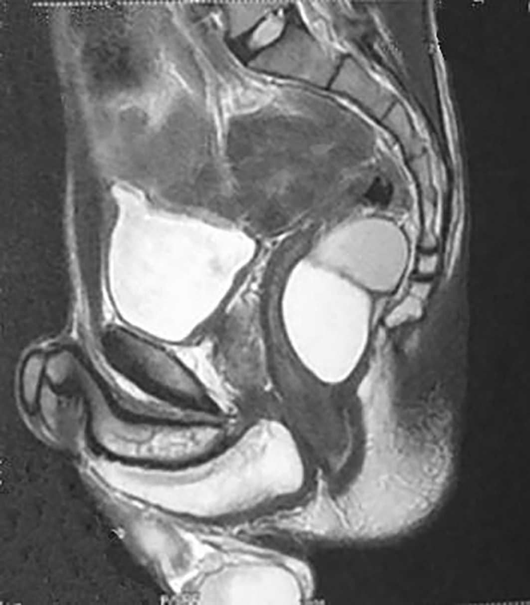
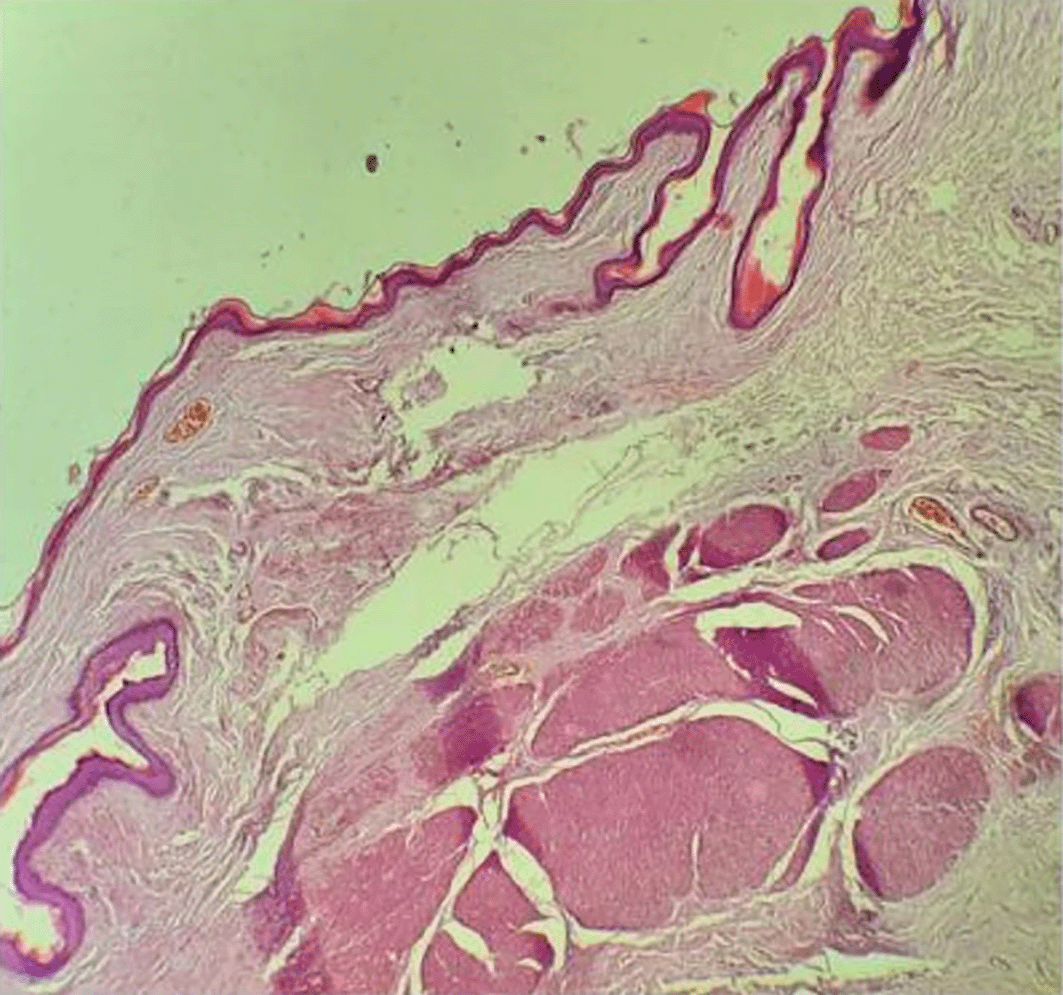
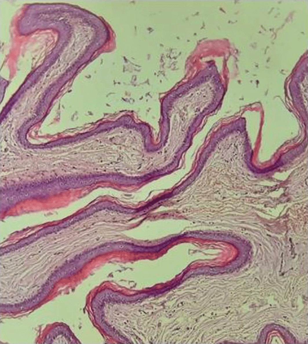
A 34-year-old man with no significant medical history was referred from an infirmary to our university hospital center for management of a post-traumatic hepatic laceration. The patient had no known allergies or hereditary diseases. Upon examination, his vital signs were found to be within the normal range. Physical examination revealed tenderness in the upper right quadrant. Biological examination revealed no abnormalities. On a full-body CT scan, a bilobed cystic mass measuring 5 cm presacral at the level of S4 displacing the rectum anteriorly and laterally measuring 67 mm was incidentally discovered. The patient reported no pain while sitting, rectal bleeding, constipation, urinary problems, or fever. Rectal examination revealed a painless, bulging posterior rectal wall with a palpable upper limit on the fingertip. Magnetic resonance imaging (MRI) revealed an 80 × 72 × 62 mm bilobed retrorectal cystic mass with a thin, regular wall, T1 hyposignal, and T2 heterogeneous signal, comprising a left paramedian portion with T2 hypersignal and a right paramedian portion with intermediate T2 signal, pushing back the rectum anteriorly and laterally without invading it (Figure 1). The patient was administered analgesics, with good clinical and radiological outcomes. After convalescence, the patient underwent laparoscopic resection. He was placed supine in the French position. Trocart placement was as follows: T12 on the right iliac fossa, T5 on the left iliac fossa, and T5 on the right flank. Upon exploration, it was found to be a bilobed retrorectal mass measuring 4 and 7 cm well defined cleavable cystic masses. At the end of the dissection, the tumor ruptured, and jelly like mucoid material was aspirated. postoperative course was uneventful. The patient was discharged on 4th postoperative day. Anatomopathological findings were as follows: cystic formation with a lumen filled with keratin strips and lined by regular squamous epithelium resting on a connective wall containing disorganized smooth muscle fibers (Figures 2 and 3). Final histologic examination indicated a tailgut cyst. The patient was scheduled for a close follow-up. The patient was doing well and was fine three months postoperatively.
The retrorectal or presacral space is bounded anteriorly by the rectum, superiorly by the peritoneal reflection, inferiorly by the Waldeyer’s fascia, posteriorly by the presacral fascia, and laterally by the ureters and iliac vessels. It is a crossroads of different histological components, of which tumorigenesis can manifest as several histological types: neural, bony, inflammatory, fatty, lymph node, vascular, or congenital. Their invasive potential is variable but often benign (92%).2
Malignancy appears to be related to male sex (p < 0.05) and young age (43 vs 54.4 years, p < 0.05).4 Tailgut cysts tops the list of these tumors, accounting for 41% of all tumors and 75% of benign tumors.2 However, it remains a rare tumor with only 53 cases in the largest series.5
These tumors manifest themselves through vague symptoms such as pain when sitting,6 defecatory pain, or rectorrhagia.1 Proctological examination revealed a well-defined soft and mobile retrorectal mass.6
The initial workup must include a CT and magnetic study.
Transparietal biopsy for histological evidence is not recommended because of the hypothetical risks of hemorrhage, infection, and tumor dissemination.7 Moreover, their interpretation may be inconclusive because they often contain only fibrous tissues, without epithelial tissues or malignant foci.8 In addition, surgical excision can be performed without difficulty in the majority of cases and therefore does not require neoadjuvant treatment for tumor downsizing. This therapeutic modality can be proposed in cases of Ewing’s sarcoma, osteosarcoma, and lymphoma3; thus, the interpretation of iconographic images is essential to guide the diagnosis.
The first diagnostic step is to rule out meningocele, as any inadvertent procedure can worsen the prognosis by causing iatrogenic meningitis. Recurrent headaches associated with bowel movements should raise suspicion of this diagnosis.7 “The scimitar sign” and signal intensity of its content parallel to the cerebrospinal fluid signal on all sequences are present in meningicele.13 The second diagnostic step is to consider malignant features such as the presence of enhancing or necrotic soft tissue, mural nodularity, post-contrast enhancement,9 intracystic vegetation, or infiltration of perilesional fat.1 On non-injected sequences, one should also look for irregular walls or loss of nodule sphericity.1 This stage is crucial for guiding the surgical procedure, anticipating tissue sacrifice, and adapting to the surgical approach. In fact, in sacrectomies, at least one side of S2 must be preserved to avoid urinary and bowel disturbance.10 No iconographic data characterize the tailguit cyst, but rather retrorectal, presacral, median topography above the plane of the levator ani is suggestive of the diagnosis. These lesions usually present as several loculi with a honeycomb appearance. The signal is variable, depending on the protein concentration; T2 hypersignals most often.1 Diffusion sequences reveal isosignal lesions associated with mucinous content.1 But many cases escape preoperative diagnosis: in a cohort of 28 patients, the diagnosis was mistaken in 35,7% of cases.11
A broad range of complications may arise during the course of the tumor, including hemorrhage, infection, fistulization, destruction or compression of the surrounding structures, and degeneration. There are many types of malignant transition in tailgut cysts, including NETs, adenocarcinoma, and squamous carcinoma.11
Resection is essential to avoid these complications, confirm the histological diagnosis, and establish milestones for subsequent follow-up. This preventive attitude is reinforced by the absence of a correlation between the presence of complaints and tumor histology,4 and complete resection does not prevent systemic tumor recurrence in cases of degenerescence.12 A wait-and-see approach should be avoided as asymptomatic lesions are at greater risk of malignancy (46,2 vs. 72,2%; p = 0.08).13
Surgery must resect the entire tumor without spilling the contents, especially if malignancy is suspected, bearing in mind that postoperative pain is not influenced by the associated bone resection (p = 0,3).14 The choice of the procedure should consider tissue sacrifice10 and tumor location. The posterior approach should be avoided for high-lying tumors (above S3), as a lack of access to the pelvic vessels in cases of intraoperative bleeding represents a real threat.15 When using a perineal approach, coccygectomy is often performed to improve surgical exposure and reduce the risk of recurrence by removing totipotent cells that may reside in the coccyx When operating via a perineal approach, coccygectomy is associated with improved surgical exposure and eliminates the possibility of recurrence from totipotent cells in the coccyx.15 When the boundary between the presacral tumor and the rectum is unclear, rectal palpation with the left hand can help delineate the interface, aiding in rectal preservation during dissection When no clear boundary was noted between the presacral tumor and rectum, rectal palpation with the left hand could be used to indicate the boundary between the presacral tumor and rectum to better protect the rectum.16 In cases of capsule rupture or when cyst decompression is required during surgery, the use of iodine tincture may help prevent seeding of cyst contents. When the capsule ruptures or cyst decompression is necessary during surgery, supplementary iodine tincture treatment could help prevent cyst content seeding.17 Moreover, in a French cohort of 270 patients who underwent surgery for retrorectal tumors, laparoscopic resection was associated with a lower incidence of intraoperative complications. Moreover, in the French cohort of 270 patients who underwent surgery for retrorectal tumors, laparoscopic resection avoided intraoperative accidents, and there was no statistical difference between the laparoscopic and Kraske approaches in terms of lesion perforation (47 vs. 47%, p = 1), rectal perforation (6 vs. 5%, p = 0,6), presacral hemorrhage (6 vs. 2%, p = 0,4) and bladder rupture (2 vs. 0%, p = 0,2).2 And as a corollary, no statistical difference in postoperative recurrence (12 vs. 3,5%, p = 0,06).2 In addition, there was no statistically significant difference in overall morbidity between the laparoscopic and Kraske approaches (28 vs. 30%, p = 0,7) neither in unplanned readmissions (9 vs. 11%, p = 0,8) nor, postoperative pain (10 vs. 20%, p = 0,3) nor, sexual dysfunction (3 vs. 1%, p = 0,5) nor urinary incontinence (3 vs. 1%, p = 0,5).2 Rectal fistula was only observed in patients who underwent laparoscopic surgery (6 vs. 0%, p = 0,01) but it should be however, the latter was preferred in cases of rectal invasion, which explains this marked difference. An important point to emphasize in this study is the importance of postoperative urinary disorders in patients undergoing laparoscopic surgery (13 vs. 0%, p = 0,006) and postoperative defecation (10 vs. 0%, p = 0,02) whereas these tumors presented more uro-defecatory disorders in patients operated on by the Kraske approach (9 vs. 11%, 6 vs. 13%).2 Severe postoperative complications (Clavien-Dindo ≥ 3) are non-negligible, reaching 7,6% in a cohort of 144 patients who underwent surgery for retrorectal tumors.18
Despite the absence of consensus guidelines on surveillance schedule, patients should be followed up closely, as the risk of recurrence is approximately 5% according to the follow-up of 73 patients in a median of 24 months, despite an R0 resection rate of 94%.19 In addition, in a cohort study of the Mayo Clinic, 8% of resected specimens harbored malignancy.19
Although no conclusions can be drawn from our case report, it can be added to the current reported cases to further widen the literature, especially by featuring its incidental presentation and surgical difficulties.
Tailgut cysts are mostly benign tumors with low malignant potential. Definitive diagnosis was based on the histological examination of the resected mass. Preoperative sampling is considered hazardous. Both surgical routes are proven procedures, with adequate short- and long-term results. The choice should be tailored to the tumoral extent and planned tissular damage.
The CARE checklist was uploaded to the Zenodo plateform. Its DOI is: 10.5281/zenodo.15872750
Data are available under the terms of the Creative Commons Attribution 4.0 International license (CC-BY 4.0).
| Views | Downloads | |
|---|---|---|
| F1000Research | - | - |
|
PubMed Central
Data from PMC are received and updated monthly.
|
- | - |
Provide sufficient details of any financial or non-financial competing interests to enable users to assess whether your comments might lead a reasonable person to question your impartiality. Consider the following examples, but note that this is not an exhaustive list:
Sign up for content alerts and receive a weekly or monthly email with all newly published articles
Already registered? Sign in
The email address should be the one you originally registered with F1000.
You registered with F1000 via Google, so we cannot reset your password.
To sign in, please click here.
If you still need help with your Google account password, please click here.
You registered with F1000 via Facebook, so we cannot reset your password.
To sign in, please click here.
If you still need help with your Facebook account password, please click here.
If your email address is registered with us, we will email you instructions to reset your password.
If you think you should have received this email but it has not arrived, please check your spam filters and/or contact for further assistance.
Comments on this article Comments (0)