Keywords
general, digestive, surgery, appendix, tumor, hernia
This article is included in the Oncology gateway.
general, digestive, surgery, appendix, tumor, hernia
Appendicular tumors represent a wide range of clinico-pathological entities.1 They are often found incidentally in appendix specimens.2 They are rarely diagnosed preoperatively due to the absence of clinical prodromes or in the open abdomen, due to the scarcity of signs of malignancy and the rarity of their incidence.3 Therefore, a high index of suspicion is necessary to take appropriate actions. Herein, we report a case of appendicular adenocarcinoma triggered by hernial strangulation. To our knowledge, this is the first reported case of an appendicular adenocarcinome inside a strangulated incisional hernia associated with a tumoral deposit inside the hernial sac.
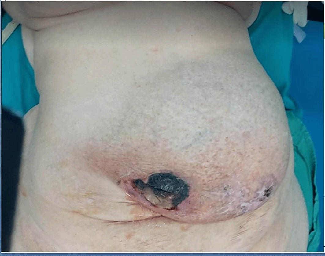
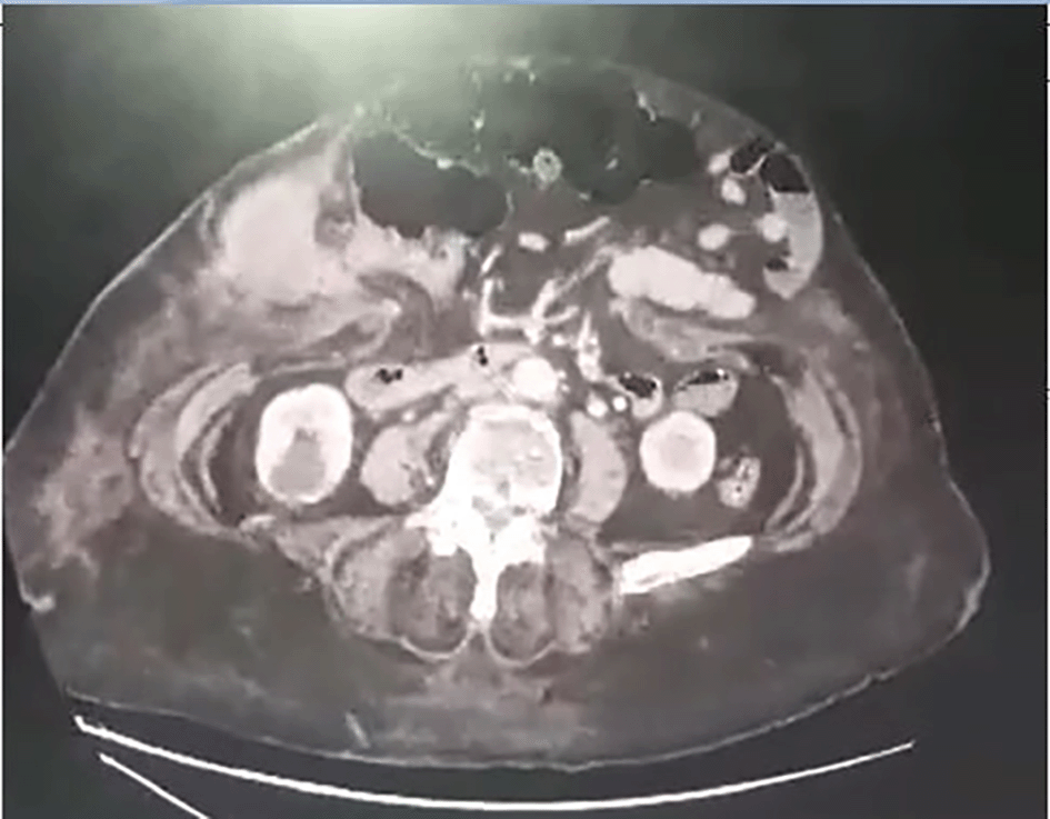
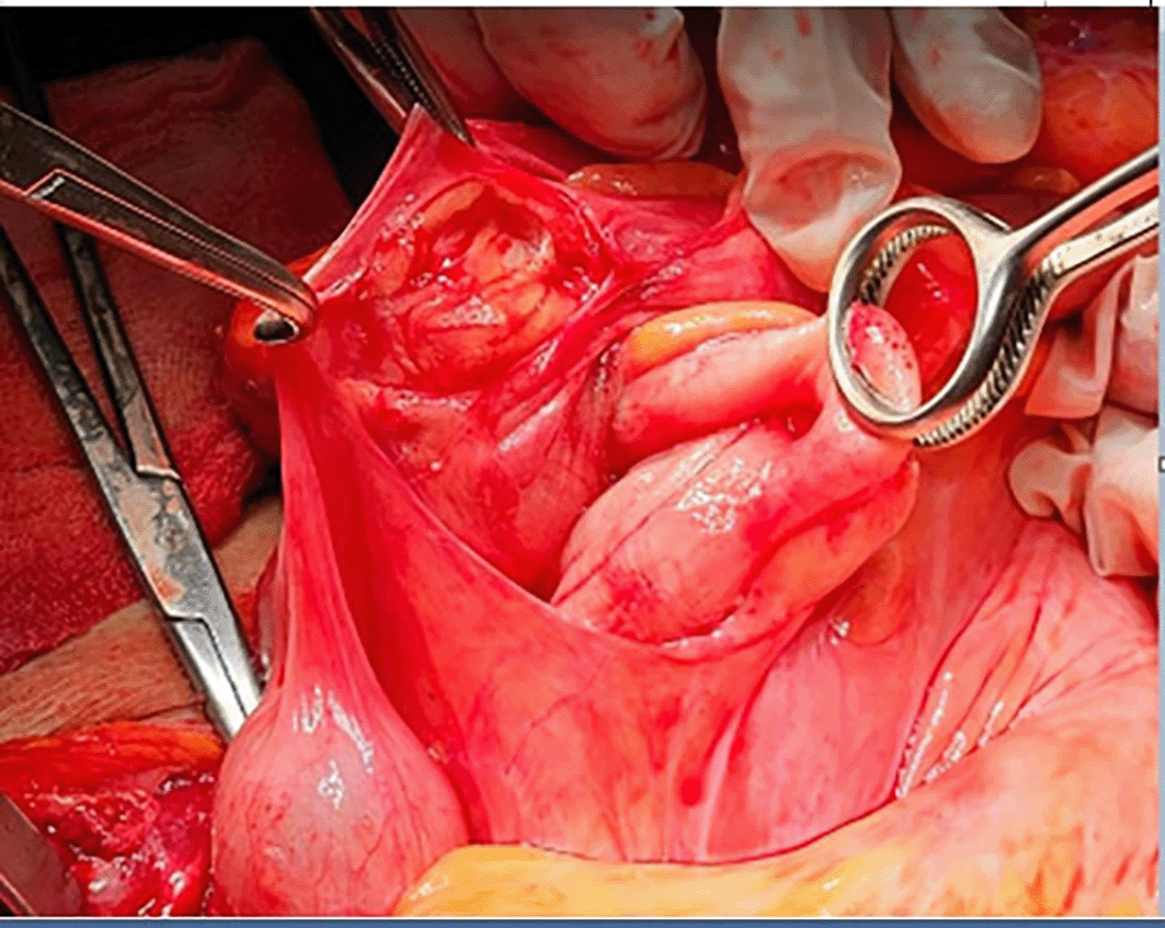
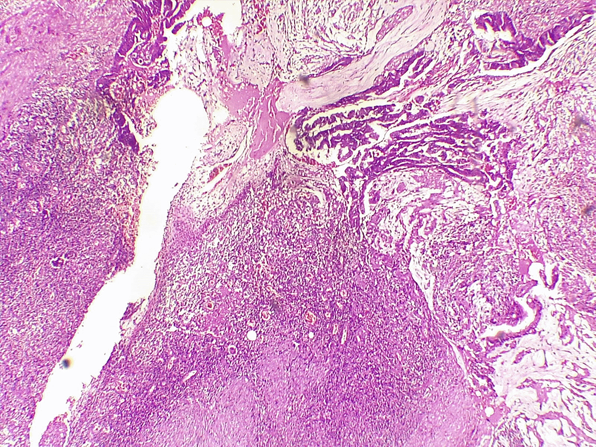
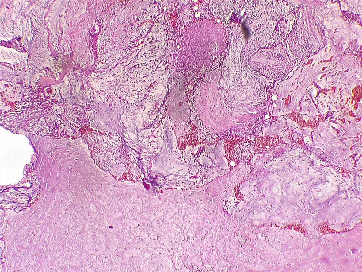
An 81-year-old woman with a history of diabetes mellitus, hypertension, arterial fibrillation, and ventral hernia repair was scheduled to undergo surgery for a recurrent hernia. She consulted an emergency department for rapidly increasing abdominal pain for 6 days. Upon examination, the umbilicus bulged with overlying sphacelous skin (Figure 1). Biology was as following: CRP = 167.3 mg/L, WBC = 12020/mm3, creatinine = 121.84 μmol/L, urea 9 mmol/L, hemoglobin = 12.3 g/dL. An abdominal CT scan revealed a herniated right colon through a 25 mm defect, an enlarged appendix measuring 18 mm, and communication with a 5 cm intrasaccular abscess (Figure 2). She then rushed to the operating theater. Upon incision, pus was evacuated from the hernial sac. The right colon was incarcerated within the aponeurotic defect, with no signs of digestive distress, and the peritoneal cavity was not contaminated due to the constriction ring at the neck of the sac. The appendix was gangrenous with a perforated tip eliciting mucus (Figure 3). Thorough lavage with appendicectomy and herniorrhaphy was performed. She died on 5th postoperative day to a pulmonary oedema. Histological analysis revealed a well-differentiated adenocarcinoma that developed within a serrated adenoma (Figure 4). It was classified pT3. Mucus was acellular (Figure 5). The hernial sac harbors similar cancerous cells.
Tumors revealed during hernia with metastases inside the sac are scarce. In fact, during 39 years of inguinal hernia repair, 0.07% of patients had metastatic tumors during herniorrhaphy.4 This situation reaches 2,2% if we consider all abdominal hernias, as concluded in a recent retrospective cohort study of 455 hernia sac specimens.5 It mostly involves incisional hernias (67%),6 as in our case.
To our knowledge, this is the first case of appendicular adenocarcinoma involving a strangulated incisional hernia with a metastatic sac. However, other cases of appendicular tumors have been published, developing inside Amyand’s hernia7 or a Spiegel hernia.8
Some cases have reported the presence of peritoneal carcinomatosis nodules in the hernial sac, mostly of the origin colon,9 and all were in the T3 or T4 stage.10 This could be explained by the drop metastases theory, where malignant cells metastasize following gravity, and inflammatory oncotaxis and chemotactic agents are probably operative mechanisms in the development of metastatic lesions in hernial sacs. Peritoneal metastasis consists of different phases. Cellular mechanisms involved in peritoneal spread can be ordered in sequential steps, such as detachment of malignant cells and increased motility, anoikis evasion, adherence to the peritoneal surface, invasion of the peritoneum, and colonization and survival in the new niche inflammatory oncotaxis and chemotactic factors are likely key mechanisms in the development of metastatic lesions within hernial sacs. The process of peritoneal metastasis involves multiple distinct phases. The cellular mechanisms underlying peritoneal dissemination can be described in a sequential manner, including: detachment of malignant cells accompanied by increased motility; evasion of anoikis; adherence to the peritoneal surface; invasion of the peritoneal tissue; and, finally, colonization and survival within the new microenvironment.11 However, extra-abdominal tumors (9.3% of metastatic tumors found during inguinal hernia repair, involving breast – thymus – pleura and hairy cell leukemia) have been reported to develop carcinosis nodules in the inguinal hernia sac.9 This could be explained by metastasis via the hematogenous route. In addition, mesothelioma of peritoneal origin can be identified as inguinal hernia.4 This statement suggests the promotion of routine examination of the hernia sac during surgery for a tumor with a co-existent hernia or any elective hernia repair, especially when gross pathology was not detected in the macroscopic examination of abdominal hernias in all cases,5 and that this situation is the initial presentation of malignancy in the third to half of cases.6,12,13 Further studies are needed to answer this question. Currently, selective microscopic examination is recommended in cases with grossly abnormal hernia sac specimens or macroscopically suspected lesions.14 Routine examination is not justified because of its high costs, with fees reaching €21,087.50, to detect a single case of malignant tumor in the hernia sac, as stated in a Spanish study.11
This situation raises both diagnostic and therapeutic challenges. First, the inflammatory process accompanying herniation may lead to swelling of its contents, making it difficult to recognize a suspected lesion tactilely. In addition, this entity should be differentiated from testicular tumors misdiagnosed as inguinal hernia.15 Second, another issue to highlight is tumor perforation, which can occur during the evolution of these intrasaccular tumors, raising the possibility of misdiagnosis of strangulated hernia and rushing subsequent management.16,17 In such cases, surgical treatment may be inadequate due to a lack of awareness of the condition. The surgeon must be vigilant when faced with intrasaccular phlegmon without signs of vascular insufficiency of the herniated organ. Third, because of the possible infiltration of adjacent structures, extensive extirpative surgery may be required. Gnas et al. reported the need for en bloc resection of sigmoid tumors infiltrating the elements of the spermatic cord.18
Regardless of the approach, the operation must achieve resection based on sound oncological principles and ensure secure hernia repair. Regardless of the surgical approach, the procedure must adhere to established oncological principles for tumor resection while ensuring a durable and secure hernia repair.
Finally, reinforcing with a mesh is also confusing because of the high risk of perforated tumors, reaching 44,8% in cases of inguinal hernia. The current general opinion is that using mesh in clean-contaminated surgical fields could be considered on a case-by-case basis; however, the use of mesh in contaminated or dirty surgical fields is still not recommended. Hernia repair using a mesh should be performed as a two-stage surgery when necessary The prevailing consensus is that mesh use in clean-contaminated surgical fields may be considered on a case-by-case basis. However, its use in contaminated or dirty fields remains generally contraindicated. When clinically indicated, hernia repair with mesh should be performed as a staged procedure.19
In summary, surgeons should be aware of the possibility of encountering malignant diseases at hernia sites Surgeons should remain vigilant for the potential presence of malignancies at hernia sites. It is essential to avoid a hasty and poorly performed examination of the hernia sac, because if we devote sufficient attention to this time, we will be able to detect suggestive signs of malignancy and then take the appropriate action: carcinological resection and long-term postoperative surveillance. Since subtle lesions may be overlooked on gross examination, caution should be exercised with hernia sacs from older patients.
Written informed consent for the publication of clinical details and images was obtained from the patients.
CARE checklist of article: Case report: An appendicular adenocarcinoma presenting in a strangulated incisional hernia with metastatic sac.
https://doi.org/10.5281/zenodo.1587267820
Data are available under Creative Commons Attribution 4.0 International
| Views | Downloads | |
|---|---|---|
| F1000Research | - | - |
|
PubMed Central
Data from PMC are received and updated monthly.
|
- | - |
Provide sufficient details of any financial or non-financial competing interests to enable users to assess whether your comments might lead a reasonable person to question your impartiality. Consider the following examples, but note that this is not an exhaustive list:
Sign up for content alerts and receive a weekly or monthly email with all newly published articles
Already registered? Sign in
The email address should be the one you originally registered with F1000.
You registered with F1000 via Google, so we cannot reset your password.
To sign in, please click here.
If you still need help with your Google account password, please click here.
You registered with F1000 via Facebook, so we cannot reset your password.
To sign in, please click here.
If you still need help with your Facebook account password, please click here.
If your email address is registered with us, we will email you instructions to reset your password.
If you think you should have received this email but it has not arrived, please check your spam filters and/or contact for further assistance.
Comments on this article Comments (0)