Keywords
Chilaiditi Syndrome, Abdominal Pain, Diagnostic Errors, Emergency Medicine,Case Report.
This article is included in the Rare diseases collection.
Chilaiditi Syndrome, Abdominal Pain, Diagnostic Errors, Emergency Medicine,Case Report.
Chilaiditi syndrome is a rare condition characterized by the interposition of a bowel loop, usually the colon, between the liver and the right hemidiaphragm.1,2 First described by Demetrius Chilaiditi in 1910,3 it differs from theptomatic Chilaiditi sign, presenting clinically with symptoms such as abdominal pain, constipation, nausea, vomiting, or respiratory distress.2,3
Epidemiological data report an incidence of about 1%, predominantly in elderly males.3 Multiple predisposing factors have been identified, including chronic constipation, colonic redundancy, ligament laxity, cirrhosis, diaphragmatic paralysis, reduced liver volume, and neurocognitive disorders.4–8
The syndrome is diagnostically challenging because it can mimic free intraperitoneal air on radiographs, leading to unnecessary surgical procedures. Imaging studies, particularly CT scans, are critical for accurate diagnosis.6–9 Although several cases have been reported, its occurrence in the emergency department remains of special clinical interest, as it may closely simulate life-threatening conditions such as hollow viscus perforation.
Here, we present the case of an elderly male with multiple comorbidities, including Alzheimer’s disease, who presented with acute abdominal pain. Prompt recognition of the radiographic features and confirmation by CT allowed conservative management and avoided unnecessary surgery. This case highlights the importance of considering Chilaiditi syndrome in the differential diagnosis of acute abdomen in the emergency setting.
A 70-year-old male with a medical history of type 2 diabetes mellitus, hypertension, and Alzheimer’s disease, without prior abdominal surgery, presented to the emergency department (ED) on February 27, 2025, with abdominal pain and constipation evolving since February 26, 2025. The patient had no history of abdominal surgery or prior medical interventions for gastrointestinal symptoms. No previous episodes of bowel obstruction, invasive procedures, or hospitalizations for similar complaints were reported.
The abdominal pain was rated at 70/100 on the Visual Analogue Scale (VAS). There was no history of vomiting, gastrointestinal bleeding, or weight loss. On examination, the patient was afebrile and hemodynamically stable. The abdomen was moderately distended and tympanic to percussion, with no signs of peritoneal irritation. Hernial orifices were free. Digital rectal examination revealed no palpable mass or fecal impaction.
Laboratory investigations, including complete blood count, electrolytes, renal function, and liver function tests, were all within normal limits.
An abdominal plain radiograph revealed a crescent-shaped air collection beneath the right hemidiaphragm. The presence of visible colonic haustrations within the gas pattern suggested colonic interposition rather than free intraperitoneal air ( Figure 1, Table 1).
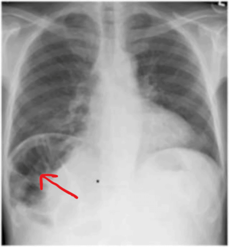
Image obtained at our institution and published with written informed consent from the patient’s legal guardian (his son).
Subsequently, an abdominal computed tomography (CT) scan was performed, which clearly demonstrated interposition of the hepatic flexure of the colon between the liver and the right hemidiaphragm, without evidence of perforation, obstruction, or ischemia. No ascites or intra-abdominal collections were detected ( Figure 2).
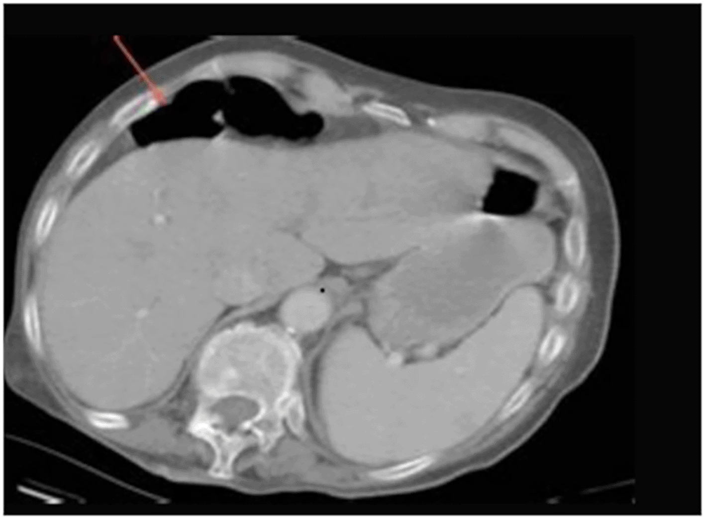
Image obtained at our institution and published with written informed consent from the patient’s legal guardian (his son).
Based on these findings, a diagnosis of Chilaiditi syndrome was established. Differential diagnoses included pneumoperitoneum and subphrenic abscess, which were excluded based on imaging.
The patient was managed conservatively with:
• Bowel rest (nil per os),
• Intravenous fluids (0.9% normal saline at 2 L/day for 48 hours),
• Analgesics (paracetamol 1 g IV every 8 hours),
• Laxatives (lactulose syrup 15 mL orally twice daily).
Clinical improvement was observed within 48 hours, with the VAS pain score decreasing from 70/100 on admission to 20/100 before discharge, along with normalization of bowel transit. At one-week follow-up, the patient remained asymptomatic.
Adherence was monitored through direct observation of medication administration during hospitalization and by patient self-report at follow-up. No adverse effects related to analgesics (e.g., hepatotoxicity, gastrointestinal intolerance) or laxatives (e.g., abdominal cramping, diarrhea) were noted. The patient reported good tolerance of the treatment regimen and full compliance with oral medications at home.
The patient did not experience any adverse drug reactions, intolerance, or unanticipated events related to the conservative management during hospitalization or follow-up.
Chilaiditi syndrome, characterized by colonic interposition between the liver and the right hemidiaphragm, is an uncommon clinicoradiologic entity with reported incidence between 0.025% and 0.28% on plain radiographs, showing a male predominance and increasing prevalence with age.1–4 Predisposing factors include increased colonic mobility (redundant colon, elongated mesentery, ligament laxity), diaphragmatic elevation (lung hyperinflation, phrenic nerve palsy), and reduced liver volume, as well as functional issues such as chronic constipation and neurocognitive disorders.3,5–7
The main difficulty lies in differentiating the syndrome from true pneumoperitoneum. Radiographic clues include:
• Fixed gas patterns beneath the diaphragm
• Presence of colonic haustrations
• Absence of Rigler sign
CT imaging is definitive, confirming colonic interposition and ruling out perforation. Conservative treatment (bowel rest, fluid therapy, analgesia, and laxatives) is effective in uncomplicated cases. Surgical intervention is reserved for complications such as obstruction, ischemia, or volvulus.
A review of the literature reveals that Chilaiditi syndrome poses significant diagnostic challenges due to its mimicry of free intraperitoneal air, often leading to unnecessary laparotomies.6–9 Radiographic clues such as fixed gas patterns with colonic haustral markings and absence of Rigler sign, confirmed by CT imaging, are critical for correct diagnosis.7–10 Cases in the literature underscore conservative management as the first-line treatment in uncomplicated cases,6,8 with surgery reserved for complications or recurrent symptoms.9–17 (Table 2)
| Author(s) | Year | Patient Age/Sex | History | Presentation | Management | Outcome |
|---|---|---|---|---|---|---|
| Vazquez et al.1 | 2023 | Two, pediatric patient: 10/F 9/F | Autism, IgA deficiency, and constipation constipation, developmental delay, and hypotonia | Abdominal pain, nausea, vomiting, constipation abdominal pain, vomiting, constipation, and decreased appetite | A bowel cleanout, compartmentalization of the sigmoid and rectum Conservative treatment rectal irrigations and catheter decompression | Recovery improvement of symptoms |
| Kamel et al.8 | 2024 | 70/M | Depression, anxiety, gastroesophageal reflux disease (GERD), and postpolio syndrome | Shoulder pain, chronic weakness, and dizziness. hypotension elevated lactic acid | Conservative (hydration, bowel rest) | Improvement |
| Mohamed et al.15 | 2024 | 72/M (COPD) | COPD | Dyspnea, abdominal discomfort | Conservative management | Recovery |
| Ettaouss et al.11 | 2024 | 54/M | Appendectomy in 2016 | Abdominal pain with obstructive syndrome | Surgey: resecting the volvulized, necrotic, and perforated old ileocolic anastomosis in Chilaiditi syndrome | Recovery the patient was discharged on postoperative day 6 |
| Kao et al.12 | 2023 | 61/M | No history | Dyspnea, abdominal discomfort | Surgical management involving a right hemicolectomy | Recovery the patient was discharged on postoperative day 8 |
| Tola et al.16 | 2024 | 65/M | No history | Abdominal pain, imaging revealing colonic interposition | Conservative management | Symptom resolution |
| Bourakkadi & Dkhissi17 | 2024 | 61/F | No history | Misdiagnosed as pneumoperitoneum | Conservative management | Recovery |
The literature confirms that most cases are managed conservatively and highlights the importance of clinician awareness to avoid unnecessary laparotomy.
Our case aligns with previous reports emphasizing that elderly patients with comorbidities, such as constipation and neurocognitive disorders, are particularly susceptible to intermittent presentations of the syndrome.4,9,11–17 This review highlights the importance of education among emergency and radiology personnel to ensure early recognition and appropriate management pathways.
In conclusion, recognizing Chilaiditi syndrome and understanding its imaging features is crucial to avoid unnecessary surgeries and optimize patient outcomes. Conservative management remains effective in most cases, with surgery reserved for complications. The inclusion of a literature review enhances the scientific value of the case.
| Views | Downloads | |
|---|---|---|
| F1000Research | - | - |
|
PubMed Central
Data from PMC are received and updated monthly.
|
- | - |
Provide sufficient details of any financial or non-financial competing interests to enable users to assess whether your comments might lead a reasonable person to question your impartiality. Consider the following examples, but note that this is not an exhaustive list:
Sign up for content alerts and receive a weekly or monthly email with all newly published articles
Already registered? Sign in
The email address should be the one you originally registered with F1000.
You registered with F1000 via Google, so we cannot reset your password.
To sign in, please click here.
If you still need help with your Google account password, please click here.
You registered with F1000 via Facebook, so we cannot reset your password.
To sign in, please click here.
If you still need help with your Facebook account password, please click here.
If your email address is registered with us, we will email you instructions to reset your password.
If you think you should have received this email but it has not arrived, please check your spam filters and/or contact for further assistance.
Comments on this article Comments (0)