Keywords
3D printing; thermal aging; nanoparticles; fracture resistance; resin teeth

This article is included in the Nanoscience & Nanotechnology gateway.
This article is included in the Health Services gateway.
3D printing; thermal aging; nanoparticles; fracture resistance; resin teeth
According to reviewer comments, all comments were addressed and modified mainly in the methods and discussion section. all major modifications were changed using track changes and highlighted through the text. In addition to the added references with references reordered in the discussion and references section.
See the authors' detailed response to the review by Emel Uzunoglu Ozyurek
See the authors' detailed response to the review by Elif Yalçın and EYYÜP ALTINTAŞ
See the authors' detailed response to the review by Yomna M. Ibrahim
See the authors' detailed response to the review by Yasser Mohamed Aly
See the authors' detailed response to the review by Büşra Tosun
Artificial teeth for removable dentures are manufactured from various materials, including acrylic resin, ceramics, 3D-printed materials, and resin composites.1,2 These artificial teeth must be durable enough to withstand fractures induced by contact with the opposing teeth and abrasive foods.2,3 Conventionally, prefabricated denture teeth are made of acrylic resin, making them prone to fracture or chipping, particularly in situations in which a complete denture opposes natural teeth or an implant-supported overdenture.4–6 With growing demand for artificial teeth with enhanced fracture resistance, various approaches have been proposed, including the use of different monomers, cross-linking agents, and organic and inorganic fillers in polymer matrices.7,8 Despite these advancements, denture-teeth fractures continue to be a persistent issue, highlighting the need for the development of denture teeth fabricated using innovative methods capable of withstanding higher loads and forces.8,9
Digital dentures can be produced by using subtractive or additive manufacturing techniques.3 In the additive process, a photopolymerized liquid resin is used to build removable prostheses layer-by-layer.3,4,10 This method offers several benefits, including the significantly lower cost of 3D printers compared with milling machines, which facilitates its broader adoption in clinical practice.3 Additionally, unlike subtractive milling, 3D-printing generates less material waste.3,11,12 Occlusal stability, function, and esthetics are all reduced when the fracture resistance and elastic modulus of denture teeth are compromised, which is why high strength is crucial.7,8,13 Moreover, 3D-printed denture teeth are typically manufactured from methacrylate-based photopolymerized resin, a material specifically designed for processing through 3D-printing technology.11–14 This method provides flexibility and precision, and improves functionality in the fabrication of removable dentures, making it a modern alternative to traditional techniques. Further advancements in digital dentistry are needed to further enhance the quality and durability of dental prostheses.8,14
Gad et al., 8 studied the fracture resistance of a specific type of 3D-printed teeth (NextDent), and showed that they exhibited greater fracture resistance than traditional prefabricated teeth. However, their strength decreased after exposure to thermal cycling. Similarly, Chun et al.,10 examined another variety of 3D-printed resin teeth (Dentca) and reported that their fracture resistance was comparable to that of prefabricated teeth. These studies8,10 identified the potential of 3D-printed resins and highlighted the need to improve their strength, especially after thermal aging (TA). The absence of nanoparticle (NP) reinforcement in previous research left a gap in the exploration of how NPs can enhance durability.13 Therefore, exploring the incorporation of different NPs may be an effective approach for improving the mechanical properties of 3D-printed resin teeth.
Owing to the capacity of 3D-printed resins for reinforcement, zirconium dioxide nanoparticles (ZNPs) and silicon dioxide nanoparticles (SNPs) have attracted considerable interest. When added to a resin matrix, ZNPs, which are well known for their antibacterial properties, increase the mechanical strength and wear resistance of dental composites.4,13–16 Similarly, SNPs enhance the hardness, fracture toughness, and elastic modulus of dental materials owing to their large surface area and compatibility with resin matrices.15 By resolving the drawbacks of conventional materials, previous studies4,13 have shown that the addition of these NPs to interim resins has yielded encouraging results, resulting in long-lasting and resilient interim restorations.
The vitro testing of NP addition, with a shape that replicates clinical use through a standardized procedure, may provide valuable insights into material-specific properties that are critical for understanding their behavior. The addition of NPs to 3D printed resins was investigated in previous studies however, there is still a lack of information in the literature about the addition effect of ZNPs and SNPs with different concentrations to 3D printed resin designated for teeth fabrication. Up to author knowledge, no previous studies have thoroughly investigated the strength of 3D-printed nanocomposite teeth in comparison with prefabricated teeth; therefore, this study aimed to assess the fracture resistance and elastic modulus of newly introduced 3D-printed nanocomposite denture teeth before and after TA.
The study tested three null hypotheses: first, that incorporating zirconium dioxide (ZrO2) and silicon dioxide (SiO2) nanoparticles into 3D-printed denture-tooth resins would have no significant effect on their fracture resistance or elastic modulus; second, that thermal aging would not significantly influence these mechanical properties in any of the tested materials; and third, the interaction effect of variables has no significant effect on tested properties.
The sample size for this in vitro investigation was determined using data gathered from a prior study8 that evaluated the mechanical characteristics of temporary crowns made of 3D-printed resin. The World Health Organization (Geneva, Switzerland) specified the formulae, and power analysis was used in the counting procedure. The study was conducted using a significance threshold of 5%, power of 80%, and marginal error of 5%. A total of 220 specimens were fabricated and subdivided as follows: 20 prefabricated teeth, 100 ASIGA, 100 NextDent. The prefabricated group (n = 20) consisted of mandibular molar teeth (Major Dent-V, MAJOR Prodotti Dentari S.P.A., Moncalieri (TO), Italy). For the printed groups, two types of photopolymerized resins were used to create the 3D-printed resin teeth (n = 100/resin): NextDent C&B MFH (NextDent, 3D Systems, Soesterberg, The Netherlands) and ASIGA (Asiga DentaTOOTH, Shade A1, ASIGA, Erfurt, Germany). Half of the prepared specimens (N = 110) were tested without thermocycling while another half (N = 110) were tested after thermocycling.
In this study, 3D-printed groups were reinforced with two types of NPs: ZNPs (Shanghai Richem International Co., Ltd., Shanghai, China) and SNPs (AEROSIL R812; Evonik-Degussa, Essen, Germany). Each NP type was incorporated into the resin fluid at two different concentrations (0.5 wt.% and 1 wt.%). The NPs were treated and silanized before being added to the resin.13,15,16 The silanized NPs were carefully weighed using a digital balance (S-234; Denver Instruments, Gottingen, Germany) to ensure precise measurements.15,16 Subsequently, the resin/NP mixtures were thoroughly mixed using a magnetic stirrer for 30 min to ensure the homogeneity of the NPs within the resin matrix.13 A total of five groups were prepared for each resin type: one control group without any reinforcement, and four experimental groups containing varying NP concentrations (0.5 wt.% ZNP, 1 wt.% ZNP, 0.5 wt.% SNP, 1 wt.% SNP). This design enabled a comprehensive evaluation of the effects of both the NP type and concentration on the mechanical performance of 3D-printed resin.
A standard tessellation language (STL) file was created by scanning the prefabricated mandibular molar teeth standardized to the dimensions of the mandibular molar (10.2 × 11.1 × 7.3 mm). Each STL file was printed in a horizontal orientation (0° build angle) with a desktop laser scanner (E3; 3Shape A/S, Copenhagen, Denmark), and the file was then exported to the respective 3D printer.8 Each specimen was manufactured in a 3D-printer chamber in accordance with the digital blueprint and required standards.8,13 The manufacturer’s guidelines for the NextDent 5100 (3D Systems, Rock Hill, SC, USA) and ASIGA MAX (Asiga, Alexandria, NSW, Australia) 3D printers were followed to accurately convert the resin material into the specified dental components. All specimens were fabricated using two different additive manufacturing systems according to the manufacturer-recommended printing protocols. The first group was printed using the ASIGA MAX, which operates using digital light processing (DLP) technology with a 385-nm UV light source. Specimens were printed with a layer thickness of 50 μm, and the exposure parameters were automatically controlled through the printer’s validated resin profile to ensure consistent polymerization for each layer. The second group was fabricated using a NextDent 5100 with a 405-nm UV LED light source. Printing was performed using a 50 μm layer thickness, and the exposure time was set at approximately 5–6 s per layer according to the manufacturer’s resin profile for denture base materials. Once printing was completed, the specimens underwent post-processing using the post-curing units of each printing system with the recommended post-curing process and conditions.13,15,16 The post-polymerization protocol for ND was using curing unit (LC-D Print Box, 3D systems Vertex Dental B.V., Soesterberg, Netherland) for 15 min with wavelength measured 360–435 nm and ASIGA underwent for (Asiga Flash UV Curing Chamber) for 20 min with wavelength 480–530 nm. After curing, a diamond disc was used to remove the support structure. Half of the specimens (N = 110) were subjected to 5000 TA cycles at 5°C and 55°C with a dwell time of 30 s (Figure 1). The TA process was performed using a Thermocycler (Thermocycler THE- 1100, Mechatronik GmbH, Feldkirchen-Westerham, Germany), simulating intraoral temperature changes for six months of clinical use.13,17
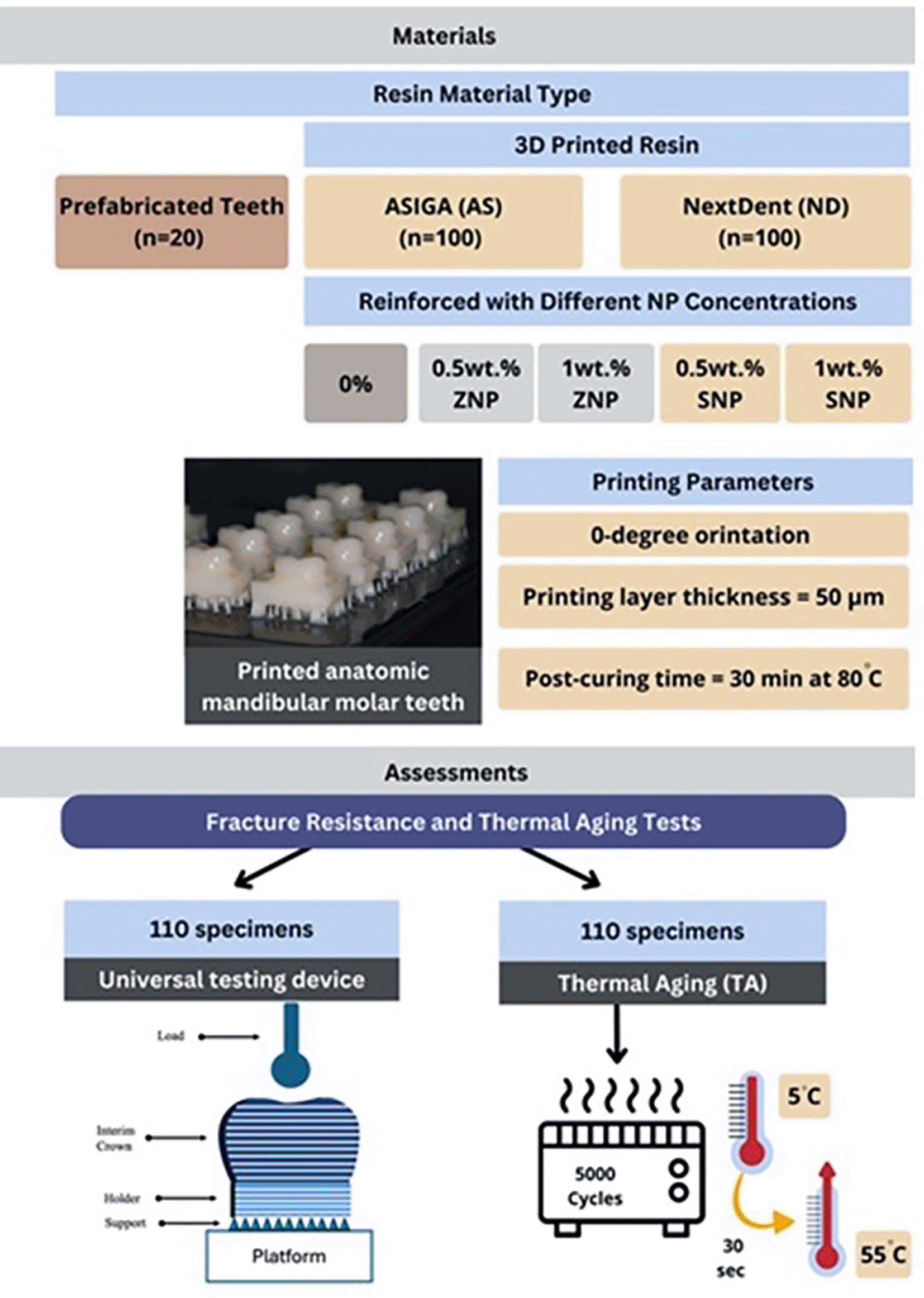
A stainless-steel ball indenter with a 7 mm radius was used to load the specimens at the occlusal surfaces using a universal testing apparatus (Instron model 5965, Massachusetts, United States) with a 5 kN load cell at a loading rate of 1 mm/min until failure occurred (Figure 1). A 1.5 mm-thick rubber sheet was positioned between the occlusal surface and indenter to reduce contact damage and aid in distributing the load.13,17
Fracture resistance was recorded as the maximum load at fracture and expressed in Newtons (N). The elastic modulus was calculated from the linear portion of the load–deflection curve obtained from the universal testing machine using the following formula: (E = FL3/4Ybd3) Where: E = elastic modulus (MPa), F = applied load (N), L = span length (mm), b = specimen width (mm), d = specimen thickness (mm), Y = deflection (mm).
Essential data on the fracture behaviour of the modified 3D-printed denture teeth were obtained through fracture-site analysis using scanning electron microscopy (SEM; TESCAN VEGA3 LM model, TESCAN Orsay Holding, Kohoutovice, Czech Republic). To ensure optimal image quality, the fractured samples were cleaned and prepared by coating nonconductive polymer or resin materials with a thin layer of conductive gold (Quorum, Q150R ES, UK). This coating, applied at an accelerating voltage of 20 kV, prevented charging effects and enhanced image clarity. The specimens were then placed in the SEM chamber for analysis, where fracture surfaces were examined at a magnification of 2000× with a scale bar of 50 μm. Key fracture characteristics such as initiation sites, crack-propagation patterns, and interactions between the polymer matrix and reinforcing particles were identified. The NPs, with an average size of 40 nm and surface area of 9 m2/g, were analyzed using both SEM and transmission electron microscopy (TEM).
Fourier transform infrared (FTIR) analysis is an essential tool for examining the bonding interactions between NPs and the polymer matrix because it offers comprehensive details on the chemical structure and functional groups present in the material. FTIR analysis assisted in identifying any changes in the molecular bonding caused by the addition of NPs. The specimens were placed inside an FTIR spectrometer for transmission spectroscopy (Hartmann & Braun, MB series), and two readings were obtained per specimen. FTIR spectra were obtained at a resolution of 4 cm−1, spanning a wavenumber range of 4000 to 400 cm−1. The resultant spectra showed distinctive peaks corresponding to certain functional groups; thus, the bond types and any matrix chemical changes caused by the presence of NPs could be identified.
The means and standard deviations of the data were calculated for a descriptive analysis by using SPSS v.23. The normality of the data was assessed using the Shapiro–Wilk test, with nonsignificant results indicating that the data followed a normal distribution. Consequently, parametric tests were applied for an inferential analysis. A two-sample t-test was used to evaluate the effect of TA on fracture resistance. To explore the effects of the NP concentration on the fracture resistance and elastic modulus, a one-way analysis of variance (ANOVA) was performed. If the ANOVA results were significant, pairwise comparisons were conducted using Tukey’s post hoc test. A three-way ANOVA was used to examine the interaction effects of the NPs, their concentration levels, and their properties. Statistical significance was set at p < 0.05.
Table 1 shows the three-way ANOVA of the three variables (NP type, material type, and impact of TA) and their interactions for fracture resistance. The combined interaction effect showed a significant interaction only between TA and the material type (P < 0.001) while no significant when combined with other variables (P > 0.05).
| Source | Type III sum of squares | df | Mean square | F | P |
|---|---|---|---|---|---|
| Intercept | 516513388.282 | 1 | 516513388.282 | 3087.313 | <0.001* |
| concentration * TA effect | 651393.526 | 5 | 130278.705 | 0.779 | 0.566 |
| concentration * material | 1224234.417 | 4 | 306058.604 | 1.829 | 0.125 |
| TA effect * material | 8297039.229 | 1 | 8297039.229 | 49.593 | <0.001* |
| concentration * TA effect * material | 1140271.465 | 4 | 285067.866 | 1.704 | 0.151 |
| Error | 33125781.671 | 198 | 167301.928 | ||
| Total | 582653174.952 | 220 |
The fracture resistance results are summarized in Table 2 and Figure 2. For ND pre-TA, in comparison to pure resin, the addition of both SNPs and ZNPs significantly increased the fracture resistance (p < 0.001) except 0.5%ZNPs (p > 0.05) which showed the lowest fracture resistance value (1047.5 ± 100.6 N). In between NPs groups, SNPs showed significant increase compared to ZNPs while SNPs groups demonstrated an insignificant difference in fracture resistance compared to the prefabricated teeth (p > 0.05). For ND post-TA, in comparison to pure resin, the addition of both SNPs and ZNPs significantly increased the fracture resistance (p < 0.001) except 0.5%ZNPs (p > 0.05) which showed the lowest fracture resistance value (984.5 ± 81.9 N). In between NPs groups, 0.5 wt.% SNPs with the highest fracture resistance value (1387.1 ± 101.4 N) significantly showed an increase in the fracture resistance compared to all reinforced groups and demonstrated an insignificant difference in fracture resistance compared to prefabricated teeth (p > 0.05). In terms of the TA effect on ND, the fracture resistance was significantly decreased (P < 0.05) except the prefabricated (p = 0.087) and 0.5%SNP (P = 0.656).
| Materials | NPs/% | Pre-TA | Post-TA | P |
|---|---|---|---|---|
| Prefabricated | - | 1517.1 ±121.9a | 1221.6 ± 103.5a | 0.087 |
| NextDent (ND) | 0 (pure) | 1097.8 ± 167.7b | 844.4 ± 136.8c | 0.032* |
| 0.5 wt.% SNPs | 1357.5 ± 110.1a | 1387.1 ± 101.4a | 0.656 | |
| 1 wt.% SNPs | 1405.9 ± 128.4a | 1102.7 ± 114.8b | <0.001* | |
| 0.5 wt.% ZNPs | 1047.5 ± 100.6b | 984.5 ± 81.9c | 0.034* | |
| 1 wt.% ZNPs | 1209.1 ± 140.9 | 1050.1 ± 75.8b | 0.012* | |
| P | <0.001* | 0.003* | ||
| Prefabricated | - | 1517.1 ± 121.9 | 1221.6 ± 93.5a | 0.087 |
| ASIGA (AS) | 0 (pure) | 2346.6 ± 162.9a | 1437.3 ± 101.7b | 0.001* |
| 0.5 wt.% SNPs | 2192.2 ± 181.5a | 1352.9 ± 116.4a | 0.004* | |
| 1 wt.% SNPs | 2250.1 ± 168.6a | 1691.3 ± 110.8b | 0.005* | |
| 0.5 wt.% ZNPs | 2185.2 ± 117.8a | 1498.6 ± 156.3b | 0.003* | |
| 1 wt.% ZNPs | 2321.6 ± 171.3a | 1492.9 ± 118.2b | 0.009* | |
| P | 0.031 | 0.042 |
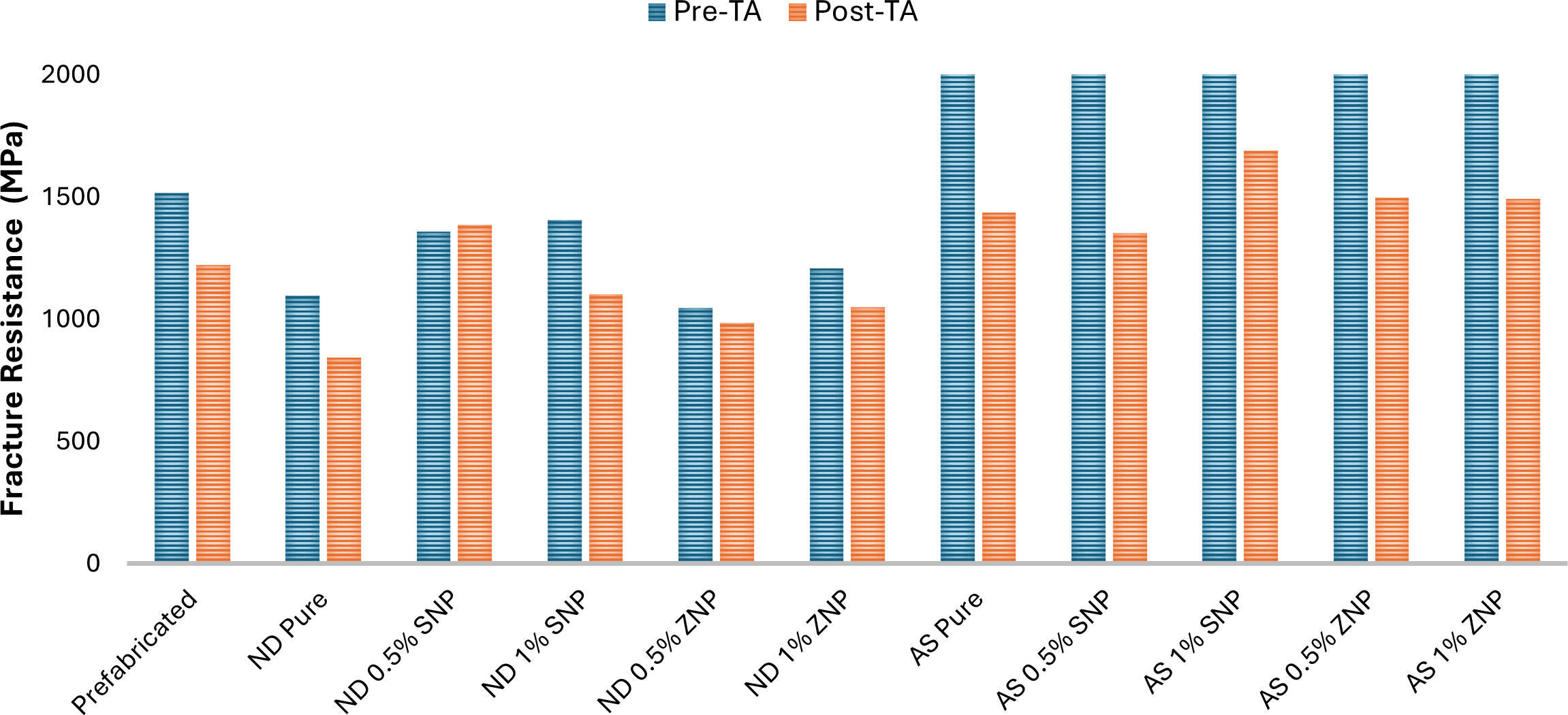
For ASIGA pre-TA, the pure resin exhibited the highest fracture resistance value (2346.6 ± 162.9 N). In comparison to pure resin, the addition of both SNPs and SNPs showed no insignificant increase in fracture resistance (p > 0.05). In between NPs groups, no significance was found between all groups (p > 0.05); however, all reinforced groups and the pure group showed a significant increase in fracture resistance compared to the prefabricated group which showed the lowest fracture resistance value (1517.1 ± 121.9 N).
For ASIGA post-TA, the results showed the same behavior before TA, no significant differences were found between groups (p > 0.05) except 0.5%SNPs which showed a decrease fracture resistance recorded the lowest value (1352.9 ± 116.4 N). In comparison to the prefabricated group, all reinforced groups showed a significant increase in fracture resistance (P < 0.001) except 0.5%SNPs (p = 0.921) and the prefabricated group showed the lowest fracture resistance value (1352.9 ± 116.4 N). In terms of the TA effect on ND, the fracture resistance was significantly decreased per respective group (P < 0.05) except for the prefabricated group (p = 0.087).
The statistical T-test analysis for fracture resistance in Table 3 highlights the material-specific differences per respective NP type and concentration. Pre-AT, ASIGA significantly showed an increase in the fracture resistance (p < 0.05). Post-TA, significant differences in the fracture resistance were observed between the ND and AS materials across all NP-incorporated groups (p < 0.05), except for the pure resin (p = 0.387) and 1 wt.% SNPs (p = 0.657) groups.
| Materials | NPs/% | NextDent (ND) | ASIGA (AS) | P |
|---|---|---|---|---|
| Pre-TA | 0 (pure) | 1097.8 ± 167.7 | 2346.6 ± 162.9 | 0.000* |
| 0.5 wt.% SNPs | 1357.5 ± 110.1 | 2192.2 ± 181.5 | 0.004* | |
| 1 wt.% SNPs | 1405.9 ± 128.4 | 2250.1 ± 168.6 | 0.001* | |
| 0.5 wt.% ZNPs | 1047.5 ± 100.6 | 2185.2 ± 117.8 | <0.001* | |
| 1 wt.% ZNPs | 1209.1 ± 140.9 | 2321.6 ± 171.3 | <0.001* | |
| Post-TA | 0 (pure) | 1221.6 ± 103.5 | 1221.6 ± 93.5 | 0.387 |
| 0.5 wt.% SNPs | 844.4 ± 136.8 | 1437.3 ± 101.7 | 0.003* | |
| 1 wt.% SNPs | 1387.1 ± 101.4 | 1352.9 ± 116.4 | 0.657 | |
| 0.5 wt.% ZNPs | 1102.7 ± 114.8 | 1691.3 ± 110.8 | <0.001* | |
| 1 wt.% ZNPs | 984.5 ± 81.9 | 1498.6 ± 156.3 | <0.001* |
Table 4 shows the three-way ANOVA of the three variables (NP type, material type, and impact of TA) and their interactions for the elastic modulus. The results showed a significant interaction between all variables (P < 0.05) and when the three variables were combined (P = 0.007).
| Source | Type III sum of squares | df | Mean square | F | P |
|---|---|---|---|---|---|
| Intercept | 619284785.339 | 1 | 619284785.339 | 4469.913 | <0.001* |
| concentration * TA effect | 10514215.811 | 5 | 2102843.162 | 15.178 | <0.001* |
| concentration * material | 2097194.635 | 4 | 524298.659 | 3.784 | 0.005* |
| TA effect * material | 918520.695 | 1 | 918520.695 | 6.630 | 0.011* |
| concentration * TA effect * material | 2031145.360 | 4 | 507786.340 | 3.665 | 0.007* |
| Error | 27431940.310 | 198 | 138545.153 | ||
| Total | 1013871903.958 | 220 |
The elastic modulus results are summarized in Table 5 and Figure 3. The prefabricated teeth significantly showed the highest elastic modulus values pre-TA (5002.0 ± 172.8 MPa) and post-TA (4255.5 ± 100.5 MPa). For ND pre-TA, the addition of SNPs and ZNPs significantly decreased the elastic modulus when compared with the pure resin (p < 0.001) and the lowest elastic modulus value was recorded with 1%ZNPs (1772.0 ± 128.8 MPa). In between NPs-reinforced groups per NPs type, there were no significant differences between SNPs groups (p = 0.064) as well as ZNPs groups (p = 0.072). When comparing NPs type, ZNPs showed a significant decrease in elastic modulus and the highest elastic modulus value was found with 1%SNPs (2048.2 ± 132.8 MPa) and the lowest values with 1%ZNP (1772.0 ± 128.8 MPa). For ND post-TA, in comparison to the prefabricated group, the elastic modulus of pure and NP-modified groups were significantly decreased (P < 0.001). While no significant differences between the pure and NP-modified groups (p < 0.05) and the pure group showed the highest elastic modulus value (376.6 ± 35.6 MPa). In terms of the TA effect, the elastic modulus of prefabricated, pure, and NP-modified groups was significantly decreased per respective NP type and concentration (p < 0.05).
| Materials | NPs/% | Pre-TA | Post-TA | P |
|---|---|---|---|---|
| Prefabricated | - | 5002.0 ± 172.8 | 4255.5 ± 100.5 | 0.002* |
| NextDent (ND) | 0 (pure) | 3097.5 ± 115.9 | 376.6 ± 35.6a | <0.001* |
| 0.5 wt.% SNPs | 1951.9 ±147.3a,b | 370.2 ± 36.4a | <0.001* | |
| 1 wt.% SNPs | 2048.2 ± 132.8a | 357.3 ± 31.9a | <0.001* | |
| 0.5 wt.%ZNPs | 1891.1 ±112.5b,c | 364.5 ± 35.1a | <0.001* | |
| 1 wt.% ZNPs | 1772.0 ± 128.8c | 359.8 ± 21.8a | <0.001* | |
| P | <0.001* | <0.001* | ||
| Prefabricated | - | 5002.0 ± 172.8 | 4255.5 ± 102.5 | 0.002* |
| ASIGA (AS) | 0 (pure) | 2725.2 ± 115.3a | 2364.1 ± 42.2a | <0.001* |
| 0.5 wt.% SNPs | 1951.1 ± 161.9b | 376.3 ± 25.6a | 0.001* | |
| 1 wt.% SNPs | 2608.1 ± 94.1a | 345.8 ± 91.5a | <0.001* | |
| 0.5 wt.% ZNPs | 2639.0 ± 93.0a | 362.0 ± 29.3a | <0.001* | |
| 1 wt.% ZNPs | 2201.8 ± 108.1b | 390.0 ± 25.1a | <0.001* | |
| P | <0.001* | <0.001* |
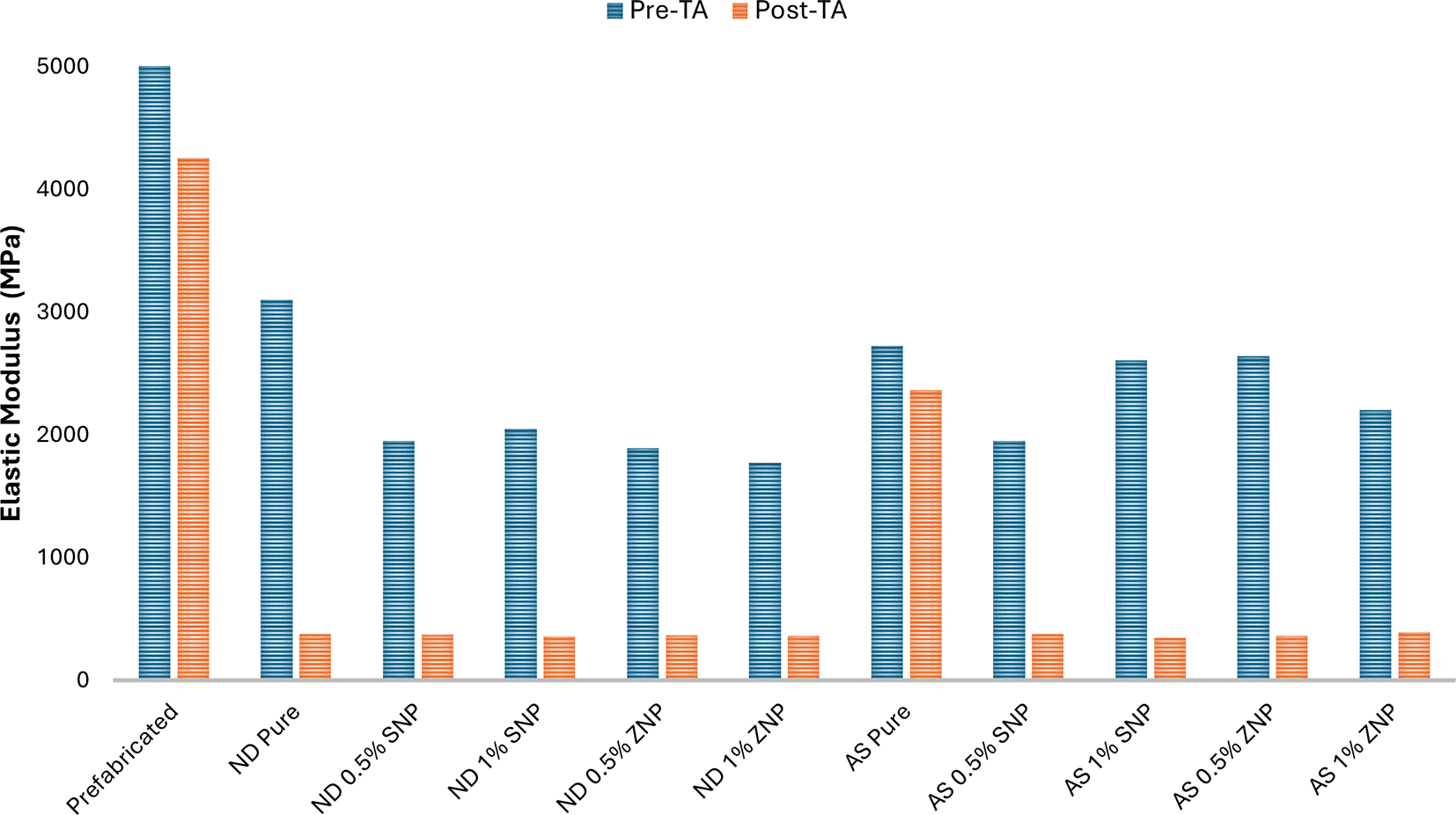
For ASIGA pre-TA, the pure resin and NP-modified groups showed a significant decrease in elastic modulus when compared with the prefabricated group (p < 0.001). In comparison to the pure group, the addition of both NPs showed a significant decrease in elastic modulus with 0.5%SNP and 1%ZNPs and 0.5%SNPs showed the lowest elastic modulus value (1951.1 ± 161.9 MPa). While no significant differences between pure resin and other NP-modified groups (pure vs. 1%SNPs and pure vs. 0.5%ZNPs, p > 0.05). in between NP-modified groups, 1%SNPs and 0.5%ZNPs significantly showed higher elastic modulus compared with 0.5%SNPs and 1%ZNPs and the highest values were recorded with 0.5%ZNPs (2639.0 ± 93.0 MPa) followed by 1%SNPs (2608.1 ± 94.1 MPa). For ASIGA post-TA, the elastic modulus was significantly decreased in pure and NP-modified groups when compared with the prefabricated group (p < 0.001). While no significant differences between the pure and NP-modified groups (P > 0.05) and 1%ZNPs showed the highest elastic modulus value (390.0 ± 25.1 MPa). In terms of the TA effect, the elastic modulus of prefabricated, pure, and NP-modified groups was significantly decreased per respective NP type and concentration (p < 0.05).
The statistical T-test analysis for elastic modulus in Table 6 highlights the material-specific differences per respective NP type and concentration. Pre-AT, pure ASIGA significantly showed a decrease in the elastic modulus compared with pure NextDent (p = 0.003). While NP-modified ASIGA groups showed a significant increase in elastic modulus except at 0.5 wt.% SNPs (p = 0.998). Post-TA and Pre-AT, pure ASIGA significantly showed a decrease in the elastic modulus compared with pure NextDent (p = 0.032*). While no significant differences in the elastic modulus were observed between the ND and AS materials across all NP-incorporated groups (p > 0.05).
| Materials | NPs/% | NextDent (ND) | ASIGA (AS) | P |
|---|---|---|---|---|
| Pre-TA | 0 (pure) | 3097.5 ± 115.9 | 2725.2 ± 115.3 | 0.003* |
| 0.5 wt.% SNPs | 1951.9 ± 147.3 | 1951.1 ± 161.9 | 0.998 | |
| 1 wt.% SNPs | 2048.2 ± 132.8 | 2608.1 ± 94.1 | <0.001* | |
| 0.5 wt.% ZNPs | 1891.1 ± 112.5 | 2639.0 ± 93.0 | <0.001* | |
| 1 wt.% ZNPs | 1772.0 ± 128.8 | 2201.8 ± 108.1 | 0.022* | |
| Post-TA | 0 (pure) | 4255.5 ± 100.5 | 2364.1 ± 42.2 | 0.032* |
| 0.5 wt.% SNPs | 376.6 ± 35.6 | 376.3 ± 25.6 | 0.531 | |
| 1 wt.% SNPs | 370.2 ± 36.4 | 345.8 ± 91.5 | 0.712 | |
| 0.5 wt.% ZNPs | 357.3 ± 31.9 | 362.0 ± 29.3 | 0.809 | |
| 1 wt.% ZNPs | 364.5 ± 35.1 | 390.0 ± 25.1 | 0.070 |
Figure 4 shows the entire range of the infrared spectrum, 4000–500 cm−1, demonstrating the bonding of AS and ND with different types of NPs at varied concentrations. The FTIR spectra of the AS and ND materials revealed characteristic bonds attributed to carbonyl groups. All the groups of specimens exhibit the C-H bond at approximately 2900 cm−1 and C=O double bond at approximately 1703 cm−1. The most significant difference between the AS and ND groups was observed in the spectral region of 1144–1034 cm−1 in the FTIR spectrum. This range corresponded to the stretching vibrations of the C–O–C (ether) functional group. The differences in this region indicate that the chemical environment or bonding characteristics of the C–O–C group vary, suggesting structural or compositional differences between the materials.
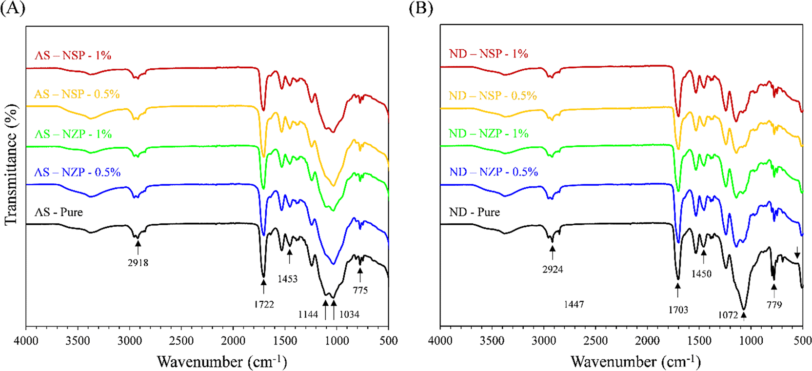
(A) Modified pure and nanoparticle-incorporated ASIGA specimens. (B) Modified pure and nanoparticle-incorporated NextDent specimens. The important bands are labelled in each set of specimens.
Figures 5–7 display the SEM images of both the occlusal and fracture surfaces. The fracture behavior of the occlusal surface showed more crack propagation in the NP-reinforced groups than in the pure-resin smooth fracture and absence of cracks. Prefabricated teeth (Figure 5) include a smooth occlusal surface and smooth fracture line. An additional note regarding 3D-printed teeth is the stepwise effect, representing printing layers on curved surfaces, such as cusp tips and cusp slopes (Figures 6 and 7). While comparing the two groups, pure ND and AS exhibited deep and multiple cracks. NP- incorporated AS are showing multiple and long propagated crack as compared to NP- incorporated ND. These SEM findings are useful while evaluating the mechanical strength of two types of specimens. Some cracks followed the line between the printed layers, which may be a reason for the weakened strength of printed teeth compared with prefabricated teeth that showed a smooth occlusal surface (absence of the staircase effect). In the cross-sectional surface analysis, most of the groups reinforced with NPs showed a rough surface with some lamellae as well as prefabricated teeth, whereas the pure teeth showed a smooth fracture side. Compared with AS groups the ND cross-sections showed higher surface roughness with several lamellar surfaces as marked with white arrows (Figure 7).
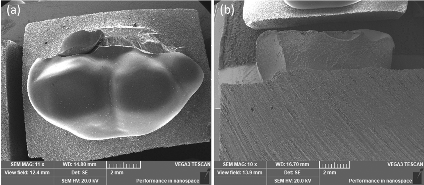
a) Occlusal surface, b) fracture surface. SEM magnifications are ×11 (a) and ×10 (b).
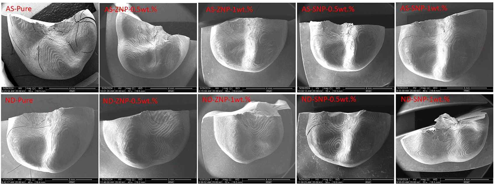
SEM magnifications from top row left to right are: ×35, ×22, ×35, ×35, ×40 and bottom row left to right are: ×40, ×40, ×32, ×38, ×33.
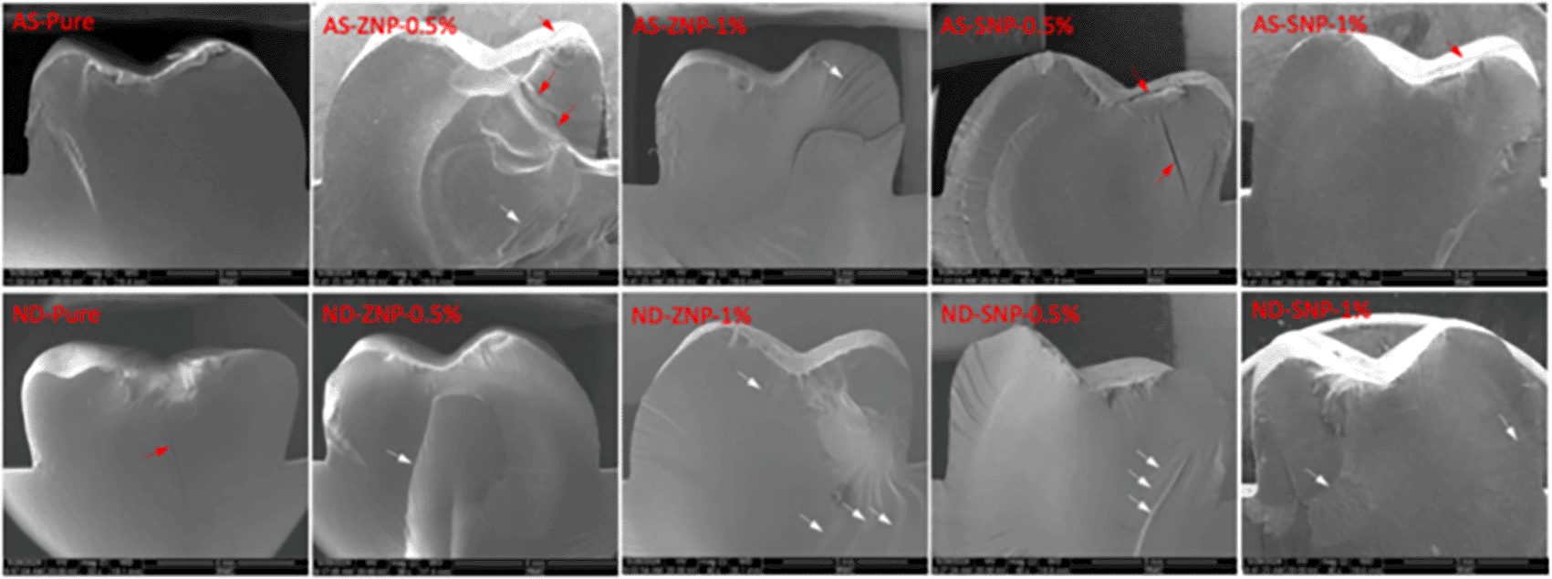
The cracks propagations are highlighted with red arrows while lamella structures are marked with white arrows. SEM magnifications top row are: ×40 and bottom row left to right are: ×33, ×39, ×40, ×40, ×40.
The aim of this study was to examine the fracture resistance and elastic modulus of 3D-printed denture-teeth pre- and post-TA with the addition of ZNPs and SNPs to the 3D-printing resins. The first and second null hypotheses were rejected because of the significant effect of NP addition and TA on the fracture resistance and elastic modulus when compared with the prefabricated teeth. For third null hypothesis, for fracture resistance, the null hypotheses of no interaction between thermal aging and material type were rejected due to the presence of a statistically significant interaction effect (p < 0.05). However, the null hypotheses related to interactions involving nanoparticle concentration were accepted, as these interactions were not statistically significant (p > 0.05). Therefore, while nanoparticle incorporation demonstrated certain effects at the main factor level, its interaction with other variables did not significantly influence the measured outcomes under the tested conditions. These findings indicate that the observed changes in material performance were primarily associated with the combined influence of thermal aging and material type, rather than synergistic interactions involving nanoparticle concentration. These findings are consistent with previous investigations that demonstrated the reinforcing effects of nanoparticle incorporation on 3D-printed dental resins. For instance, Alshamrani et al.22 and Alhotan et al.23 reported that adding silica or zirconia nanoparticles significantly enhanced the mechanical and thermal stability of photopolymer resins. In contrast, Yan et al.24 noted that higher nanoparticle concentrations could disrupt polymer cross-linking, reducing the elastic modulus. Our results align with these reports, highlighting the importance of maintaining optimal nanoparticle concentrations (0.5–1 wt%) to achieve a balance between strength and flexibility. Furthermore, compared with Gad et al.,8 who examined the strength of unmodified 3D-printed denture teeth, our findings show superior fracture resistance due to nanoparticle reinforcement, confirming the benefit of composite modification.
In this study, the incorporation of SNPs at varying concentrations into 3D-printed denture teeth increased the fracture resistance at immediate testing prior to TA in 3D-printed ND materials. This enhancement occurred by reinforcing the polymer matrix, which increased stiffness, reduced crack propagation, and boosted toughness through mechanisms such as crack deflection and energy dissipation.18–20 The findings of the present study agree with those of previous studies13,17,20 that have examined the potential benefits of integrating low concentrations of SNPs into 3D-printed interim resins to enhance their flexural properties. These studies reported that the addition of SNPs can effectively strengthen the mechanical properties of 3D-printed ND materials. In addition, previous studies have investigated the fracture resistances of 3D-printed resins containing 0.5 and 1 wt.% SNP concentrations.20 The results indicate that the addition of 1 wt.% SNP significantly improved the fracture resistances of both the ND and AS 3D-printed resins.
Based on previous studies,13,16,17 ZNPs have been recommended for incorporation into dental composites to improve their mechanical qualities, as ZNPs are well known for their remarkable hardness and capacity to reinforce resin composites. This investigation demonstrates that ZNPs improve the fracture resistance of 3D-printed ND only at 1 wt.% before TA. The improvements in fracture resistance resulting from ZNP integration align with previous studies, demonstrating the ability of ZNPs to enhance the mechanical properties of polymer-based materials.13,16 The primary mechanism proposed in previous studies is the ability of ZNPs to interact with polymer chains, distribute applied stress, and inhibit fracture formation. 16 For the ND material, the enhancement in fracture resistance at 1 wt.% concentrations is attributed to the stiffening effect of zirconia, which reinforces the polymer matrix by promoting crack deflection and energy dissipation.21,22
Before TA, the ND resin exhibited a greater increase in fracture resistance than the AS resin when the same amount of SNPs was added. While both resins benefited from SNP addition, ND significantly increased the fracture resistance with 1 wt.%SNP. In comparison to previous study, the fracture resistance of AS was reported to be 1305.7 ± 197.4.8. In contrast, significantly higher values were observed in the current study, with AS showing a fracture resistance of more than 2,000 N with and without NPs. Similarly, the fracture resistance of ND in earlier study8 was 867.8 ± 108.4, whereas in the present study, it was 1097.8 ± 167.7. This difference may be due to the stronger intrinsic mechanical qualities of AS, a potentially higher cross-linking density, and variations in matrix-nanoparticle compatibility, making it less dependent on external reinforcements than ND.12–14 This finding also supported by FTIR analysis with shifts in C–O–C regions suggesting polymer–filler bonding differences between AS and ND.
No significant differences were observed (p = 0.981) in a comparison between the prefabricated teeth and ZNP-AS. In contrast, although ZNPs were incorporated into the AS resin, the overall improvement in fracture resistance was not as significant as that in the ND. This is because of the better baseline mechanical characteristics of the AS resin, which inherently has a higher fracture resistance. Previous studies investigating the fracture strength of different dental materials have shown similar findings, in which resin materials with higher intrinsic fracture strengths demonstrate less improvement when reinforced with NPs.19–23
It is important to investigate the strength of dental materials following artificial aging as many products experience decay in their strength after aging. In this study, the effect of thermal stress on the mechanical properties of the materials revealed significant differences between the pure and NP-modified groups in their fracture resistance. In pure groups, thermal stress generally led to a substantial reduction in the fracture resistance. This reduction in fracture resistance is attributed to the development of micro-cracks and other defects arising from repeated expansion and contraction under temperature changes.24 These internal stresses lead to material weakening, making the material more prone to fracture. Owing to the addition of the NPs, the thermal stress caused thermal stability in the nanocomposite materials to some extent, thereby sustaining the properties of the materials and maintaining their elastic modulus.22–24
Following TA, the 3D-printed resins with 1 and 0.5 wt.%SNP maintained superior mechanical properties and were comparable to the performance of traditional prefabricated resins and higher than pure control in ND 3D-printed resins. In AS 3D-printed resins, 0.5 wt.% of SNP revealed higher strength than the prefabricated teeth and the pure control. These findings indicate that the incorporation of an optimal amount of 0.5 wt.% concentrations can enhance the ability of 3D-printed interim restorations from the ASIGA and NextDent manufacturers to endure thermal stresses in the oral environment, positioning them as a feasible alternative to conventional prefabricated resins.23 These findings suggest that while developing nanocomposite 3D-printed resins for dental applications, attention must be given to the concentration of incorporated ZrO2 nanoparticles, as higher levels may compromise color stability following thermal aging and beverage exposure, potentially affecting both esthetic durability and clinical performance.21,25 Such findings agree with previous studies indicating that the addition of SNP increased the fracture resistances of 3D-printed resins even after TA.17,20 After TA in the ZNPs groups, the incorporation of 1 and 0.5 wt.% of ZNPs in ND 3D-printed resin was associated with higher fracture resistance than pure control but not prefabricated teeth. While in AS 3D-printed resins, ZNPs incorporation did not offer additional strength following TA compared to pure control, but the fracture resistance was higher than prefabricated teeth. Such findings may suggest that NPs in general as reinforcing particles could be preferred to improve the strength of dental materials. The results found in this study may indicate that the incorporated NPs were more effective in maintaining the fracture resistance of the 3D-printed resins when compared to the pure control and prefabricated teeth.
For the elastic modulus, all the groups experienced significant reduction in their elastic modulus pre-TA compared to the prefabricated teeth, suggesting that the addition of NPs may alter the stiffness of the polymer network. The highest elastic modulus in the ND control group (3097.5 ± 115.9 MPa) indicates that the base resin formulation provides higher rigidity, which is reduced upon NP incorporation.12,22 This decline aligns with the findings who reported that, while ZNPs enhanced flexural strength, excessive concentrations may disrupt polymer cross-linking, leading to reduced stiffness. Also, there is frequently a nonlinear relationship between the mechanical properties of composite materials and their filler content.22 Such an explanation could be applied as well when the elastic modulus values of the SNPs groups are observed. However, our findings contraindicate other studies showing that the addition of 1 wt.% SNP significantly improved the elastic modulus of 3D-printed resins compared with the control groups without the NPs.17,20 This may suggest that other parameters could affect how NPs may influence the elastic modulus of dental materials. This study revealed that TA significantly reduced the elastic modulus for pure control and experimental groups of 3D-printed resins, and prefabricated teeth. This significant reduction is mainly related to the 3D-printed resins rather than the incorporated NPs. The significant reduction in elastic modulus observed after thermal aging can be attributed to several material-related mechanisms. The reduction in elastic modulus observed after thermal aging can be attributed to several degradation mechanisms affecting resin-based dental materials. Exposure to a moist environment and repeated thermal fluctuations promotes water diffusion into the polymer matrix, leading to plasticization of the resin network and a consequent reduction in stiffness.26 Water molecules penetrate the polymer structure and act as internal plasticizers, weakening intermolecular forces and increasing chain mobility, which ultimately lowers the elastic modulus of the material. In addition, hydrolytic degradation and swelling of the resin matrix may generate stresses at the filler–matrix interface, facilitating filler debonding or microstructural deterioration, which further compromises the mechanical integrity of the material.24,26,27 It has been widely reported that artificial aging methods, such as thermocycling and long-term water immersion can significantly alter the mechanical properties of dental resins, including reductions in elastic modulus due to these physicochemical changes. Moreover, water sorption has been shown to negatively affect the mechanical behaviour of denture polymers by reducing their stiffness and structural stability over time.26,27 In summary, the repeated thermal cycling induces microstructural stress and hydrolytic degradation within the resin matrix, leading to plasticization and the gradual breakdown of polymer cross-links.24 Similar findings were reported by Yan et al.,24 and Alhotan et al.,23 who observed that prolonged exposure to alternating hot and cold temperatures compromises the molecular integrity of polymer-based materials. In addition, the incorporation of nanoparticles at higher concentrations may interfere with polymer chain mobility and cross-linking efficiency, resulting in a more brittle and less elastic network.22 These findings suggest that both intrinsic polymer degradation and filler–matrix interactions contribute to the observed decline in stiffness after aging, highlighting the need for further optimization of nanoparticle content to balance strength and flexibility.
Another factor that could affect the strength of 3D printed resin within the fabrication procedures is the post-polymerization conditions.28–30 Post-polymerization conditions are critical determinants of the final physicochemical and mechanical properties of photopolymerized dental resins.29 Parameters such as light intensity, exposure duration, wavelength, and post-curing temperature significantly influence the degree of conversion (DC) and cross-link density of the polymer network.29–31 An insufficient or suboptimal post-curing protocol may result in a lower DC and higher residual monomer content, which can compromise the stiffness and long-term mechanical stability of the material. In additively manufactured dental resins, variations in manufacturer-recommended post-curing protocols between materials may therefore contribute to differences in their mechanical behaviour. Furthermore, materials with lower cross-link density are more susceptible to water sorption and hydrolytic degradation during thermal aging, which can lead to plasticization of the polymer matrix and a consequent reduction in elastic modulus.29–31 Accordingly, optimized post-curing procedures improve polymerization efficiency and mechanical performance of 3D-printed resins, whereas inadequate post-polymerization may accelerate degradation and reduction in stiffness after aging.29,31,32 Therefore, differences in post-curing protocols between the evaluated materials may represent a potential confounding factor affecting the observed changes in elastic modulus following thermal aging.
In this study, two fracture modes are identified: brittle and ductile. The fracture mode serves as a guide for the material strength and microstructural properties.8,9 According to SEM findings, the fracture resistance of denture teeth with a ductile fracture pattern before breakage was greater than that of teeth without this pattern, such as in the AS group. The weakened structure of the teeth, which enables them to support their weight for an extended period, appears to be the cause of such outcomes.8 Notably, the NextDent groups exhibited more pronounced structural degradation and surface defects following thermal cycling and staining, indicating lower resistance to aging compared to other tested resins.25 As the material ages or is subjected to elevated temperatures, its degradation rate significantly increases. This accelerated deterioration is often due to thermal-oxidative processes and molecular changes within the polymer matrix. The primary consequence is a progressive weakening of the crucial interfacial bond between the reinforcing fibers and the surrounding matrix. This compromised adhesion leads to a shift in failure mode, where fibers tend to pull out effortlessly from the matrix rather than remaining firmly anchored and fracturing within the material.8,9 This phenomenon was empirically confirmed by microscopic observations revealing smooth fiber surfaces, notably lacking the typical embedded nanoparticles or matrix material. Such an appearance is a clear indicator that the interface between the fiber and the matrix has catastrophically failed, suggesting a loss of chemical bonding, mechanical interlocking, and overall load transfer capability at the critical interface.13 Similar to the findings of the current study, Chung et al.,9 showed that quasi-plastic materials exhibited a more gradual loss of strength than stiff materials. Furthermore, Gad et al., 8 illustrated that the scattered fractures in AS demonstrated that these materials have a stronger fracture resistance, which is further corroborated by their higher strengths. However, the ND materials showed a fracture mode that was characterized by an early failure dominated by brittle fractures and a lack of dispersed fractures. This implies that the strength threshold of the ND material is lower, resulting in early breaking without the progressive deformation observed in stronger quasi-plastic materials. The existence of brittle fracture modes in the ND material emphasizes the necessity of strengthening its mechanical characteristics to increase its resistance to fractures.
In general, SNPs demonstrated greater strength in ND than ZNPs, but both NPs (SNPs and ZNPs) had similar ASIGA values per concentration. This highlighted the significance of NP type selection and the type of resin to be reinforced. Based on the findings of this study, the use of the investigated nanocomposites in clinical applications has both potential benefits and limitations. The practical importance of this project lies in its potential to enhance the durability and lifetime of 3D-printed denture teeth. As this is the one of primary project investigated 3D printed nanocomposite resin for denture teeth, the results should be interpreted with caution. The observed improvements in fracture resistance suggest that these nanocomposites may reduce the risk of denture fractures in high-stress areas. However, the decline in the elastic modulus post-TA indicates potential limitations in structural stiffness and durability over time. Therefore, further optimization is recommended to mitigate the effects of TA.
The findings of this study suggest several potential clinical applications for nanocomposite 3D-printed resins in removable prosthodontics. The enhanced fracture resistance of the nanoparticle-reinforced materials—particularly those containing 0.5–1 wt.% SNP or ZNP indicates their suitability for fabricating denture teeth in situations where high occlusal loads are expected. Such cases include complete dentures opposing natural dentition or implant-supported overdentures, where conventional acrylic teeth are more prone to fracture and wear. Additionally, these reinforced 3D-printed resins could be beneficial for partial denture prostheses or interim restorations that require improved mechanical strength and dimensional stability over time. The lower elastic modulus observed, may contribute to a more favorable stress distribution under masticatory forces, potentially reducing the risk of debonding or microfracture at the tooth–base interface. From a clinical standpoint, the combination of digital fabrication precision and improved material durability positions these nanocomposite resins as a promising option for custom-made prostheses, particularly when individualized esthetics and load-bearing performance are priorities. Further in vivo studies are recommended to evaluate long-term outcomes, wear behavior, and bonding integrity under functional conditions.
Despite the important findings of this study, some limitations exist. First, the study focused solely on two types of NPs and did not investigate additional potentially helpful NPs that may improve the mechanical characteristics of 3D-printed materials. Second, the study evaluated a narrow range of NP concentrations, which may not have captured the complete range of interactions between the NPs and polymer matrix. Furthermore, the long-term impacts of environmental elements such as saliva and oral-temperature fluctuations were not assessed, which may affect the longevity of materials in real-world clinical settings. Besides, the absence of long-term aging and fatigue testing in this study presents significant limitations. Without long-term aging tests, critical data on the materials’ performance over extended periods is missing, which can lead to an overestimation of their lifespan and reliability. Additionally, fatigue testing is essential for identifying failure modes that may only emerge under repeated stress. Therefore, further investigations on the effects of different NPs with different percentages added to different tooth resins are required. In addition, performing all tests under similar oral conditions and environments. At the printing-technology level, further research is needed to fully understand the synergistic interactions between the NP content, 3D-printing parameters, and fracture resistance of these materials in dental applications.
In conclusion, this study demonstrates that the incorporation of ZNPs and SNPs enhances the fracture resistance of the tested materials. TA significantly decreased the fracture resistances and elastic moduli of most of the NP-incorporated groups, except at certain NP concentrations in both materials. AS exhibited the most favorable fracture resistance profile under the tested conditions, suggesting the need for further in vivo investigation. Further optimization is recommended to mitigate the effects of TA.
| Views | Downloads | |
|---|---|---|
| F1000Research | - | - |
|
PubMed Central
Data from PMC are received and updated monthly.
|
- | - |
Provide sufficient details of any financial or non-financial competing interests to enable users to assess whether your comments might lead a reasonable person to question your impartiality. Consider the following examples, but note that this is not an exhaustive list:
Sign up for content alerts and receive a weekly or monthly email with all newly published articles
Already registered? Sign in
The email address should be the one you originally registered with F1000.
You registered with F1000 via Google, so we cannot reset your password.
To sign in, please click here.
If you still need help with your Google account password, please click here.
You registered with F1000 via Facebook, so we cannot reset your password.
To sign in, please click here.
If you still need help with your Facebook account password, please click here.
If your email address is registered with us, we will email you instructions to reset your password.
If you think you should have received this email but it has not arrived, please check your spam filters and/or contact for further assistance.

Comments on this article Comments (0)