Keywords
O-RADS, MRI, ADC, adnexal mass, gynecologic oncology, radiology, diffusion imaging.
This article is included in the Oncology gateway.
Ovarian tumors remain one of the most lethal gynecological malignancies due to delayed diagnosis. Differentiating benign from malignant adnexal masses is crucial to guide clinical management. While ultrasound is the first-line imaging modality, its limitations in certain indeterminate cases justify the use of MRI, particularly through the O-RADS classification (Ovarian-Adnexal Reporting and Data System). However, challenges remain in interpreting solid components and enhancement curves.
This retrospective study included 89 patients with 116 adnexal masses deemed indeterminate on ultrasound. All patients underwent pelvic MRI using a standardized protocol, including diffusion-weighted imaging (DWI) and ADC mapping. Adnexal masses were classified according to O-RADS, and ADC values were used to refine classifications between categories 3–4 and 4–5. Final diagnoses were confirmed by histopathological analysis after surgical resection.
Integration of ADC values improved diagnostic accuracy. Six masses initially classified as O-RADS 3 were upgraded to O-RADS 4 based on ADC cut-offs, and 18 O-RADS 4 masses were reclassified as O-RADS 5. Additionally, eight lesions initially classified as O-RADS 4 were reclassified to O-RADS 3. The integrated O-RADS/ADC strategy demonstrated excellent diagnostic performance, with sensitivity reaching 100%, specificity of 95.1%, positive predictive value (PPV) of 97.4%, and negative predictive value (NPV) of 100%. The diagnostic accuracy, calculated using Youden’s index, was 0.95. Furthermore, the use of non-contrast-enhanced MRI sequences (T2-weighted imaging, DWI, and ADC mapping) alone also provided robust diagnostic performance, achieving a sensitivity of 98.67% and a specificity of 90.48%. Conclusions:
Incorporating ADC values into the O-RADS classification enhances the diagnostic performance of MRI in evaluating adnexal masses. This refined approach enables more accurate stratification of malignancy risk, improves clinical decision-making, and may help avoid unnecessary surgery, particularly in women where fertility preservation is a concern.
O-RADS, MRI, ADC, adnexal mass, gynecologic oncology, radiology, diffusion imaging.
Malignant ovarian tumors rank sixth in frequency and fifth in mortality among malignant neoplasms in women.1 Most adnexal masses are benign. However, when faced with an adnexal mass, it is crucial to rule out malignancy through meticulous clinical examination and appropriate radiological imaging.
Transabdominal and transvaginal pelvic ultrasound is the first-line examination for characterizing adnexal masses. Despite the application of International Ovarian Tumor Analysis (IOTA) group criteria, ultrasound examination continues to present limitations, with approximately 18–31% of ovarian masses classified as indeterminate.2,3 MRI is the imaging modality of choice for characterizing ultrasound-indeterminate adnexal masses. The Ovarian-Adnexal Reporting and Data System (O-RADS), recently validated by the American College of Radiology, has high positive and negative predictive values for malignancy. It enables the reclassification of adnexal masses into benign, indeterminate-risk, and malignant categories, thereby guiding therapeutic management.4
However, a 2020 study revealed that 9.2% of adnexal masses were misclassified, primarily due to errors in interpreting the solid component and enhancement curve analysis.5 Additionally, the classification presents certain limitations.
The first constraint lies in the inability to obtain an enhancement curve if the MRI protocol is not properly followed, preventing classification into O-RADS categories 3, 4, and 5. The second limitation concerns the difficulty in distinguishing between intermediate-risk and high-risk enhancement curves in patients who have undergone hysterectomy.6
Several studies have demonstrated the value of the apparent diffusion coefficient (ADC) in improving tumor characterization by establishing a threshold value (cut-off ). Based on the work of Lucia Manganaro,6 a modification of the O-RADS classification was proposed, integrating threshold values obtained from ADC mapping.
The objective of our study is to enhance the performance of the O-RADS classification by incorporating diffusion-weighted imaging (DWI) and ADC values within the solid tissue portion of adnexal masses classified as O-RADS 3, 4, and 5.
A cross-sectional, single-center study with retrospective data collection was conducted at the Department of Diagnostic and Interventional Radiology, CHU La Rabta, over a period of 15 months from January 2022 to April 2024.
This study was conducted in accordance with the STARD (Standards for Reporting Diagnostic Accuracy Studies) guidelines to ensure comprehensive reporting of diagnostic accuracy.
This retrospective study was approved by the local Ethics Committee of the Maternity and Neonatology Center of Tunis (approval number 15/2022) on January 1, 2022.
Patients who underwent pelvic MRI for the characterization of an adnexal mass in our department were referred from the Department of Gynecology-Obstetrics C of the Maternity and Neonatology Center of Tunis (CMNT) after ultrasound revealed an indeterminate ovarian mass according to IOTA criteria7 (Appendix 1).
In addition to clinical examination, all patients underwent pelvic ultrasound, serum tumor marker assays, and pelvic MRI for adnexal mass characterization performed in our department.
Patients who underwent preoperative pelvic MRI using a standardized imaging protocol, followed by surgical resection of ovarian masses at the Gynecology C Department of the Center for Maternity and Neonatology of Tunis (CMNT), were included. Histopathological examination of the specimens was performed by the Pathology Department. Only cases where the time interval between MRI and surgical resection was less than one year were considered for inclusion.
Exclusion criteria comprised cases with non-interpretable MRI due to artifacts, patients with a history of hysterectomy, and ovarian masses classified as O-RADS 3 without a solid tissue component. Exclusion criteria included adnexal masses containing fat, hemorrhagic or fibrous material without enhancing solid tissue, as these were considered benign. Additionally, patients presenting with acute symptoms at the time of MRI acquisition and those without an available pathology report were excluded from the study. This study was conducted following the STARD (Standards for Reporting Diagnostic Accuracy Studies) guidelines to ensure comprehensive and reproducible reporting of diagnostic performance.
The sample size was determined based on the availability of cases meeting the inclusion criteria during the study period. No formal sample size calculation was performed.
MRI examinations were performed using a General Electric Healthcare 1.5 Tesla Signa Artist MRI scanner.
Appropriate patient preparation was ensured before the examination:
• Fasting for 4–6 hours was recommended for most patients.
• The bladder was maintained in a semi-filled state during the scan.
• A phased-array surface coil was used to optimize imaging quality.
• No antispasmodics were available to reduce motion artifacts caused by bowel peristalsis.
A standardized MRI protocol was implemented according to the recommendations of the European Society of Urogenital Radiology ( Table I).
All MRI examinations were interpreted by two radiologists—one resident and one senior—using dedicated post-processing consoles. They were blinded to histopathological results. Diagnostic assessments were conducted collegially to ensure consistency, and in cases of discordance, the final classification was determined through consensus. No formal inter-reader or intra-reader reproducibility analysis was performed. All adnexal masses were classified according to the O-RADS MRI classification system, published in 2020, based on the lexicon established by the American College of Radiology (ACR).
After confirming the ovarian origin of the mass and performing a comprehensive semiological analysis of the entire lesion, ovarian tumors were categorized using the O-RADS classification.5
Two radiologists, one resident and one senior, analyzed the ADC mapping obtained from diffusion-weighted imaging (DWI) sequences.
• A lesion was classified as benign if it exhibited a hyperintense solid component on ADC mapping and a hypointense signal on high b-value DWI.
• A lesion was classified as malignant if it presented a hypointense solid component on ADC mapping and hyperintensity on high b-value DWI.
• Fibrous lesions were characterized by low signal intensity on DWI, reduced ADC values, and a distinctly hypointense appearance on T2-weighted sequences, indicating their benign nature.
• Adnexal masses containing fat, hemorrhagic, or fibrous components, without evidence of enhancing solid tissue, were considered benign and excluded from the ADC analysis ( Table II).
ROI (Region of Interest) Placement criteria:
• Excluding necrotic areas, hemorrhagic zones, fatty regions, and susceptibility artifacts.
• Avoiding adjacent structures.
• Diameter > 5 mm.
• If multiple solid components were present, 4 to 6 ROIs were placed, and the ROI with the lowest ADC value was recorded.
For anatomical reference, T2-weighted images and dynamic contrast-enhanced (DCE) sequences following gadolinium injection were used.
The histological diagnosis, considered the gold standard, was established after complete surgical excision. Lesions were classified as benign, borderline, or malignant.8,9 The interval between MRI examination and histopathological analysis was typically less than one month. No clinical interventions were performed between imaging and surgical resection.
The pathology reports provided the following information:
The statistical analysis was conducted using SPSS® software (version 26.0). Descriptive statistics for categorical variables were presented as frequencies and percentages, while continuous variables were summarized using means, medians, standard deviations, and extreme values. Comparisons between categorical variables were performed using Pearson’s chi-square test or Fisher’s exact test, depending on the sample size and expected frequencies.
The diagnostic performance of the O-RADS/ADC classification system was assessed through the calculation of sensitivity, specificity, positive predictive value (PPV), negative predictive value (NPV), and likelihood ratios (LRs). These metrics were complemented by estimates of 95% confidence intervals (CIs) using the Clopper-Pearson exact method for proportions.
Malignancy risk was calculated for each O-RADS category, and an ADC threshold was determined to distinguish between O-RADS 3–4 and O-RADS 4–5 categories. To establish the most appropriate cutoff for differentiating benign from malignant lesions, a Receiver Operating Characteristic (ROC) curve analysis was performed. The area under the curve (AUC) was calculated with 95% CIs to evaluate the discriminative power of the diagnostic model. Youden’s index was also determined to identify the optimal threshold.
In addition, the diagnostic performance of non-contrast-enhanced MRI sequences—specifically T2-weighted imaging, diffusion-weighted imaging (DWI), and ADC mapping—was assessed independently to evaluate their potential for accurate lesion characterization.
A total of 140 patients with ultrasound-indeterminate adnexal masses (according to IOTA simple rules) underwent pelvic MRI for lesion characterization.
A total of 51 patients were excluded.
Ultimately, between July 1, 2021, and April 1, 2024, we included 89 patients with 116 ovarian masses.
26 patients presented bilateral adnexal masses ( Figure 1).
The analysis of patient history and clinical examination revealed that 44% of patients were postmenopausal. The median age of the study population was 45 years, with a mean age of 46 years and extremes ranging from 16 to 89 years. Half of the study population was aged between 34 and 59 years. A history of pelvic surgery was reported in 7% of cases, while 6% had a previous ovarian tumor. No patients carried a BRCA1 or BRCA2 mutation, nor had a history of ovarian stimulation. Regarding clinical symptoms, pelvic pain was the most frequently reported complaint, affecting 69% of patients. Abnormal uterine bleeding was observed in 14% of cases, while 12% presented with abdominal distension or a general deterioration in health. Urinary and digestive symptoms were reported in 8% and 4% of patients, respectively. On physical examination, a pelvic mass was palpable in 41% of cases, and lymphadenopathy was detected in 20% of patients.
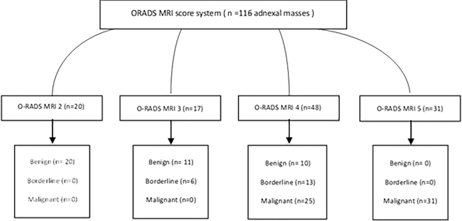
Ovarian masses were classified according to the O-RADS lexicon. Among the 116 adnexal masses analyzed, 20 were classified as O-RADS 2, 17 as O-RADS 3, 48 as O-RADS 4, and 31 as O-RADS 5. Histopathological examination revealed that 35% of the lesions were benign, 16% were classified as borderline, and 48% were malignant (Table III).
| PPV O-RADS | PPV O-RADS/ADC | |
|---|---|---|
| O-RADS 3 | 35% | 0% |
| O-RADS 4 | 79% | 92,9% |
| O-RADS 5 | 100% | 100% |
In the O-RADS 2 category, histopathological examination confirmed the benign nature of all masses. In the O-RADS 3 category, 11 lesions were found to be benign, 6 were identified as borderline, and none were malignant. For the O-RADS 4 category, 10 lesions were diagnosed as benign, 13 were classified as borderline, and 25 were malignant. Finally, in the O-RADS 5 category, all masses were confirmed as malignant upon histopathological examination ( Figure 2).
This analysis enabled the calculation of the positive predictive value (PPV) and the positive likelihood ratio (PLR) for malignancy associated with each O-RADS MRI score. The PPV and PLR increased progressively with higher O-RADS categories, reaching 0% and 0 for O-RADS 2, 35% and 0.5 for O-RADS 3, 79% and 2.2 for O-RADS 4, and 100% and infinity for O-RADS 5, respectively. Overall, the O-RADS MRI classification system demonstrated a sensitivity of 92.0% and a specificity of 75.6% in characterizing adnexal masses. The PPV and negative predictive value (NPV) for malignancy were 87.3% and 83.3%, respectively, with a PLR of 3.77 and a negative likelihood ratio (NLR) of 0.11. The overall diagnostic accuracy, as measured by Youden’s index, was 0.68.
Two ROC curves were generated to determine an optimal ADC threshold between the O-RADS 3-4 and O-RADS 4-5 categories. This analysis allowed us to establish two cut-off values based on ADC mapping across the different classes. Consequently, in certain cases, it was possible to up-classify, or down-classify ovarian masses initially categorized as O-RADS 3 and 4. For O-RADS scores 3 and 4, the area under the curve (AUC) was 0.986 with an ADC cut-off value of 1.4 ( Figure 3).
For adnexal masses classified as O-RADS 3, if the ADC value was below 1.4, the lesion was upgraded to O-RADS 4. Six ovarian masses initially classified as O-RADS 3 were reclassified accordingly. Histopathological analysis confirmed these new classifications, identifying five borderline serous tumors and one borderline mucinous tumor ( Figures 4 and 5).
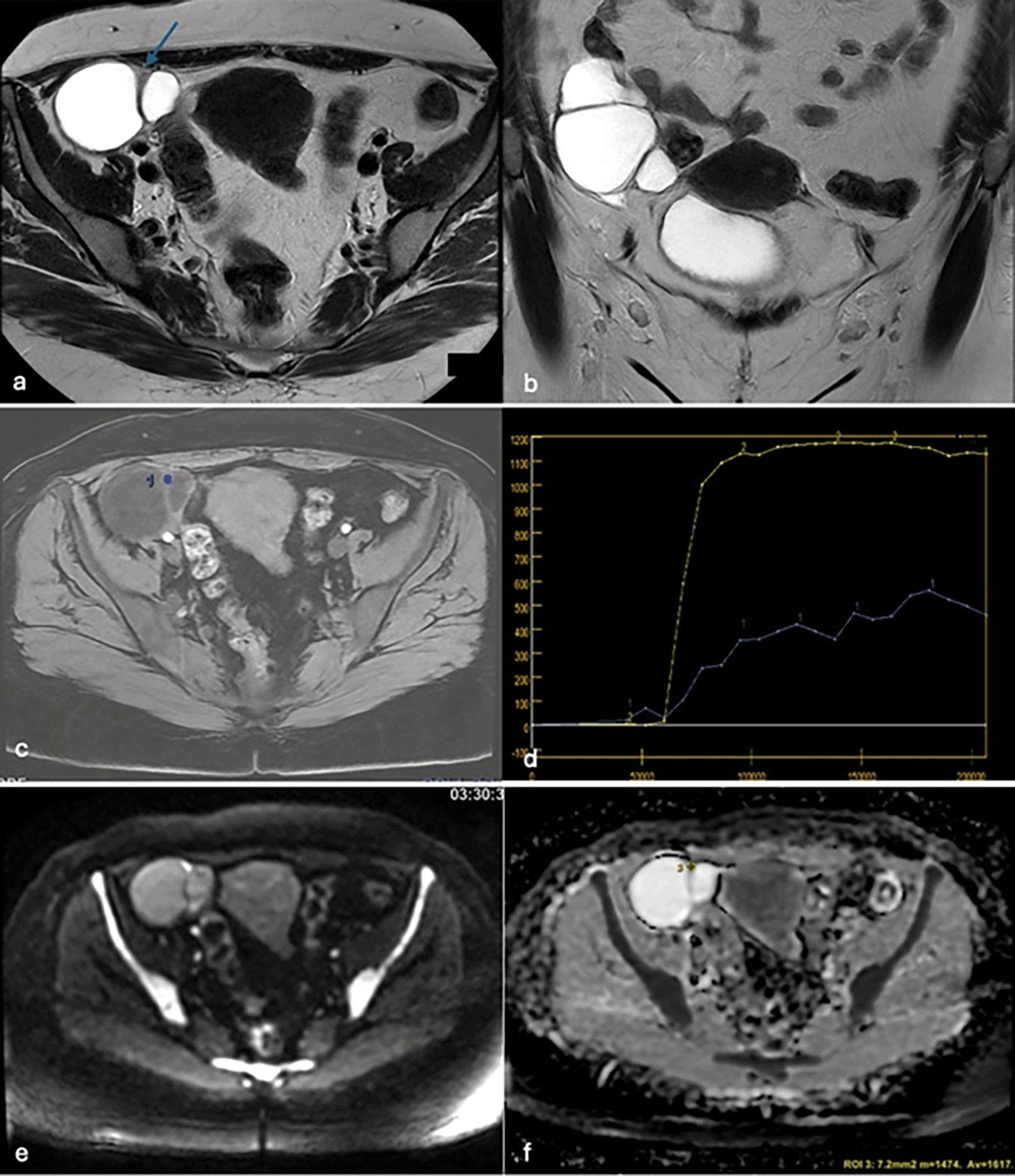
Axial (a) and coronal (b) T2-weighted images show a right adnexal cystic mass with thick septations (arrows). The perfusion sequence demonstrates a low-risk enhancement curve (c, d). DWI and ADC mapping show diffusion restriction with an ADC value of 1.617 (e, f ). The lesion remained O-RADS 3. Histology confirmed a benign serous cystadenoma.
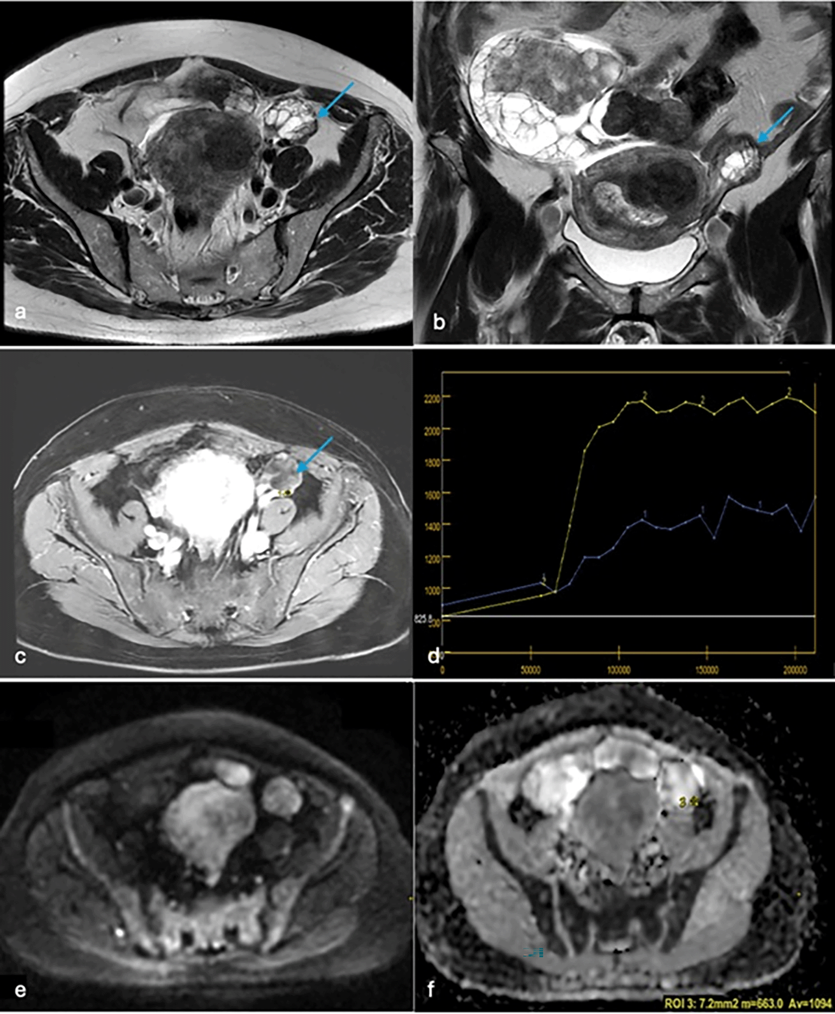
Axial (a) and coronal (b) T2-weighted images reveal two bilateral solid-cystic masses. The left mass is multilocular with irregular thickened walls (arrows). The perfusion sequence shows a low-risk enhancement curve (c, d). DWI and ADC mapping reveal diffusion restriction with an ADC value of 1.094 (e, f ). The lesion was upgraded to a higher category. Histology revealed a borderline mucinous tumor.
For adnexal masses classified as O-RADS 4, if the ADC value was greater than 1.4, the lesion was downgraded to O-RADS 3. Eight ovarian masses initially classified as O-RADS 4 were reclassified accordingly. Histopathological analysis confirmed these new classifications, identifying four benign serous cystadenomas, one seromucinous cystadenoma, one mucinous cystadenoma, one ovarian fibroma, and one ovarian fibrothecoma ( Figures 6 and 7).
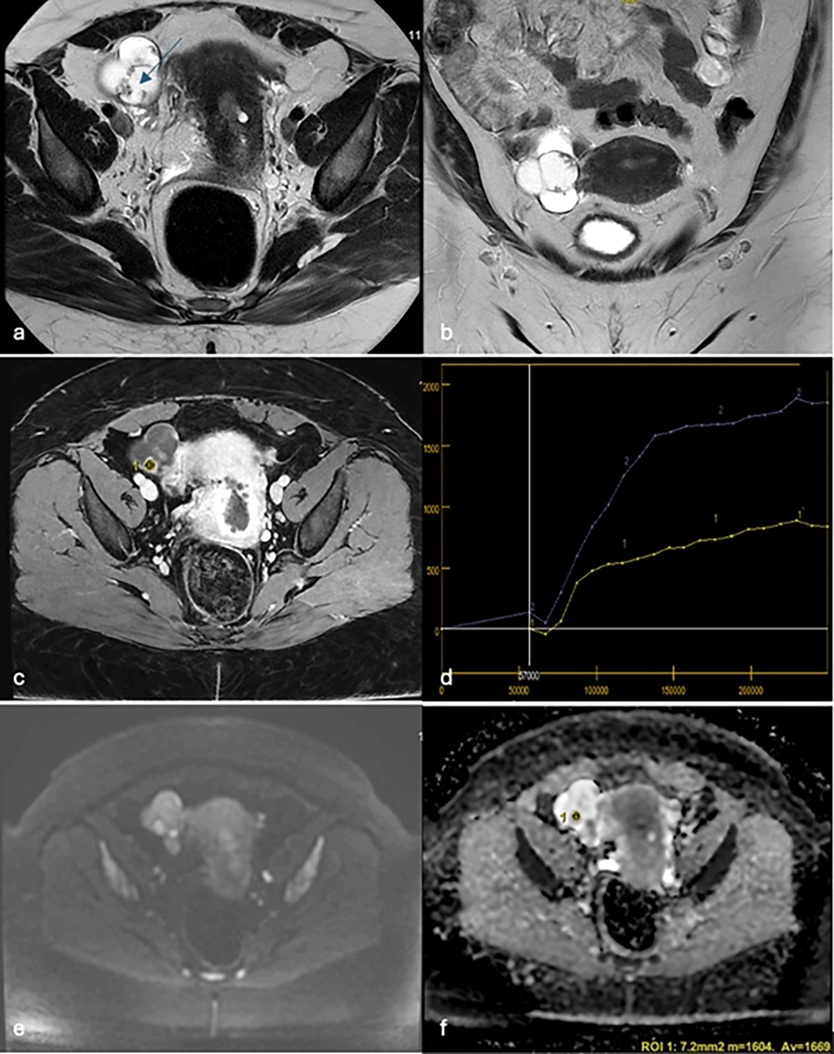
Axial (a) and coronal (b) T2-weighted images show a right solid-cystic adnexal mass with vegetations (arrows). Perfusion imaging shows an intermediate-risk enhancement curve (c, d). DWI and ADC mapping reveal diffusion restriction with an ADC of 1.669 (e, f ). Histology confirmed a benign papillary serous cystadenoma.
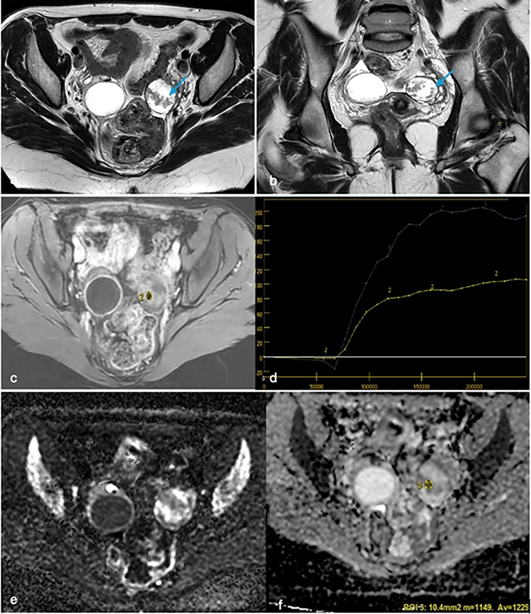
Axial (a) and coronal (b) T2-weighted images show a predominantly cystic adnexal mass with internal vegetations (arrows). The perfusion sequence demonstrates an intermediate-risk enhancement curve (c, d). DWI and ADC mapping reveal restriction with an ADC of 1.229 (e, f ). Histology confirmed a borderline serous tumor.
For adnexal masses classified as O-RADS 4-5, they remained within the same category, with histological analysis confirming the malignant nature of all lesions in this group. For O-RADS 4 and 5 scores, the area under the curve (AUC) was 0.927, with an optimal cutoff value of 0.976 ( Figure 8).
For adnexal masses classified as O-RADS 4, lesions were upgraded to O-RADS 5 if the apparent diffusion coefficient (ADC) value was below 0.976. A total of 18 adnexal masses initially classified as O-RADS 4 were reclassified. Histopathological analysis confirmed these new classifications, identifying 10 serous cystadenocarcinomas, 5 granulosa cell tumors, 1 endometriotic cystadenocarcinoma, 1 mixed endometrioid and serous cystadenocarcinoma, and 1 mucinous cystadenocarcinoma ( Figures 9 and 10).
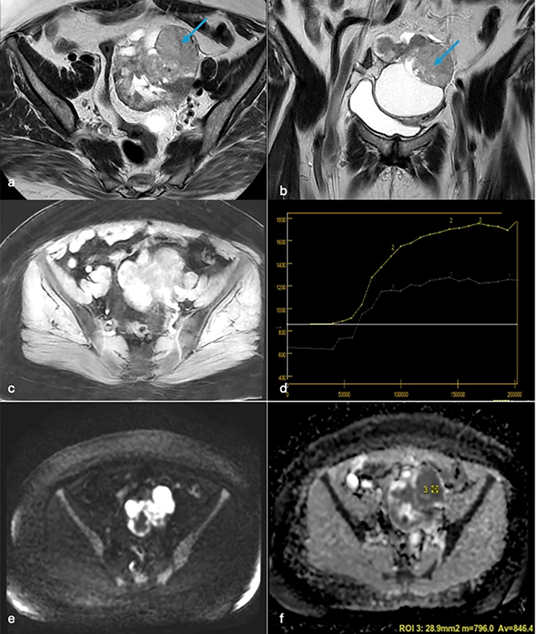
Axial (a) and coronal (b) T2-weighted images demonstrate a solid-cystic mass with tissue budding (arrows). Intermediate-risk enhancement curve on perfusion (c, d). DWI and ADC mapping show diffusion restriction with an ADC of 0.846 (e, f ). Histopathology confirmed a serous cystadenocarcinoma.
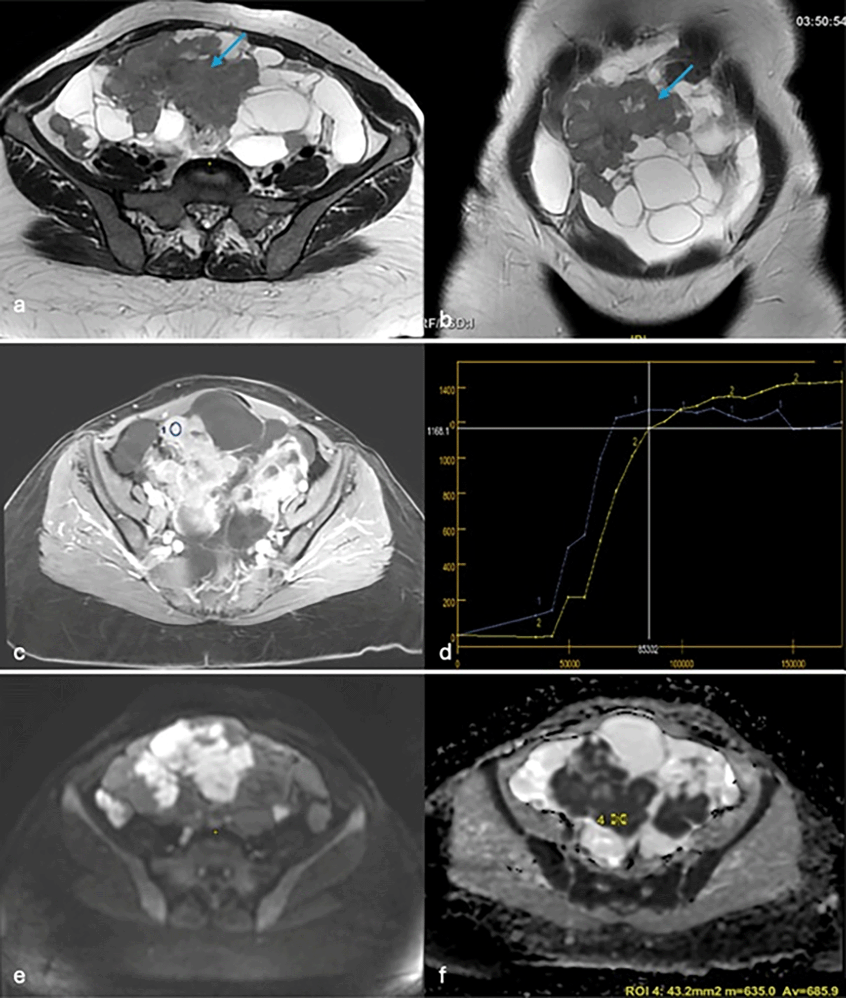
Axial (a) and coronal (b) T2-weighted images show a solid-cystic adnexal mass with a tissue bud (arrows). The perfusion sequence reveals an intermediate-risk curve (c, d). DWI and ADC mapping show diffusion restriction with an ADC value of 0.685 (e, f ). Histology confirmed a high-grade serous cystadenocarcinoma.
The introduction of two ADC cut-off values, 1.4 and 0.976, for the O-RADS 3-4 and O-RADS 4-5 categories, respectively, allowed for the reclassification of certain adnexal masses.
In summary, 24 adnexal masses were upgraded: 6 masses from category 3 to category 4 and 18 masses from category 4 to category 5. Additionally, 8 adnexal masses were downgraded from category 4 to category 3. The new results are detailed in Tables III and IV:
The final step of our study aimed to classify adnexal masses using MRI without considering enhancement curves and to correlate the obtained results with histopathological findings. This approach relied on T2-weighted sequences, diffusion-weighted imaging, and ADC map values. The method demonstrated high diagnostic performance, with sensitivity and specificity measured at 98.67% and 90.48%, respectively ( Table V).
Ovarian tumors represent one of the most severe and lethal forms of cancer affecting the female genital tract.8 Their frequently delayed diagnosis poses a significant challenge for clinicians.9 Early detection of adnexal masses is therefore essential to assess malignancy risk and determine the most appropriate therapeutic strategy.10 Magnetic Resonance Imaging (MRI) has proven to be effective in this assessment, particularly using the Ovarian-Adnexal Reporting and Data System (O-RADS), which provides a standardized risk stratification tool. However, limitations remain, notably in terms of false positives and false negatives, which may lead to diagnostic errors.5
This study has several limitations. First, the retrospective design may introduce selection bias. Second, the sample size is limited, particularly for certain O-RADS categories, which may affect the reliability of statistical comparisons. Third, the lack of formal inter-reader reproducibility analysis could influence the consistency of results. Furthermore, the ADC thresholds used were derived from the current sample and may not be universally applicable. Finally, the study was conducted at a single center, which may limit the generalizability of the findings. Interpretation of Findings
The investigators analyzed the diagnostic performance of the O-RADS classification by evaluating its ability to stratify different categories of adnexal masses. The methodological approach followed the design used in previous studies, including the EURADS 2020 validation study by Thomassin-Naggara. The findings of the present study confirm the overall effectiveness of the O-RADS classification, with a sensitivity and specificity of 92% and 75.6%, respectively.
Results for O-RADS 2 and O-RADS 5 categories were consistent with existing literature, showing malignancy rates of 0% and 100%, respectively.11 However, for O-RADS 3 (intermediate-risk) masses, the study revealed a positive predictive value (PPV) of 35%, considerably higher than values reported in earlier studies by Thomassin-Naggara,12 Aslan,13 and Hottat,14 which found PPVs of 6%, 1%, and 2%, respectively. This discrepancy may be attributed to the limited number of cases in this category and the exclusion of masses without solid components.
Regarding O-RADS 4 lesions, the study showed a malignancy PPV of 79%, exceeding the expected 49% reported by Thomassin-Naggara in 2020. Literature data on this category show significant variability, with reported PPVs ranging from 25%15 to 84%.16 Such discrepancies may result from interpretative errors, particularly in cases involving collision tumors5 or pelvic inflammatory processes.
To address these inconsistencies, recent studies have proposed the integration of Apparent Diffusion Coefficient (ADC) values derived from diffusion-weighted MRI as a complementary tool to enhance O-RADS classification accuracy. Malignant lesions typically exhibit lower ADC values than benign ones, offering an additional discriminative parameter.
For instance, the study by Nathalie A. Hottat14 demonstrated that among 42 ovarian masses classified as O-RADS 4, 72% of those with ROI-ADC < 1.7 × 10⁻³ mm2/s or WL-ADC < 2.6 × 10⁻³ mm2/s were histologically confirmed as malignant. This integration improved the sensitivity and specificity of the O-RADS system from 95.5% and 86.6% to 95.7% and 93.3%, respectively.
In the present study, using the methodology described by Lucia Manganaro’s team, the investigators identified ADC threshold values between O-RADS categories 3–4 and 4–5 at 1.4 × 10⁻³ mm2/s and 0.976 × 10⁻³ mm2/s, respectively. Incorporating these thresholds helped reduce false positives in the O-RADS 3 and 4 categories, thereby improving overall diagnostic sensitivity and specificity ( Figure 11).
These findings underscore the value of diffusion-weighted MRI in the management of adnexal masses, particularly in younger patients for whom fertility-preserving surgery may be considered.17 By combining T2-weighted sequences with diffusion-weighted imaging and ADC values, the investigators achieved a sensitivity and specificity of 98.67% and 90.48%, respectively. This imaging approach is especially relevant for patients who have undergone hysterectomy or those with contraindications to gadolinium-based contrast agents ( Figure 12).
Despite the high diagnostic performance of the O-RADS score published in 2020 and described by Thomassin-Naggara, the O-RADS/ADC system based on MRI demonstrated superior diagnostic accuracy, with improved sensitivity and specificity. This study highlighted the diagnostic value of ADC thresholds between O-RADS categories 3–4 and 4–5, contributing to a more standardized radiological assessment and more precise characterization of adnexal masses. This novel approach holds the potential to significantly enhance clinical and therapeutic management by avoiding unnecessary surgical procedures, preventing surgical menopause, and guiding appropriate treatment for malignant adnexal lesions.
This retrospective study was approved by the local Ethics Committee of maternity and neonatology center of Tunis (approval number 15/2022) on January 1, 2022. The requirement for written informed consent was waived due to the retrospective design and anonymized nature of the patient data.
All raw data underlying the study, including clinical, imaging, and histopathological findings, are provided in the dataset deposited on Zenodo:
Zenodo: Dataset of raw data from the study “O-RADS MRI Classification and ADC Values in the Characterization of Adnexal Masses”.
DOI: https://doi.org/10.5281/zenodo.1508622418
This dataset includes the following file:
- ORADS_ADC_dataset.csv: includes patient age, menopausal status, initial O-RADS score, ADC value, histology, and malignancy status.
Data are available under the terms of the Creative Commons Zero “No rights reserved” data waiver (CC0 1.0 Public domain dedication):
The following extended data are also available on Zenodo:
Zenodo: Extended data from the study “O-RADS MRI Classification and ADC Values in the Characterization of Adnexal Masses”.
DOI: https://doi.org/10.5281/zenodo.1508622319
These include:
- Fiche_Collecte_ORADS_ADC.xlsx: Structured data collection sheet
- Guide_Interpretation_ORADS_ADC.pdf: Internal interpretation guide
- STARD_Checklist.pdf: Completed checklist
- Fiche_de_renseignement.pdf: Clinical and imaging information sheet
- MRI_protocol.pdf: MRI acquisition protocol
- Courbe_ROC_Anapath_Cut_Off.xlsx, Sortie_partie_descriptive.xlsx: Analytic spreadsheets
- ZIP folders with representative MRI images: 3C1_BENIN.zip, 4C2BORDER.zip, 5.zip, etc.
Extended data are also shared under the CC0 1.0 Public Domain Dedication license.
No formal study protocol was pre-registered for this retrospective diagnostic accuracy study.
The authors wish to thank the radiology and gynecology teams at La Rabta University Hospital and the Maternity and Neonatology Center of Tunis for their valuable support in patient care and data acquisition.
| Views | Downloads | |
|---|---|---|
| F1000Research | - | - |
|
PubMed Central
Data from PMC are received and updated monthly.
|
- | - |
Is the work clearly and accurately presented and does it cite the current literature?
Yes
Is the study design appropriate and is the work technically sound?
Yes
Are sufficient details of methods and analysis provided to allow replication by others?
Yes
If applicable, is the statistical analysis and its interpretation appropriate?
Yes
Are all the source data underlying the results available to ensure full reproducibility?
Yes
Are the conclusions drawn adequately supported by the results?
Yes
Competing Interests: No competing interests were disclosed.
Reviewer Expertise: radiology and imaging
Alongside their report, reviewers assign a status to the article:
| Invited Reviewers | |
|---|---|
| 1 | |
|
Version 1 18 Sep 25 |
read |
Provide sufficient details of any financial or non-financial competing interests to enable users to assess whether your comments might lead a reasonable person to question your impartiality. Consider the following examples, but note that this is not an exhaustive list:
Sign up for content alerts and receive a weekly or monthly email with all newly published articles
Already registered? Sign in
The email address should be the one you originally registered with F1000.
You registered with F1000 via Google, so we cannot reset your password.
To sign in, please click here.
If you still need help with your Google account password, please click here.
You registered with F1000 via Facebook, so we cannot reset your password.
To sign in, please click here.
If you still need help with your Facebook account password, please click here.
If your email address is registered with us, we will email you instructions to reset your password.
If you think you should have received this email but it has not arrived, please check your spam filters and/or contact for further assistance.
Comments on this article Comments (0)