Keywords
Oesophageal foreign body, Congenital Abnormalities, difficult airway, videolaryngoscopy, rigid oesophagoscopy, interdisciplinary approach, cognitive impairment, hiatal hernia, case report.
Managing esophageal foreign bodies in cognitively impaired adults with multiple congenital anomalies presents different complex challenges, particularly when anatomical abnormalities, airway anomalies, and behavioral disturbances are present at the same time. These underlying factors can complicate both diagnosis and treatment, necessitating a highly individualized and multidisciplinary approach. This case shows the importance of a multidisciplinary approach involving videolaryngoscopic intubation, flexible endoscopy, and rigid oesophagoscopy.
A 32-year-old male with multiple congenital anomalies—including microcephaly, cognitive impairment, epilepsy, severe kyphoscoliosis, and lower limb muscle atrophy—presented with acute dysphagia. CT imaging revealed a calcified foreign body lodged within a multidiverticular oesophagus in its cervical portion, along with a sliding hiatal hernia. Due to behavioural unpredictability and risk of aspiration, awake fibreoptic intubation was considered unsafe. A modified rapid sequence induction using propofol and C-MAC® videolaryngoscopy enabled successful first-pass intubation. Flexible endoscopy was initially misdirected into a large proximal diverticulum, leading the team to proceed with rigid oesophagoscopy. The ENT team successfully retrieved multiple bone fragments. Postoperative respiratory distress was managed conservatively, without reintubation.
This case underlines the significance of interdisciplinary coordination and the limitations of flexible endoscopy in altered oesophageal anatomy. Videolaryngoscopy proved essential in managing a complex airway scenario.
Tailored, proactive strategies involving anaesthetists, ENT, and gastroenterology teams are essential in similar high-risk settings to ensure procedural success.
Oesophageal foreign body, Congenital Abnormalities, difficult airway, videolaryngoscopy, rigid oesophagoscopy, interdisciplinary approach, cognitive impairment, hiatal hernia, case report.
Foreign body ingestion is common in paediatric, elderly, and intellectually disabled populations. Management becomes particularly challenging in adults with multiple congenital anomalies, who often present with a combination of behavioural unpredictability, anatomical deformities, and impaired communication. In the present case, complex spinal and cranial malformations, distorted oesophageal anatomy with multiple diverticula, and a sliding hiatal hernia created a uniquely challenging scenario for airway control and endoscopic retrieval. These features required deviation from conventional strategies such as awake fibreoptic intubation or sedation-based endoscopy.
This report underlines the important role of close coordination between anaesthesia, gastroenterology, and ENT teams to ensure both safe airway management and successful oesophageal clearance. Moreover, it aims to share a practical and reproducible anaesthetic strategy that can be applied to similarly high-risk, non-cooperative patients.
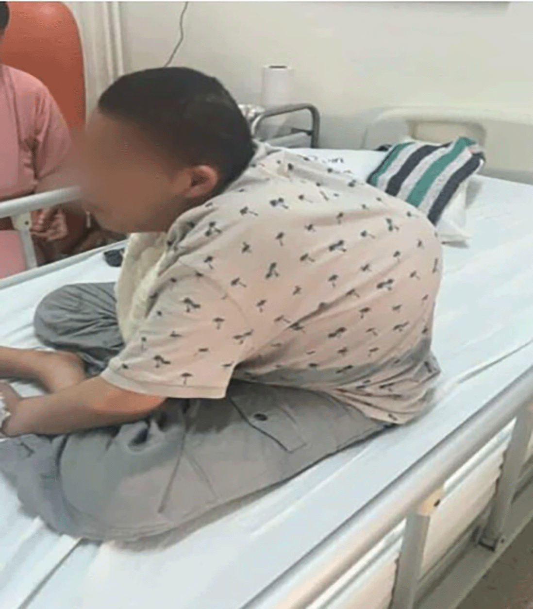
A 32-year-old patient with multiple congenital anomalies was admitted to the ENT department for acute dysphagia and hypersalivation after suspected ingestion of chicken bones. He had a medical history of microcephaly, profound cognitive delay, nonverbal status, epilepsy treated with sodium valproate, severe kyphoscoliosis, lower limb muscular atrophy necessitating the use of a wheelchair, and recurrent aspiration episodes with baseline bronchial congestion. Notably, his behavioural profile included episodes of aggressiveness and biting, mentioned explicitly by his mother. There was no relevant family history. Figure 1 illustrates the patient’s clinical morphology, including severe kyphoscoliosis, microcephaly, and generalized muscular atrophy.

At our first encounter, clinical examination revealed an alert but non-cooperative patient who moved all four limbs spontaneously to avoid being touched. Auscultation revealed bilateral rhonchi and wheezing, and oxygen saturation was 94% on room air. With the help of his brother, we managed to assess his intubation criteria. He had poor dentition, a short thyromental distance (6 cm), and severely limited neck mobility due to spinal deformity. The chest X-ray showed a severe thoracic deformity, asymmetrical lung expansion, and left-sided alveolar opacities, likely related to poor cooperation and pre-existing pulmonary pathology.
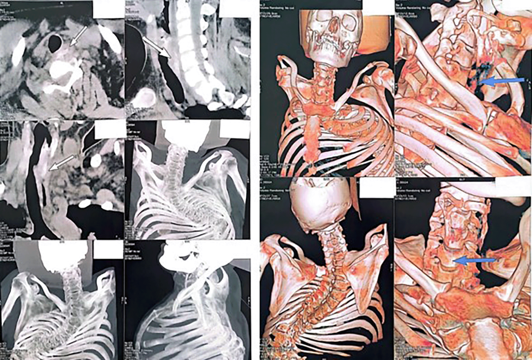
The radiology team identified in the CT imaging a high-density object consistent with a calcified foreign body lodged within the cervical portion of a multidiverticular oesophagus at the level of C7, along with oesophageal wall thickening and a sliding hiatal hernia with no evidence of strangulation. As shown in Figure 2, the calcified foreign body was located in the cervical oesophagus, just anterior to the vertebral level C7.

During our pre-anaesthetic consultation, we identified multiple predictors of difficult intubation: short thyromental distance, poor dental hygiene, cervical spine immobility, severe kyphoscoliosis, chronic airway congestion, and possible myopathy. Most importantly, the patient was not fasting due to the oesophageal impaction, and the presence of a sliding hiatal hernia further increased the risk of inhalation. These features are summarised in Table 1, which outlines the predictive criteria identified during the pre-anaesthetic assessment. Because of his severe cognitive impairment, history of agitation and biting, and full-stomach status, any form of awake intubation or sedation with spontaneous ventilation using a nasofibroscope was considered unsafe. We feared that topical anaesthetics, nasal packing, or airway manipulation under minimal sedation would provoke combative behaviour, airway trauma, or vomiting.
We solicited the presence of his brother to help minimise the patient’s agitation during positioning on the operating table. The patient appeared more at ease with a familiar figure by his side, which allowed us to proceed calmly with the induction sequence. His brother remained present during both positioning and preoxygenation. As a result, we were able to perform a full three-minute preoxygenation with a tightly fitted facemask, achieving an end-tidal oxygen concentration (EtO2) greater than 90%—a target rarely reached in patients with behavioral dysregulation.
Additionally, 8 mg of intravenous dexamethasone was administered at induction for its antiemetic properties, to attenuate bronchial inflammation given the patient’s chronic airway congestion and copious preoperative expectorations, and to prevent postoperative laryngeal pain in the absence of intra-induction opioids.
We did not have access to rocuronium or sugammadex, which narrowed our options to succinylcholine. Nevertheless, due to concerns about possible underlying myopathy, we avoided succinylcholine, and a modified rapid sequence induction (RSI) was performed using propofol only, with the patient in a semi-upright position. Beyond its hypnotic effect, propofol was selected for its inherent muscle-relaxant properties, which can favourably impact intubating conditions even without neuromuscular blockers.
Anticipating a high-risk airway scenario, the whole ENT team was present in the operating room before induction. This included two senior otolaryngologists and a dedicated scrub nurse. In addition to preparing the C-MAC® videolaryngoscope, the team ensured immediate access to a complete set of rigid direct laryngoscopy instruments and a surgical cricothyrotomy kit. This proactive setup, built on close collaboration between anaesthesia and ENT teams, was designed to minimize delays in the event of airway obstruction or intubation failure, critical in a patient with both complex anatomy and unpredictable behaviour.
The use of the C-MAC® videolaryngoscope allowed us to obtain an excellent view of the glottis with minimal airway manipulation. Under direct vision, we used an angled flexible stylet to guide a size six endotracheal tube into the trachea. The tube was secured at 20 cm at the lips, and all clinical and instrumental criteria confirmed a proper, non-selective tracheal intubation. The airway was secured safely and efficiently on the very first attempt.
After tracheal intubation, anaesthesia was maintained using a titrated infusion of propofol, supplemented by alfentanil, initiated at a low dose and adjusted as needed. Neuromuscular blockade was provided with intermittent doses of atracurium, which offers a short and predictable duration of action, with onset within 2–2.5 minutes and recovery in 20–45 minutes, allowing titration and intermittent dosing without accumulation. This regimen ensured adequate anaesthetic depth and muscle relaxation throughout the procedure, without excessive sedation or haemodynamic instability. The patient remained haemodynamically stable, well adapted to the ventilator, with satisfactory oxygen saturation, appropriate airway pressures, and no spontaneous respiratory activity, allowing safe and controlled oesophageal instrumentation.
After successful intubation, the procedure began with the intervention of the gastroenterology team. The flexible endoscope was inadvertently advanced into a large proximal diverticulum, terminating at 15 cm from the dental arch. At first, the team suspected a severe oesophageal stenosis. Then, the ENT team proceeded with rigid oesophagoscopy. This time, they successfully accessed the true oesophageal lumen and recaptured multiple fragments of chicken bone.
A second FOGD was performed to confirm the absence of residual foreign bodies. It revealed a non-strangulated hiatal hernia and a narrowed oesophagus with diffuse mucosal ulcerations, but no additional complications were noted.
We thoroughly aspirated tracheal secretions, administered salbutamol via inhalation, and waited for the return of spontaneous ventilation without assistance, eye opening on command, and full neuromuscular recovery (TOF 4/4). Then, the patient was extubated in the endoscopy room. He exhibited mild respiratory distress, including tachypnoea, sibilant rhonchi, and brief desaturation, which gradually improved with high-flow oxygen and physiotherapy. He was admitted to the ICU and received empiric antibiotics: cefotaxime, gentamicin, and metronidazole, targeting potential inhalation pneumonia.
We prescribed postoperative nebulisation with Bricanyl and Atrovent, along with intensive physiotherapy. Non-invasive ventilation could not be used due to behavioural disorders and the patient’s inability to tolerate the interface. However, he remained haemodynamically stable and did not require reintubation. His respiratory status and level of alertness gradually improved. He was then transferred to the Otorhinolaryngology department after several days. The patient’s parents expressed profound gratitude and relief upon his discharge from the intensive care unit. Nevertheless, it was essential to thoroughly inform them about the endoscopic findings and their implications, to raise awareness and help prevent similar incidents in the future.
Table 2 provides a structured summary of the main anaesthetic decisions taken during the perioperative period, along with their respective clinical justifications.
The chronological sequence of key interventions, from pre-anesthetic evaluation to postoperative monitoring, is summarized in Table 3. This timeline highlights the multidisciplinary contributions and the progression of care.
Flexible endoscopy is the standard first-line technique for upper gastrointestinal foreign body extraction, as recommended by the American Society of Gastrointestinal Endoscopy.1 Success rates can reach up to 90% when performed under optimal conditions. Nevertheless, anatomical variants such as diverticula, luminal narrowing, or altered oesophageal alignment may hamper access and limit the effectiveness of flexible endoscopy.
Sugawa et al.2 suggest switching promptly to rigid oesophagoscopy in such situations to avoid prolonged attempts and further complications. In the present case, the early flexible oesophagoscopy was misdirected into a diverticulum, prompting a transition to a rigid technique. The approach proved more relevant, as it enabled safe and complete extraction of the foreign body. The oesophageal mucosa was fragile, ulcerated, and anatomically distorted, making rigid instrumentation more suitable than flexible tools.
Furthermore, the diagnosis in our case was guided more by indirect signs—hypersalivation and behavioural changes—than by classic symptoms. Wyllie3 emphasises that patients with intellectual disabilities may present atypically, often lacking overt complaints, which can delay diagnosis.
It is commonly recommended to use awake fibreoptic intubation when complex airway management is anticipated. However, this approach is contraindicated in non-cooperative, combative, and cognitively impaired patients, as highlighted by Langford et al.4 In such cases, alternative strategies must be considered.
Videolaryngoscopy, particularly with the C-MAC® system, is increasingly preferred due to its superior glottic visualisation, minimal neck extension requirements, and reduced upper airway trauma. These advantages are well described by Lewis et al. and Apfelbaum et al.5,6 In our patient, whose anatomy and behavioural profile precluded fibreoptic intubation, the use of C-MAC® was the most appropriate and effective option.
Moreover, the 2022 ASA guidelines recommend a multi-layered airway plan when intubation is expected to be difficult, including preparation for surgical airway access.6 Our team anticipated this challenge by ensuring ENT presence at induction, readiness for cricothyrotomy, and availability of second-generation airway devices.
Weingart and Levitan7 warn against the use of sedated fibreoptic intubation in unfasted patients, citing the risk of silent aspiration and rapid desaturation. In our case, the coexistence of a hiatal hernia, baseline bronchial congestion, and behavioural unpredictability supported the decision to proceed with videolaryngoscopic intubation under general anaesthesia.
The benefit of videolaryngoscopy in patients with multiple congenital anomalies has also been demonstrated in a paediatric case series involving syndromes such as Pierre–Robin, Beckwith–Wiedemann, and Hurler, where successful intubation was achieved after failed direct attempts.8 Our experience aligns with these findings, reinforcing the role of videolaryngoscopy in syndromic airway scenarios.
In addition, a tailored pharmacological strategy was used to maximise safety and comfort. A modified rapid sequence induction with propofol alone was performed due to the unavailability of rocuronium and concerns regarding possible underlying myopathy. Although not routinely recommended, this approach is supported by literature showing that propofol at 2.5 mg/kg can, on its own, provide acceptable intubating conditions without neuromuscular blockers, with no significant haemodynamic compromise in healthy adults with normal airways.9 Despite our patient’s complex anatomy, the favourable intubation outcome—achieved without people with paralysis—suggests that in selected high-risk scenarios, the myorelaxant properties of propofol, combined with optimal conditions and videolaryngoscopy, may offer a viable alternative.
We also administered 8 mg of intravenous dexamethasone at induction, for its triple benefit: prevention of postoperative laryngeal pain in the absence of opioids,10 antiemetic effects,11 and suppression of bronchial hypersecretion, supported by its ability to downregulate MUC5AC gene expression in human airway epithelial cells.12
Anaesthesia was then maintained with titrated propofol, low-dose alfentanil and intermittent doses of atracurium, selected for its short and predictable duration of action, allowing fine titration and reducing the risk of accumulation—an important consideration in fragile or polymorbid patients.13 This regimen ensured haemodynamic stability, adequate muscle relaxation, and smooth ventilator synchrony throughout the procedure, enabling safe and controlled oesophageal instrumentation.
Finally, while most evidence comes from paediatric settings, non-pharmacological methods such as parental or caregiver presence have consistently been shown to reduce preoperative anxiety and improve cooperation during induction.14 In the present case, the presence of the patient’s brother significantly lowered agitation, enabling a calm 3-minute preoxygenation and achieving EtO2 > 90%, which would likely have been impossible without his support. This emphasizes the potential benefit of including a familiar caregiver during induction in cognitively impaired adults.
• Awake fibreoptic intubation is unsafe in non-cooperative patients with severe behavioral disorders and distorted airway anatomy.
• Videolaryngoscopy (C-MAC®) offers excellent glottic visualization with minimal cervical manipulation in syndromic patients.
• In the presence of esophageal diverticula, flexible endoscopy can fail and should be replaced early by rigid oesophagoscopy.
• Modified RSI using propofol alone can be a valid alternative when neuromuscular blockers are contraindicated.
• Inclusion of a familiar caregiver during induction can significantly improve patient cooperation and safety.
This report is limited by its single-case nature, which restricts the generalizability of the conclusions. Long-term follow-up data were not available, and some aspects of the patient’s complex condition—such as potential genetic factors or detailed pulmonary function testing—could not be fully assessed. Nevertheless, the case remains instructive as it illustrates practical strategies for airway and endoscopic management in a highly challenging clinical scenario.
This case highlights the complexity of managing esophageal foreign bodies in cognitively impaired patients with multiple congenital anomalies. Our patient’s behavioural unpredictability, severe spinal deformity, and baseline respiratory fragility rendered conventional approaches—such as awake fibreoptic intubation or delayed induction—unsafe. In this context, selecting the C-MAC® videolaryngoscope as a first-line strategy proved essential. It allowed excellent visualisation of the glottis with minimal airway manipulation and enabled a smooth, first-pass intubation despite multiple anatomical predictors of difficulty.
Beyond the technical choices, this case highlights how clinical success hinges on dynamic, real-time coordination between teams. From early radiological identification and anaesthetic planning to the dual endoscopic approach led by gastroenterology and ENT, each specialty played a vital role. The proactive presence of the ENT team—fully prepared to perform an emergency tracheotomy in case of failed ventilation or intubation—was an integral part of this anticipatory strategy. Ultimately, this case underscores that in high-risk, anatomically complex patients, especially in resource-limited settings where agents like sugammadex are unavailable, multidisciplinary preparation is the true cornerstone of safety.
Ethical approval for this case report was obtained from the Habib Thameur Hospital Ethics Committee (Tunisia) on 08 September 2025. Written informed consent was obtained from the patient’s legal guardian for participation and publication. The study complies with the Declaration of Helsinki.
Written informed consent was obtained from the patient’s legal representative for publication of this case report and all accompanying clinical images. A copy of the signed consent is available upon request.
The case presented in this report is authentic and based on the real-life management of a patient in our anaesthesia department. All clinical analysis, reasoning, and scientific content were produced independently by the authors. To enhance the clarity of the manuscript, we used ChatGPT (OpenAI) for grammar correction and linguistic refinement.
The CARE checklist supporting this case report has been deposited in Figshare: Arfaoui S, Rouaissi H, Nefzaoui S, Romdhane N, Sliti H, Jlassi H, et al. CARE checklist for C-MAC Case Report. Figshare. 2025. doi:10.6084/m9.figshare.29958554.v2.15
This project contains the following extended data:
• CARE Checklist: Completed CARE checklist supporting the case report, detailing the reporting standards adhered to.
Data are available under the terms of the Creative Commons Zero “No rights reserved” data waiver (CC0 Public domain dedication).
| Views | Downloads | |
|---|---|---|
| F1000Research | - | - |
|
PubMed Central
Data from PMC are received and updated monthly.
|
- | - |
Is the background of the case’s history and progression described in sufficient detail?
Yes
Are enough details provided of any physical examination and diagnostic tests, treatment given and outcomes?
Yes
Is sufficient discussion included of the importance of the findings and their relevance to future understanding of disease processes, diagnosis or treatment?
Yes
Is the case presented with sufficient detail to be useful for other practitioners?
Yes
References
1. Baker P, Depuydt A, Thompson J: Thyromental distance measurement – fingers don’t rule. Anaesthesia. 2009; 64 (8): 878-882 Publisher Full TextCompeting Interests: No competing interests were disclosed.
Reviewer Expertise: Anesthesiology, Airway Management
Alongside their report, reviewers assign a status to the article:
| Invited Reviewers | |
|---|---|
| 1 | |
|
Version 1 22 Sep 25 |
read |
Provide sufficient details of any financial or non-financial competing interests to enable users to assess whether your comments might lead a reasonable person to question your impartiality. Consider the following examples, but note that this is not an exhaustive list:
Sign up for content alerts and receive a weekly or monthly email with all newly published articles
Already registered? Sign in
The email address should be the one you originally registered with F1000.
You registered with F1000 via Google, so we cannot reset your password.
To sign in, please click here.
If you still need help with your Google account password, please click here.
You registered with F1000 via Facebook, so we cannot reset your password.
To sign in, please click here.
If you still need help with your Facebook account password, please click here.
If your email address is registered with us, we will email you instructions to reset your password.
If you think you should have received this email but it has not arrived, please check your spam filters and/or contact for further assistance.
Comments on this article Comments (0)